Case Report
Austin ENT Open Access. 2025; 5(1): 1020.
Fatal Misdiagnosis of Isolated Cervicofacial Myeloid Sarcoma in a Young Adult: Case Report
Amine HM¹*, Najout H², Walid A³ and Seddiki R³
1ENT Departement, Hassan II Military Teaching Hospital, Laayoune, Morocco
2Anesthesiology and Intensive Care Unit, Mohamed V Military Teaching Hospital, Rabat, Morocco
2Anesthesiology and Intensive Care Unit, Hassan II Military Teaching Hospital, Laayoune, Morocco
*Corresponding author: Hanine Mohamed Amine, ENT Departement, Hassan II Military Teaching Hospital, Laayoune, Morocco Email: hanmohami@gmail.com
Received: June 02, 2025 Accepted: June 19, 2025 Published: June 23, 2025
Abstract
Introduction: Myeloid sarcoma is a rare extramedullary manifestation of acute myeloid leukemia. Its isolated cervicofacial presentation is exceptional and often misdiagnosed.
Case Presentation: We report a case of a 19-year-old male with no significant medical history presenting with a rapidly growing cervicofacial mass initially treated as a dental abscess. Despite multiple dental interventions, there was no improvement. Histopathological examination of a biopsy specimen revealed myeloid sarcoma. Unfortunately, due to financial constraints and delayed hematological management, the patient died before initiation of chemotherapy.
Discussion: This case underscores the diagnostic challenge posed by atypical head and neck presentations of hematological malignancies. Immediate biopsy and immunophenotyping are essential. Socioeconomic barriers can have fatal consequences in such aggressive neoplasms.
Conclusion: Awareness of rare manifestations of hematological diseases is essential for early diagnosis and timely intervention.
Keywords: Cervicofacial; Myeloid sarcoma; Leukemia; Fatal prognosis
Introduction
Myeloid sarcoma (MS), also known as granulocytic sarcoma or chloroma, is a rare extramedullary tumor composed of immature myeloid cells [1]. It is most commonly associated with acute myeloid leukemia (AML), either concomitantly, during relapse, or as a harbinger of disease [2]. Isolated MS without marrow involvement is rare and may present a diagnostic dilemma, particularly in unusual locations such as the head and neck region [3].
We report a rare and fatal case of isolated cervicofacial myeloid sarcoma in a young adult, initially misdiagnosed as a dental abscess, illustrating the critical importance of early recognition and intervention.
Case Presentation
A 19-year-old male with no prior medical history presented to the Otolaryngology Department of the Military Hospital of Laayoune, Morocco, with a painful, progressively enlarging cervicofacial mass evolving over one month. The patient initially sought dental care and was treated empirically for a presumed dental abscess without improvement.
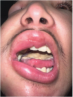
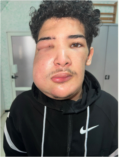
On examination, there was a firm, tender, and diffusely swollen mass predominantly involving the left cheek, extending to the periorbital region, causing complete occlusion of the left eye. Intraoral examination revealed a large, necrotic, whitish mass involving the upper vestibule and hard palate.
The patient’s general condition was deteriorated with marked asthenia and weight loss. Laboratory tests showed mild anemia but no blasts on peripheral blood smear. A biopsy was performed under local anesthesia. Histopathological examination revealed a dense infiltrate of medium-sized blastic cells with irregular nuclei. Immunohistochemistry was positive for myeloperoxidase (MPO), CD33, and CD117, confirming the diagnosis of myeloid sarcoma (Figure 1).

    Figure 1: Extensive intraoral infiltration involving the upper vestibule and
hard palate, covered by a necrotic pseudomembrane.
The patient was referred urgently to the Hematology Department for systemic chemotherapy. Unfortunately, due to lack of medical insurance and financial resources, the initiation of therapy was delayed, and the patient succumbed to his disease within two weeks after diagnosis (Figure 2).

    Figure 2: Frontal view demonstrating severe cervicofacial swelling causing
facial asymmetry and complete closure of the left eye due to tumor
infiltration.
Discussion
Myeloid sarcoma is an uncommon entity, representing about 2–8% of cases of acute myeloid leukemia [4]. It may occur at any site, but involvement of the head and neck region is rare, accounting for approximately 12–24% of cases [5]. In isolated MS, where bone marrow is initially unaffected, diagnosis becomes particularly challenging and is often delayed.
The differential diagnosis in cervicofacial swellings typically includes odontogenic infections, benign tumors, lymphomas, sarcomas, and metastatic lesions [6]. As in our patient, misdiagnosis as a dental abscess is common, especially when oral symptoms predominate.
Histological evaluation is essential but often inconclusive without immunohistochemistry. Markers such as MPO, CD68, CD33, and CD117 are critical for distinguishing MS from lymphoid malignancies and other small round blue cell tumors [7].
The prognosis of isolated MS is poor if systemic AML-type chemotherapy is not promptly initiated. Delayed treatment often leads to rapid progression to overt AML, with median survival less than 8 months in untreated patients [8]. Early systemic chemotherapy improves survival dramatically and may delay or prevent leukemic transformation [9].
Socioeconomic factors, as tragically highlighted in our case, play a major role in outcomes. Limited access to healthcare, delayed diagnosis, and inability to initiate timely chemotherapy contribute significantly to mortality, especially in resource-limited settings [10].
This case emphasizes the importance of maintaining a high index of suspicion for malignancy in atypical cervicofacial lesions, particularly when initial therapy fails. Biopsy and immunohistochemical analysis must be performed without delay.
Conclusion
Isolated cervicofacial myeloid sarcoma is an extremely rare and aggressive disease, prone to misdiagnosis. Early identification and initiation of AML-type chemotherapy are essential to improve prognosis. In developing countries, enhancing access to specialized care remains a critical need to prevent avoidable deaths.
Ethics
Ethical conditions are approved.
The Work was Reported in Accordance with the Scare Guidelines
During the preparation of this work the author used HANINE MOHAMED AMINE / ENT in order to VERIFY THE QUALITY OF WORK. After using this tool, the author reviewed and edited the content as needed and takes full responsibility for the content of the publication.
References
- Pileri SA, Ascani S, Cox MC, et al. Myeloid sarcoma: Clinico-pathologic, phenotypic and cytogenetic analysis of 92 adult patients. Leukemia. 2007; 21: 340–350.
- Avni B, Koren-Michowitz M. Myeloid sarcoma: current approach and therapeutic options. Ther Adv Hematol. 2011; 2: 309–316.
- Tsimberidou AM, Kantarjian HM, Estey E, et al. Outcome in patients with nonleukemic granulocytic sarcoma treated with chemotherapy with or without radiotherapy. Leukemia. 2003; 17: 1100–1103.
- Neiman RS, Barcos M, Berard C, et al. Granulocytic sarcoma: a clinicopathologic study of 61 biopsied cases. Cancer. 1981; 48: 1426–1437.
- Paydas S, Zorludemir S, Ergin M. Granulocytic sarcoma: 32 cases and review of the literature. Leuk Lymphoma. 2006; 47: 2527–2541.
- Audouin J, Comperat E, Le Tourneau A, Camilleri-Broet S, Molina T. Myeloid sarcoma: Clinical and morphologic features of 16 cases treated at a single institution. Ann Pathol. 2003; 23: 376–380.
- Bakst RL, Tallman MS, Douer D, Yahalom J. How I treat extramedullary acute myeloid leukemia. Blood. 2011; 118: 3785–3793.
- Yamauchi K, Yasuda M. Comparison in treatments of nonleukemic granulocytic sarcoma: report of two cases and a review of 72 cases in the literature. Cancer. 2002; 94: 1739–1746.
- Antic D, Elezovic I, Milic N, et al. Is there a “gold” standard treatment for patients with isolated myeloid sarcoma? Biomed Pharmacother. 2013; 67: 72–77.
- Velez JCQ, Petroianu A, de Faria Andrade FJ, et al. Disparities in cancer outcomes in low- and middle-income countries: A call for equity. Lancet Oncol. 2021; 22: 451–452.