
Research Article
Austin ENT Open Access. 2025; 5(1): 1021.
A Challenging Osteoma Limiting the Endoscopic Approach
Arkoubi Z*, Benjilali A, Bensaid Y, El Hafi Z, Bencheikh R, Benbouzid A and Essakalli L
Department of ENT, Head and Neck Hospital of Rabat, Morocco
*Corresponding author: Arkoubi Z, Department of ENT, Head and Neck Hospital of Rabat, Morocco Email: zakaria.arkoubi@gmail.com
Received: July 29, 2025 Accepted: August 14, 2025 Published: August 18, 2025
Summary
Osteoma is a bony benign tumor. The clinical expression is based on the size of the mass like most of the other benign masses. When it’s confined in the paranasal sinus, it could take time growing before the appearance of clinical signs. The treatment relies on surgery which can be challenging depending on the situation of the tumor. That’s the case in naso-sinusien localization, especially ethmoidal site.
Due to proximity of orbital cavities and skull base, this kind of tumors could be difficult to manage. The consistency also doesn’t help.
Frequently, it’s more solid than normal bone, which need some advanced technics to preserve the non-pathological structures.
Introduction
Most of the benign tumors has some common aspects in treatment management. The naso-sinusien localization can be challenging for surgery, but thanks to the evolution of endoscopic surgery technics, this kind of tumors are actually extremely simplified with minimizing the damages at a very low degree. The consistency of some tumors remains challenging in terms of surgical treatment.
The histopathological structure of an osteoma is quite different than a normal bone, which affects the surgical management and can be challenging to remove, even by combining endoscopic and external technics. In this paper, we are going to expose these difficulties in managing surgical treatment through a case report of fronto-orbital osteoma.
Case Presentation
50 years old man with an exophtalmia due to the development for almost 1 year of a tumefaction in the internal canthus. The patient reported also unilateral nasal obstruction and hyposmia. But the patient wasn’t annoyed, until it was complicated with orbital cellulitis. That’s what made him go to the emergency. A CT-scan shows an ossified mass localized in the left ethmoidal and frontal sinus. The tumor is extended to the homolateral orbital cavity (Figure 1, Figure 2).
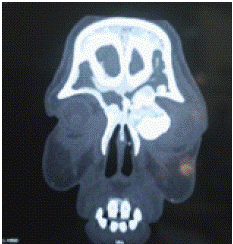
Figure 1: Coronal CT-Scan of the osteoma.
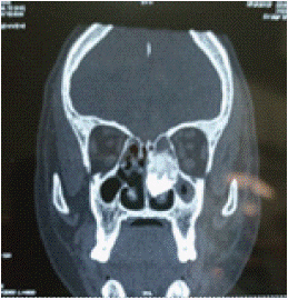
Figure 2: CT-Scan showing ethmoidal localization.
The endoscopic examination shows a solid mass in palpation covered with a normal mucosa reducing the nasal cavity.
After medical treatment of the cellulitis using antibiotics. Within 14 days, we decided to perform an endoscopic and external surgery to remove the mass (Figure 3).
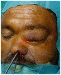
Figure 3: Surgical view.
We have started by the endoscopic technique. After realizing a middle meatotomy, and the spotting of frontal canal, we started the excision of the mass which was challenging due to the consistency of the structure. We used the endonasal burr to fragmentize the tumor (Figure 4, 5).
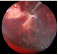
Figure 4: Endoscopic surgical view.
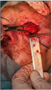
Figure 5: Surgical view of the removing of the orbital portion.
After the surgery, the symptomatology disappeared, and the left eye find its place progressively one month after surgery (Figure 6).
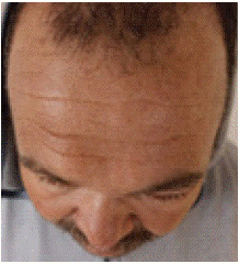
Figure 6: 1 month after surgery.
Discussion
Some tumoral lesions could be really problematic in term of surgical management referring to the site of the tumor. We usually face such situations in head and neck pathologies. The anatomy is so important and contains some precious structures that should be preserved.
The development of an osteoma could take place where even the bone is present. Most frequently the osteoma is located in the paranasal sinus [1], but still misdiagnosed for the simple reason that in the paranasal sinus it still has a room to grow easily. The osteoma generally is discovered after a radiography because it’s usually asymptomatic.
If it’s symptomatic, it means that it took time to grow and the tumor is big enough to cause functional disorder depending on its site or it’s responsible of inaesthetic deformations that make the patients consult. When it’s located and confined in the paranasal sinus, it’s often asymptomatic, and the only way to diagnosis is the imagery. Either it’s discovered after a radiography or a CT-Scan.
The osteoma can be diagnosed at any age, mostly it’s about adults between 30 and 50 years old. It occurs more often in women than men [2].
The osteoma remains unknown, but it’s supposed that traumatisms could be responsible for the development of the osteomas [1].
The osteoma is an osteogenic benign tumor by well-organized mature bone. The trigger of the construction of this mature bone is still unknown until these days. We distinguish three histological patterns of osteomas, compact or ivory osteomas, cancellous or mature osteomas and a mixed type [3].
The treatment consists on the surgery which have to be planned. The osteoma is a slow growing tumor, and usually the functional or aesthetic disorders are the motivations that makes the patient look for treatment. The size of the mass can complicate the management of the surgery, and sometimes it could implicate ENT surgeons and neurosurgeons. The type of surgery depends on the location of the tumor and the proximity to the important structures. It is not a malignant tumor, so the goal is to remove the mass with minimal damage, which can be challenging especially when the tumor is located in the orbit or in the skull base. As described in the presentation, the location in the orbit for our patient, is the main reason why we have chosen the external way, to control the excision and preserve important structures.
The treatment generally consists on en bloc resection or grinding of the tumor using a high-speed drill [4].
The follow-up remains necessary. However, even with non-total excision, it could take 2 to 8 years for the tumor to grow [4].
The endoscopic technics for paranasal sinus locations was revolutionary. But due to the consistency of the mass, it’s sometimes not easy to remove. Using the burring technic with the endoscope remains easier to manipulate and safer, and also facilitate the fragmentation of the mass. Sometimes, the endoscopic technic could not be enough, depending on the size and the location.
Conclusion
Benign tumors could be challenging when the only treatment is surgery. The goal is to preserve the anatomy and the function of the organs. Osteoma is one good example; its consistency can be really challenging for surgeons. The best deal is to catch the tumor in the early stages of growing, but it’s not always possible due to the fact that it’s rarely symptomatic and take place in paranasal sinuses.
References
- Fernando Kendi Horikawa. Peripheral osteoma of the maxillofacial region: a study of a 10 cases. Brazilian journal of otorhinolaryngology. 2012; 78: 38-43.
- Carlos Eduardo De Andrea. Bone: Osteoma. Atlas of genetics and cytogenetics in oncology and haematology. 2009.
- Carina Marques. Tumors of bone. Ortner’s Identification of pathological conditions in human skeletal remains. 2019; 639 -717.
- Farid Yudoyono. Surgical management of giant skull osteomas. Asian j Neurosurg. 2017; 12: 408–411.