Special Article - Heavy Metal Pollution
Austin J Environ Toxicol. 2017; 3(1): 1018.
Health Issues and Heavy Metals
Bhargava P1, Gupta N1, Vats S1 and Goel R2*
1Institute of Biosciences and Technology, Sri Ramswaroop Memorial University, India
2Department of Microbiology, G.B.Pant University of Agriculture and Technology, India
*Corresponding author: Goel Reeta, Department of Microbiology, G.B.Pant University of Agriculture and Technology, India
Received: August 02, 2017; Accepted: October 09, 2017; Published: October 16, 2017
Abstract
Several health hazards have been associated with heavy metals for a long time. The risk is continuously increasing though emissions have declined in developed countries over the last century. It is because of the fact that the heavy metals are very difficult to recycle and the new therapies used for treatment of many neurogenetic disorders involve the intake of heavy metals. Cigarette smoking whether active or passive is also an important source of exposure to these metals. The process of biomagnifications has given fatal diseases to many people where the pregnant women and the foetus are at maximum risk. They sometimes act as a pseudo element of the body while at certain times they interfere with the basic metabolic processes. Various measures have been taken by different countries at policy level and public level to control, prevent and treat metal toxicity occurring at various levels, such as occupational exposure, accidents and environmental factors. However, metal toxicity depends upon the amount of absorbed dose, the route of exposure and duration of exposure, i.e. acute or chronic. This can lead to various disorders and can also result in excessive damage due to oxidative stress induced by free radical formation. This review gives details about some heavy metals and their toxicity mechanisms, along with their health effects.
Introduction
One of the most negative consequences of the industrialization is the proliferation of the heavy metal pollution in air, water and land. Rising pollution, changing life style, and disturbed circadian cycles has led to rise in health-related disorders. There are phenotypic and genotypic factors which affects the health of an individual. And environment is the most important phenotypic effect affecting genotypes also. Environmental exposure of air, water and land contaminated with heavy metals like Lead (Pb), Cadmium (Cd), Arsenic (As), Mercury (Hg), Manganese (Mn), Nickel (Ni), Zinc (Zn), Chromium (Cr), Cobalt (Co), Copper (Cu), Molybdenum (Mo), Antinomy (Sb), etc have posed serious threat to the well being of humans. According to the U.S geological survey 1133, 1995 heavy metals were classified into various categories like nontoxic, low toxic moderate and highly toxic. Below is the (Table 1) depicting the classification of heavy metals based on their toxicity? Heavy metals are essential only as trace elements for normal metabolic functioning but, at higher concentration, heavy metals are toxic [1]. Once the heavy metals are consumed they persist for indefinite time and cannot be biodegraded. They keep on accumulating and at higher concentration they form complex compounds within the cells and tissues, leading to diseases. There are number of diseases outcome of heavy metals exposure. But it is also important to keep in mind that diagnosis is not always the disease [2].
Heavy metals and immune system
One of the important aspect related to the immune system is that exposure to heavy metals can suppress the immune system and increase production of toxic products in the body [3].
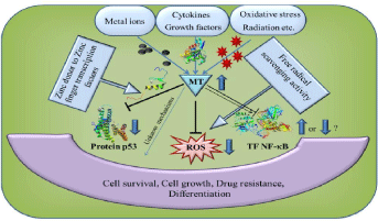
Heavy metals are stable, xenobiotic and are non-biodegradable, once taken they persist in the body, tissues and cells. Exposure to the toxic environment is done by inhalation of air contaminated with metal dusts, fumes and small particle generated by combustions, intake of contaminated food, eating at contaminated site, eating without washing hands. On intake, the heavy metals become integral part of some body parts like bones. Kidney, liver, brain and accumulate with many years half-life [4]. Toxic metals are thrown out of the body by kidney and gastrointestinal tract. There are some proteins which play an important role in their detoxification. Metallothionein (MT) a cysteine rich protein, MW ranging from 500-14000 Da, exist in ten closely related expressed proteins form in human body (Table 2 and Figure 1).
    S.    No.
    Nontoxic
    Low    toxic
    Moderately    toxic
    Highly    toxic


    1.
    Aluminum
    Tin
    Antimony
    Uranium


    2.
    Bismuth
    Scandium
    Beryllium
    Vanadium


    3.
    Calcium
    Barium
    Boron
    Zinc


    4.
    Iron
    Germanium
    Actinium
    Zirconium


    5.
    Magnesium
    Gold
    Cadmium
    Tungsten


    6.
    Manganese
    Erbium
    Chromium
    Radium


    7.
    Lithium
    Gallium
    Hafnium
    Ruthenium


    8.
    Sodium
    Holmium
    Copper
    Thorium


    9.
    Rubidium
    Neodymium
    Indium
    Thallium


    10.
    Strontium
    Terbium
    Lead
    Titanium


    11.
    Potassium
    Thulium
    Mercury
    Silver


    12.
    Molybdenum
    Tin
    Nickel
    Polonium


    13.


    Ytterbium
    Platinum


    samarium
    Palladium
(Source: U.S. GEOLOGICAL SURVEY CIRCULAR 1133, 1995)
Table 1: Heavy metals classification based on toxicity.
    S.No.
    Heavy    metals
    Effect    (Koller, 1973, 1980; Hultman et al., 1994)


    1.
    Hg
    Oldest    pollutant, decrease the production of white blood cells and T-cells, inhibit    immune responses (primary, secondary and memory), fount in dental implants,    pesticides, cosmetics products.


    2.
    Al
    Food    additives, antacids, foils, pots and pans leading to constipation,    Alzheimer's disease, cancer etc.


    3.
    Cd
    Found    in air we inhale, water we drink, and land we live on, gasolines, cigarette    smoke, coffees. Affects liver, kidney, blood cells and T-cells.
Table 2: Few very common heavy metals and negative effect.

Figure 1: Mechanisms of action of MT protein in detoxifying the body.
Synthesized in liver and kidney majorly, exists in four main isoforms MT1, MT2, MT3, and MT4. In 2001, outcome of a scientific study carried out by William Walsh of Health Research Institute, Warrenville, IL, inferenced that Metallothionein (MT) protein is critical for removal of toxic heavy metals from the body. 503 patients with autism spectrum disorder were compared with the non-autistic individuals of the same respective age group. Most of the patients who had autism were suffering from dysfunctional metallothionein. This dysfunctionality of MT was either by genetic factors or by environmental insult at early developmental stage of baby. There are many diseases which are the outcome of exposure to heavy metal contamination. This chapter focuses on the role of heavy metals toxicity and its negative effects on human well-being. However, some of the well-studied common diseases related to heavy metal pollution like Mina-mata (Hg), Itai-Itai (Cd), dental fluorosis (Fe), skeletal deformity (Fe) etc. have been excluded. This chapter has focused on the heavy metal induced disorders like congenital, neurodegenerative, cytotoxic etc. some of them have been discussed below.
Attention deficit hyperactive disorder (ADHD): ADHD is a global childhood behavioral disorder. It is the most common neurogenetic behavior among the children contributed by factors like genetic factors, environmental factors, interaction among environment and genes, exposure to heavy metals like lead etc. There is a correlation of heavy metals toxicity and ADHD. It is the most studied pediatric condition in the USA [2]. 5-10% of the Americans, 4-5% gulf countries &UAE, and 0.03% of British suffer from ADHD. A behavioral study was conducted on the students of gulf region and UAE and was observed that there is a relationship between blood level of heavy metals like lead (Pb), manganese (Mn) and zinc (Zn) to the ADHD [5]. It is also important to note that children are exposed to heavy metal pollution during outdoor play, paints, mouthing with the toys, dusts etc. There are Regio-culture specific pollutants, and their exposure to the children can be responsible for functional, behavioral and developmental impairments. According to the Center for Disease Control (CDC) concentration of lead should less than <100 µg l-1 in the blood. There are some cases where presence of heavy metals like lead even below the permissible limit has negative impact on the intellectual, thinking, learning and developmental behavior. A report prepared by Yousef, et al. [5] differs from the previous study where deficiency of zinc was responsible for ADHD, in this study higher concentration of zinc, manganese, chromium, molybdenum, mercury, and cobalt is associated with ADHD. From the study, it was found that higher blood level of lead, zinc and manganese in the ADHD group as compared with normal individuals as control. It was also found that there was a rise in case of ADHD by 5.2% with increase in lead concentration increased by 1 ppb. There was an increased case of ADHD by 30% and 80.1% with rise in zinc concentration by 1000 ppb and manganese concentration by 1 ppb respectively. ADHD makes it hard to have balanced social, emotional, per formal and behavioral adjustments. Children are the future of any nations and considering the seriousness of the situation, prevalence of the conditions and impact of the disease on the family, society and the patients, it is important to prevent environmental risk factors associated with heavy metals pollution. Heavy metals like lead are important component of paints.
Parkinson’s disease: Parkinson's Disease (PD) is a progressive neurodegenerative disorder. The disease is related to destruction of neurons in the Substantia Nigra Pars Compacta (SNpc) of the basal ganglia. The combination of high concentration of iron and the neurotransmitter, dopamine contributes to the selective vulnerability of the SNpc. Dopamine can auto-oxidize to produce free radicals particularly in the presence of iron and other heavy metals [6,7]. There is a geographic association between PD mortality and the industrial use of heavy metals and their exposure like iron, zinc, copper, mercury, magnesium, and manganese [8-10]. Studies have suggested that a disturbance of the plasmatic rate of Cu could be a marker of PD or at least, a risk factor for the development of this disease. Although zinc participates to the reduction of oxidative stress and the antioxidant role of the selenium, their implication in the onset of PD is not clearly established [11]. Zhao et al. [12], generated a model to predict PD patients based on the plasma concentrations of trace elements (Se, Fe, Zn and Cu), which achieved an accuracy of 80.97±1.34% using 10-fold cross-validation. It was found that plasma Se and Fe concentrations were significantly increased whereas Cu and Zn concentrations decreased in PD patients as compared with controls. Meta-analysis studies were done to evaluate whether circulating Zn levels in the serum, plasma, and Cerebrospinal Fluid (CSF) are altered in PD which suggested that reduced Zn levels in the serum and plasma are associated with an increased risk for PD [13].
Insomnia: Insomnia is the most common sleep disorder affecting millions of people as either a primary or comorbid condition. The prevalence of insomnia has varied widely, from 10-40 % in which insomnia symptoms may be estimated at 30% and specific insomnia disorders at 5-10 [14]. One study found elevated Blood Lead Levels (BLL) at ages 3-5 y was associated with increased risk for sleep problems in early adolescence, particularly among children with BLL ≥ 10 μg/dL compared to those with BLL < 10 μg/dL [15]. The accidental inhalation of nickel carbonyl causes immediate acute toxic effect in symptoms of insomnia [16]. The peoples whose are taking water, having high level of arsenic, showed insomnia symptoms [17].
Autism spectrum disorder: Autism spectrum disorder is the umbrella term various neurological disorders like Childhood disintegrative disorder, Asperger's disorder, Rett's disorder, Autistic disorder for it is a neurological disorder where a person suffers with repetitive and restricted behavior, faces problem in communication have social and reciprocity deficits [18]. Autism begins at very early i.e. first three years of age, in infants. By the study conducted by the Frith and Happe, [18], it was found that most of the patients of autism have retarded mental growth and language delay but in aspergers disorder there is no delay in language and not any mental disorders. Families with Asperger’s Disorder (ASD) more likely to have normal children like any other family. In ASD, mental retardation is not the necessary part of the autistic disorder. Language delays, difficulties in communications, language impairment and other disorders are overlapping but may or may not occur with autism. ASD is the outcome of full mutation of the gene, the Fragile X (FRAXA) FMR1. There is substantial minority of patients which have shown the full mutations. This mutation is the single gene mutation and under expression of this gene is responsible for mental retardation and reduced social-communicative abilities, with these symptoms there is a kind of overlap with symptoms of autism. With the research, it has been observed that there is a relationship between heavy metals and autism spectrum disorder. Copper toxicity and zinc deficiency. Faber, et al. [19] had a retrospective analysis of plasma zinc, and copper from serum, and their ratio as a biomarker for heavy metals among the autistic children. With zinc deficiency and copper toxicity there was a decrease in metallothionein system functioning. In this study zinc content in plasma/ serum copper content ratio is used as a biomarker/indicator for metals toxicity based ASDs.
Multiple sclerosis: Multiple Sclerosis (MS) is a complex disease with an etiology that is hypothesized to involve both environmental and genetic factors [20]. The cellular accumulation of lead and mercury has been associated with the development of auto antibodies against neuronal cytoskeletal proteins, neurofilaments, and myelin basic protein in humans and animals [21,22]. Overexposure to lead and mercury ions is known to be neurotoxic, particularly to motor neurons [23]. Low-to-moderate levels of lead exposure can cause functional alterations in T-lymphocytes and macrophages that lead to increased hypersensitivity and can alter cytokine production, which increases risk o f inflammation-associated tissue damage [24]. MS cases were more likely to report exposure to lead (adjusted OR (AOR) = 2.03; 95% CI: 1.07, 3.86) or mercury AOR=2.06; 95% CI: 1.08, 3.91) than controls [25]. Studies have shown that the mercury exposure in genetically susceptible animals, even at low doses accelerates autoimmune disease and leads to disruption in cytokine production [22]. These studies support an etiologic role of metals in autoimmune disease and suggest the importance of interactions between gene and environment, or between environmental factors in understanding metal toxicity. Recently Napier, et al. [25] explored the relationship between environmental exposure to lead, mercury, and solvents and 58 Single Nucleotide Polymorphisms (SNPs) in MS-associated genes. MS cases were more likely than controls to report lead (odds ratio (OR) = 2.03; 95% Confidence Interval (CI): 1.07, 3.86) and mercury exposure (OR=2.06; 95% CI: 1.08, 3.91).
Arthritis: Arthritis is an inflammation of the joints. Studies prove that long-drawn-out exposure to certain heavy metals, such as, iron, copper, lead, cadmium and mercury, can be causing rheumatoid arthritis-like symptoms. These metals can be found in foods we use daily. Osteoarthritis (OA) is a highly prevalent disease affecting bone and cartilage. A metal that is the cause of OA is Lead (Pb) which known to affect bone, and recent evidence suggests that it has effects on cartilage as well [26]. Rheumatoid Arthritis (RA) is related to high blood copper levels. Moreover; the blood copper levels in RA patients were higher than those in osteoarthritis patients and healthy person [27]. These findings mean that copper can be an important factor to determine the levels of inflammation in RA patients.
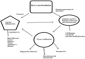
Autoimmune Disorders (AID): During the evolution to provide protection against the pathogens immune system evolved. The immune system protects against the diseases. But one of the important diseases related to the immune system itself is autoimmune disease, the condition where immune system, fails to differentiate between self and foreign cells and treats healthy cells are foreign and attacks healthy cells. Auto immune disease can be of the type depending on the types of body tissue. There are near about 80 types of AID. It has been proved by research that exposure to heavy metals or chemicals can cause, trigger or enhance the condition of AID. Heavy metals have very basic and elementary role to play at the molecular and chemical structure level during complex formation in the immune system. A study was carried out by Rowley and Monestier, [28] and they observed the inducement of genetic susceptibility among rats and mice to produce polyclonal antibodies and specific auto antibodies, when subjected to subtoxic mercury. Research has also confirmed the presence of Auto reactive T-cells in every individual. These T-Cells can be stimulated by adjuvant from microbes or by xenobiotic compounds like heavy metals and chemicals. And these heavy metals can help these autoreactive T-cells to differentiate for a pathogenic molecular pathway [29]. Heavy metals like Hg and gold (Au), were used in causing immunological disorders. Invitro study has found Hg and Au both activated the pathways related to signal transduction and expression of cytokines like IFNγ and IL-4. In some certain environmental condition, heavy metals promote activation of pathways with autoimmune reactions. In this situation, genetic makeup and background plays a significant role. In rodents, it is the overlapping chromosomal regions that are responsible for the control of the immunological disorders activated by Au. It is the Au that also regulates the balance of CD45RChigh/CD45RClowCD4+T cells and autoimmunity based encephalomyelitis. It is important to understand there are many vaccines where aluminum hydroxide and thiomersal C9H9HgNaO2S (organ mercury compound) based antimicrobial and antifungal agents can trigger not just autoimmune disorder [29]. There are other studies also which have suggested the negative effects of heavy metals like (cadmium, gold and mercury) in promotion of AID. Cadmium is also found to be one of the reasons for autoimmunity based renal pathologies (Figure 2).

Figure 2: Relationship between heavy metals and health related disorders.
    S.    No.
    Share    (%)
    Significance
    Treatment
    Treatment    (Time)


    1.
    Heavy    Metals
    2-Jan
    Detoxification
    Many    Months


    2.
    Foci
    10-May
    Surgical,    Controlling the Inflammation
    Multiple    Months


    3.
    Disorder    in Bile Duct Functioning
    20-Oct
    Bitter    Edibles
    1-3    Months


    4.
    Dysbiosis    Of Intestine
    20-30
    Antimicrobial    (Fungi), Symbiotic Bacteria
    1-3    Months


    5.
    Stress
    20-30
    Anti-Stress
    3-4    Weeks


    6.
    Emotional    Conflicts
    98
    Homeopathic
    8-12    Months
Source: Dr. Med. Reimar Banis (MD), Germany (Slide share)
Table 3: Share of Heavy metals disease and treatment strategy.
Gold slat which is an integral component of medicines used in the treatment of arthritis (rheumatoid) has also been found to cause autoimmune disorders, autoimmune complex mediated glomerulus, autoimmune thrombocytopenia etc. Hg induces autoimmune disorders related to genetics, etiology, pathogenesis and structure based activity disorders.
Asthma: Asthma is one of the most common chronic health conditions, affects an estimated 334 million individuals worldwide [30]. Several metals have been reported to be associated with childhood asthma. The elevated urinary levels of metals such as Cr, Cu, As, Se, Sr, Mo, Cd, Sn, Sb, W and U were significantly associated with the prevalence of asthma [31]. High levels of chromium entered by breathing, cause irritation to the nasal cavity, asthma and cough. A study suggested that the low concentration of cobalt is relatively less toxic compared with many other metals but toxic effect in higher concentrations affects mainly the lungs, leading to asthma, pneumonia and wheezing [32,33]. A study was to investigate the levels of heavy metals in PM 2.5 and in blood, the prevalence of respiratory symptoms and asthma, and the related factors to them [34].
Kidney disease: Lead (Pb), mercury (Hg), and cadmium (Cd) are common heavy metal toxins and cause toxicological renal effects at high levels [35]. Toxicity by cadmium gives a major setback to tissues of kidney, skeleton and lungs. Approximately 50% of the accumulated cadmium dose is stored in the kidneys [36]. Presence in urine of Low Molecular Weight Proteins (LMWP) such as b2- Microglobulin (b2M) and Retinol Binding Protein (RBP), and enzymes like N-acetyl-b-D-Glucosaminidase (NAG) are the earliest signs of renal damage which are freely filtered at the glomerulus, and then completely reabsorbed along the early proximal tubule by a megalin-dependent, receptor-mediated, endocytic uptake mechanism. Continued Cd exposure causes glomerular damage, leading to albuminuria and a progressive decline in Glomerular Filtration Rate (GFR), eventually causing end-stage renal failure. Cd exposure has been proved to be associated with Chronic Kidney Disease (CKD), especially in adults with hypertension or diabetes. Zinc is considered to be relatively non-toxic, especially if taken orally. However, excess amount can cause system dysfunctions that result in impairment of growth and reproduction. The clinical signs of zinc toxicos is have also been reported as to cause kidney failure [37].
Chronic fatigue syndrome (CFS): Presence of toxic metals in air, water and land with direct involvement with humans has a role in metal induced autoimmunity. Chronic fatigue syndrome is one of the AID. It is the exposure and genetics which are two decisive forces that lead to meal pathogenesis. And it is the sensitivity and detoxifying ability of an individual towards the metals that controls the inducement of diseases like Rheumatoid Arthritis (RA), Lou Gehrig’s Syndrome (ALS), Chronic Fatigue Syndrome (CFS), and Multiple Sclerosis (MS) [21]. It is obvious that with increased in knowledge related to the genotype based sensitivity and phenotypic variations, there is a scope for using them as a biomarker for the diagnosis of any individual’s susceptibility and mechanisms of operation. To assess the metal induced sensitization toxicologist used to perform organ biopsies, body fluid analysis, brain autopsies but the data obtained was insufficient to give sufficient data to prove a connection for metal pathogenesis [21]. Serious fatigue is one of the outcomes of AID. CFS has some specific conditions like mild hypocortisolemia by down regulation of hypothalamus pituitary adrenal axis. In autoimmune diseases, the important findings were related to decreased NK cells and its functions [38] while increased in cytotoxic T-cells (CD8+) [39]. There were two separate studies one conducted by Japanese scientists which was in line of the study conducted by the Swedish scientist, where it was found that dental implants containing heavy metals led to the CFS like symptoms [40] (Table 3).
Epilepsy: Epilepsy is one of the most prevalent non communicable neurologic conditions and an important cause of disability and mortality affecting individuals of all ages [41]. It is estimated to affect almost 70 million people worldwide [42]. The prevalence of epilepsy in low- and middle-income countries (LMIC) is about twice that of high-income countries [41]. The higher incidence of head trauma and of infections and infestations of the CNS such as malaria, neurocysticercosis, and invasive bacterial infections may be important causes. Autoimmune neurological condition caused by mercury can lead epilepsy. A major factor in epilepsy has been found to be essential mineral deficiencies and imbalances, such as magnesium, zinc, calcium, etc.
Schizophrenia: Microelements or trace metals are very important for proper functioning of immune system and optimal function of a variety of physiological processes [43]. Deficiencies or alterations in the levels of these trace metals adversely affect the ability to withstand oxidative stress-mediated cell damage. Schizophrenia is an oxidative stress induced mental illness as indicated by low levels of antioxidant defense enzymes (glutathione peroxidase, superoxide dismutase and catalase) and antioxidant activity [44,45]. Selenium is an important constituent of glutathione peroxidase enzyme. Manganese, copper, and zinc are important components of Superoxide Dismutase (SOD) and while iron is found in catalase [46]. Thus, deficiencies of these nutritionally essential metals may be suggested in schizophrenic patients. Nutritional deprivation in the early stage of life increases the risk of developing schizophrenia. Oxidative stress, disturbed thinking and irrational behavior which are common to schizophrenic patients may be a result of changes in the levels of certain trace metals. Levels of certain nutritionally essential trace metals (Fe and Se) are reduced while levels of certain heavy metals (Pb, Cr and Cd) are raised in schizophrenic patients [47].
Fibromyalgia: Fibromyalgia disorder is a widespread and chronic disease characterized by extensive pain, diffuse tenderness. The word “fibromyalgia” comes from the latin term for fibrous tissue (fibro) and the Greek ones for muscle (myo) and pain (algia).
Gulf war syndrome (GWS): Gulf war took place in the year 1991, where Canadian, US and British army deployed their soldiers in south west Asia. Gulf region is rich in various natural resources like oil and other metals. During the conflict, it is mandatory to have rapid mobilization of combat troops and the soldiers get exposed to smoke from burning oil wells, extremely high weathers, high and low temperature, hot airs, petroleum products fumes, pesticides, infections, depleting radioactive metals etc leading to stress for the soldiers [48]. According to the report of U.S Department of Veterans Affairs, During the gulf war and post war a serious condition which is a combination of various medical conditions with chronic symptoms like fatigue, joint pain, headaches, dizziness, disorders of respiratory tract and memory related issues are observed. This disease was named “undiagnosed illness” and “chronic multi-symptom illness” or in general term “gulf war syndrome”. Study conducted by Jhonson and Atchison, [49] have noted that it is the environmental heavy metals like Hg, Pb and pesticides which on exposure lead to the diseases related to muscles. Out of total case nearly 10% cases have genetic factors responsible for the disease while 90% cases of muscle disease are environmental exposure [49]. Depleted Uranium (DU), is the byproduct of uranium enrichment process. This by product is considered as the one of the causative agents for GWS. Natural uranium and DU, both have same chemo toxicity but the problem occurs when the dusts of DU generated by the hard hit of DU to any target, during ammunition and blast. Aerosols generated, on inhalation can be exposed to respiratory tract. Also these small dusts get entry into the ground water and good causing gulf war syndrome and Balkan syndrome [50].
Hypertension: Mercury, cadmium, and other heavy metals have a high affinity for sulfhydryl (-SH) groups, inactivating numerous enzymatic reactions, amino acids, and sulfur-containing antioxidants (NAC, ALA, GSH), with subsequent decreased oxidant defense and increased oxidative stress. Both bind to metallothionein and substitute for zinc, copper, and other trace metals reducing the effectiveness of metalloenzymes. Mercury induces mitochondrial dysfunction with reduction in ATP, depletion of glutathione, and increased lipid peroxidation; increased oxidative stress is common. Selenium antagonizes mercury toxicity. The overall vascular effects of mercury include oxidative stress, inflammation, thrombosis, vascular smooth muscle dysfunction, endothelial dysfunction, dyslipidemia, immune dysfunction, and mitochondrial dysfunction. The clinical consequences of mercury toxicity include hypertension, CHD, MI, increased carotid IMT and obstruction, CVA, generalized atherosclerosis, and renal dysfunction with proteinuria.

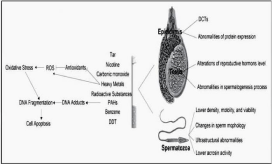
Figure 3: Schematic representation of the effects of tobacco smoking on male fertility.
Alzheimer’s diseases (AD): It is a chronic neurodegenerative disease that slowly destroys memory and thinking skills, and eventually the ability to carry out normal functions of daily need and worsens over the period. Heavy metals and dioxins show their toxic effects on the enzymes involved in phase 1 and 2, like superoxide dismutase, cytochrome P450 and glutathione peroxidase, responsible for cellular proliferations, growth controls and cell death. Heavy metals like cobalt, cadmium, and copper modulates gene expression, reduces the activity of proteins, affects signal transduction, generate ROS/RNS, alter cellular proliferations and differentiations and death, damage cells of brain, DNA damage of brain tissues, leading to neurodegenerative diseases like Parkinson disease, Alzheimer disease, and amyotrophic lateral sclerosis [51]. A study was conducted on more than 80 districts of wales and England to study the rate of Alzheimer’s disease on people below 70 years of age with the help of computerized tomographic. The metal of focus was aluminium. There were a 150 percent times higher chances of people falling to Alzheimer’s disease where the concentration of aluminium in drinking water was above 0•11 mg/l [52]. Miu and Benga, [53] studied the role of metals in Alzheimer’s disease. In metal ion homeostasis, the role of amyloid beta protein precursor and its fragment (proteolytic) amyloid beta have an important role (AβPP and Aβ) respectively. But if these proteins stop their function they can trigger various diseases like Alzheimer’s disease. Heavy metals like Cu and Zn reacts with these proteins and promotes aggregation of these normally active proteins and ROS/RNS generation. Heavy metals also interact with other pathways of AD like that of cleavage of AβPP by secretase and degradation of Aβ, and neurofibrillary tangle formation. All these activities based on disturbance in metal ion homeostasis can cause the creation of an environment that promotes and accelerate neurodegenerative properties [54].
Infertility: The World Health Organization (WHO) estimates that 60 to 80 million couples worldwide currently suffer from infertility, varies across regions of the world and is estimated to affect 8 to 12 per cent of couples [55]. Globally, most infertile couples suffer from primary infertility (inability to conceive within two years) [56]. The numerous external causes of infertility include exposure to heavy metals such as Cd, Hg, Pb, Ar, may be highly involved in impaired human fertility [57,58]. Tobacco and smoking is the primary source of Cd and lead (Pb) intake, observed in serum and semen of infertile smokers as shown in (Figure 3) [59]. Basal cadmium excretion was significantly higher in the infertile women compared with the pregnant women [60]. A study reported blood cadmium level <1.5 μg L, and decreased sperm density, number of sperm per ejaculate, decreased semen volume, forms immature sperm and directly injure the testes [61]. Exposure to cadmium, lead and inorganic arsenic may also contribute to prostate cancer development [62]. Mercury and Copper can interfere in spermatogenesis and also affects the epididymis. A number of studies reported that total mercury levels >8 μg L in blood or >8 ng L in seminal fluid are associated with abnormal sperm count, motility and lowers the sperm concentration [63]. Epidemiological and animal research suggests that different concentrations of lead (Pb) has a wide spectrum of toxicity on the male reproductive system, including spermatogenesis, sperm functional parameters and reproductive hormones, mostly via the HPT hormonal axis reduces sperm production in seminiferous tubules of the testes [64]. Exposure to inorganic lead is harmful to human semen quality because it is a reproductive toxicant. The studies showed that workers exposed to lead at equal or higher than 400 µg L-1 concentrations in blood had lower sperm count, poor semen motility, abnormal sperm morphology particularly of the sperm head and higher teratozospermia [65,66]. The study suggested the blood arsenic levels higher than 5.8 μg L were associated with increased the risk of low luteinizing hormone level and low sperm motility [67-69].
Conclusion
Overexposure to heavy metal such as lead, arsenic, mercury, cadmium, chromium is serious public health problems that harmfully affect human health including cardiovascular diseases, developmental abnormalities, neurologic and neurobehavioral disorders, diabetes, hearing loss, hematologic and immunologic disorders, and various types of cancer. The use of implants in dentals, skin surgeries and grafts in bone formation also contribute in exposure to heavy metal toxicity. Effective laws, strategy are necessary to control the heavy metals pollution. The detection of the areas where there are higher levels of heavy metals is necessary to fulfill the task. Failure to control the exposure of these heavy metals will result in severe complications not only in human health but also in the future environment, plant health and well being of all organisms.
References
- Muhammad S, Shah MT, Khan S. Health risk assessment of heavy metals and their source apportionment in drinking water of kohistan region, northern Pakistan. Microchem J. 2011; 98: 334-343. Leo J. Attention deficit disorder. Skeptic. 2000; 8: 67-70.
- Koller LD. Immunotoxicology of heavy metals. International J of immunopharmacology. 1980; 2: 269-279.
- Hu H. Exposure to metals. Primary care: clinics in office practice. 2000; 27: 983-996.
- Yousef S, Adem A, Zoubeidi T, Kosanovic M, Mabrouk AA, Eapen V. Attention deficit hyperactivity disorder and environmental toxic metal exposure in the United Arab Emirates. J of tropical pediatrics. 2011; 57: 457-460.
- Montgomery EB Jr. Heavy metals and the etiology of Parkinson's disease and other movement disorders. Toxicology. 1995; 97: 3-9.
- Racette BA, Nielsen SS, Criswell SR, Sheppard L, Seixas N, Warden MN, et al. Dose-dependent progression of parkinsonism in manganese-exposed welders. Neurology. 2017; 88: 344-351.
- Rybicki BA, Johnson CC, Uman J, Gorell JM. Parkinson's disease mortality and the industrial use of heavy metals in Michigan. Mov Disord. 1993; 8: 87-92.
- Fukushima T, Tan X, Luo Y, Kanda H. Relationship between blood levels of heavy metals and Parkinson's disease in China. Neuroepidemiology. 2010; 34: 18-24.
- Hare DJ, Kay LD. Iron and dopamine: a toxic couple. Brain. 2016; 139: 1026-1035.
- Younes-Mhenni S, Aissi M, Mokni N, Boughammoura-Bouatay A, Chebel S, Frih-Ayed M, et al. Serum copper, zinc and selenium levels in Tunisian patients with Parkinson's disease. Tunis Med. 2013; 91: 402-405.
- Zhao HW, Lin J, Wang XB, Cheng X, Wang JY, Hu BL, et al. Assessing plasma levels of selenium, copper, iron and zinc in patients of parkinson's disease. PLoS One. 2013; 8.
- Du K, Liu MY, Zhong X, Wei MJ. Decreased circulating Zinc levels in Parkinson's disease: a meta-analysis study. Sci Rep. 2017; 7: 3902-3925.
- Ancoli-Israel S, Roth T. Characteristics of insomnia in the United States: results of the 1991 National Sleep Foundation Survey. Sleep. 1999; 22: 347-353.
- Liu J, Liu X, Pak V, Wang Y, Yan C, Pinto-Martin J. et al. Early blood leads levels and sleep disturbance in preadolescence. Sleep. 2015; 38: 1869-1874.
- Das KK, Das SN, Dhundasi SA. Nickel, its adverse health effects & oxidative stress. Indian J Med Res. 2008; 128: 412-425.
- Guo JX, Hu L, Peng ZY, Tanabe K, Miyatalre M, Chen Y. Chronic arsenic poisoning in drinking water in Inner Mongolia and its associated health effects. J of Environmental Science and Health. 2007; 12: 1853-1858.
- Frith U, Happé F. Autism spectrum disorder. Current biology. 2005; 15: 786-790.
- Faber S, Zinn GM, Kern JC, Kingston HM. The plasma zinc/serum copper ratio as a biomarker in children with autism spectrum disorders. Biomarkers. 2009; 14: 171-180.
- Marrie RA. Environmental risk factors in multiple sclerosis aetiology. Lancet Neurol. 2004; 3: 709-718.
- Stejskal VD, Danersund A, Lindvall A, Hudecek R, Nordman V, Yaqob A, et al. Metal-specific lymphocytes: biomarkers of sensitivity in man. Neuroendocrinology Letters. 1999; 20: 289-298.
- Hansson M, Djerbi M, Rabbani H, Mellstedt H, Gharibdoost F, Hassan M, et al. Exposure to mercuric chloride during the induction phase and after the onset of collagen-induced arthritis enhances immune/autoimmune responses and exacerbates the disease in DBA/1 mice. Immunology. 2005; 114: 428-437.
- Callaghan B, Feldman D, Gruis K, Feldman E. The association of exposure to lead, mercury, and selenium and the development of amyotrophic lateral sclerosis and the epigenetic implications. Neurodegener Dis. 2011; 8: 1-8.
- Dietert RR, Piepenbrink MS. Lead and immune function. Crit Rev Toxicol. 2006; 36: 359-385.
- Napier MD, Poole C, Satten GA, Ashley-Koch A, Marrie RA, Williamson DM. Heavy metals, organic solvents, and multiple sclerosis: An exploratory look at gene-environment interactions. Arch Environ Occup Health. 2016; 71: 26-34.
- Amanda E Nelson, Xiaoyan A Shi, Todd A Schwartz, Jiu-Chiuan Chen, Jordan B Renner, Kathleen L Caldwell, et al. Whole blood lead levels are associated with radiographic and symptomatic knee osteoarthritis: a cross-sectional analysis in the Johnston County Osteoarthritis Project. Arthritis Research & Therapy. 2011; 13: R37.
- Yang Tao-H, Yuan Tzu-H, Hwang Yaw-H, Lian Ie-B, Meng M, Su Che-C. Increased inflammation in rheumatoid arthritis patients living where farm soils contain high levels of copper. J of the Formosan Med Association. 2016; 115: 991-996.
- Rowley B, Monestier M. Mechanisms of heavy metal-induced autoimmunity. Molecular immunology. 2005; 42: 833-838.
- Fournié GJ, Mas M, Cautain B, Savignac M, Subra JF, Pelletier L, et al. Induction of autoimmunity through bystander effects. Lessons from immunological disorders induced by heavy metals. J of autoimmunity. 2001; 16: 319-326.
- Kiboneka A, Levin M, Mosalakatane T, Makone I, Wobudeya E, Makubate B, et al. Prevalence of asthma among school children in Gaborone, Botswana. Afr Health Sci. 2016; 16: 809-816.
- Huang X, Xie J, Cui X, Zhou Y, Wu X, Lu W, et al. Association between concentrations of metals in urine and adult asthma: A case- control study in Wuhan, China. PLOS ONE. 2016; 11: 1-18.
- Jomovaa K, Valkob M. Advances in metal-induced oxidative stress and human disease. Toxicology. 2011; 283: 65-87.
- Gal J, Hursthouse A, Tatner P, Stewart F, Welton R. Cobalt and secondary poisoning in the terrestrial food chain: data review and research gaps to support risk assessment. Environ Int. 2008; 34: 821-838.
- Zeng X, Xu X, Zheng X, Reponen T, Chen A, Huo X. Heavy metals in PM 2.5 and in blood, and children's respiratory symptoms and asthma from an e-waste recycling area. Environ Pollut. 2016; 210; 346-356.
- Kim NH, Hyun YY, Lee KB, Chang Y, Ryu S, Oh KH, et al. Environmental heavy metal exposure and chronic kidney disease in the general population. J Korean Med Sci. 2015; 30: 272-277.
- Sabath E, Robles-Osorio ML. Renal health and the environment: heavy metal nephrotoxicity. Nefrologia. 2012; 32: 279-286.
- Fosmire GJ. Zinc Toxicity. Am J Clin. Nutr. 1990; 51: 225 -227.
- Caligiuri M, Murray C, Buchwald D, Levine H, Cheney P, Peterson D, et al. Phenotypic and functional deficiency of natural killer cells in patients with chronic fatigue syndrome. The J of Immunology. 1987; 139: 3306-3313.
- Landay AL, Jessop C, Lennette ET, Levy JA. Chronic fatigue syndrome: clinical condition associated with immune activation. Lancet. 1991; 338: 707-712. Stejskal V. Immunological effects of amalgam components: MELISA - a new test for the diagnosis of mercury allergy. In Proceedings of the International Symposium Status Quo and Perspectives of Amalgam and other Dental Materials. Europaeum, European Academy, Otzenhausen, Germany. 1994.
- Ngugi AK, Bottomley C, Kleinschmidt I, Sander JW, Newton CR. Estimation of the burden of active and life- time epilepsy: a meta-analytic approach. Epilepsia. 2010; 51: 883-890.
- WHO report on Epilepsy: epidemiology, aetiology and prognosis WHO. World Health Organization. WHO Factsheet. 2001b. Foster HD. The geography of schizophrenia: possible links with selenium and calcium deficiencies, inadequate exposure to sunlight and industrialization. J Orthomolecular Med. 1988; 3: 135-140.
- Dadheech G, Mishra S, Gautam S, Sharma P. Evaluation of antioxidant deficit in schizophrenia. Indian J Psychiatry. 2008; 50: 16-20.
- Uma Devi P, Chinnaswamy P. Oxidative injury and enzymic antioxidant misbalance in schizophrenics with positive, negative and cognitive symptoms. Afr J Biochem Res. 2008; 2: 92-97.
- Johnson S. Micronutrient accumulation and depletion in schizophrenia, epilepsy, autism and Parkinson's disease? Med Hypotheses. 2001; 56: 641-645.
- Arinola G, Idonije B, Akinlade K, Ihenyen O. Essential trace metals and heavy metals in newly diagnosed schizophrenic patients and those on anti-psychotic medication. J Res Med Sci. 2010; 15: 245-249.
- Murphy FM. Gulf war syndrome. BMJ. 1999; 318: 274-275.
- Johnson FO, Atchison WD. The role of environmental mercury, lead and pesticide exposure in development of amyotrophic lateral sclerosis. Neurotoxicology. 2009; 30: 761-765.
- Bleise A, Danesi PR, Burkart W. Properties, use and health effects of Depleted Uranium (DU): a general overview. Journal of environmental radioactivity. 2003; 64: 93-112.
- Mates JM, Segura JA, Alonso FJ, and Márquez J. Roles of dioxins and heavy metals in cancer and neurological diseases using ROS-mediated mechanisms. Free Radical Biology and Medicine. 2010; 49: 1328-1341.
- Martyn CN, Barker DJ, Osmond C, Harris EC, Edwardson JA, Lacey RF. Geographical relation between Alzheimer's disease and aluminium in drinking water. Lancet. 1989; 1: 61-62.
- Miu AC, Benga O. Aluminum and Alzheimer’s disease: A new look. J Alzheimer Dis. 2006; 10: 179-201.
- Adlard PA and Bush AI. Metals and Alzheimer's disease. Journal of Alzheimer's Disease. 2006; 10: 145-163.
- Rutstein Shea O, Shah Iqbal H. Infecundity, infertility, and childlessness in developing countries. DHS Comparative Reports No.9. World Health Organization. 2004.
- Sciarra J. Infertility: an international health problem. Int J Gynaecol Obstet. 1994; 46: 155-163.
- Queiroz EKR, Waissmann W. Occupational exposure and effects on the male reproductive system. Cad Saúde Pública. 2006; 22: 485-493.
- Pizent A, Tariba B, Zivkovic T. Reproductive toxicity of metals in men. Arh Hig Rada Toksikol. 2012; 63: 35-46.
- Dai JB, Wang ZX, Qiao ZD. The hazardous effects of tobacco smoking on male fertility. 2015; 17: 954-960.
- Rzymski P, Tomczyk K, Rzymski P, Poniedzialek B, Opala T, Wilczak M. Impact of heavy metals on the female reproductive system. Annals of Agricultural and Environmental Medicine. 2015; 22: 259-264.
- Xu B, Chia SE, Tsakok M, Ong CN. Trace elements in blood and seminal plasma and their relationship to sperm quality. Reprod Toxicol. 1993; 7: 613-618.
- Iavicoli I, Fontana L, Bergamaschi A. The effects of metals as endocrine disruptors. J Toxicol Environ Health B Crit Rev. 2009; 12: 206-223.
- Leung TY, Choy CM, Yim SF, Lam CW, Haines CJ. Whole blood mercury concentrations in sub-fertile men in Hong Kong. Aust N Z J Obstet Gynaecol. 2001; 41: 75-77.
- Vigeh M, Smith DR, Hsu PC. How does lead induce male infertility? Iranian J of Reproductive medicine. 2011; 9: 1-8.
- Lerda D. Study of sperm characteristics in persons occupationally exposed to lead. Am J Ind Med. 1992; 22: 567- 571.
- Wu HM, Tan DTL, Wang ML, Huang HY, Lee CL, Wang HS et al. Lead level in seminal plasma may affect semen quality for men without occupational exposure to lead. Reproductive Biology and Endocrinology. 2012; 10: 1-5.
- Meeker JD, Rossano MG, Protas B, Diamond MP, Puscheck E, Daly D, et al. Cadmium, lead, and other metals in relation to semen quality: human evidence for molybdenum as a male reproductive toxicant. Environ Health Perspect. 2008; 116: 1473-1479.
- Koller LD. Immunosuppression produced by lead, cadmium, and mercury. Am J Vet Res. (United States). 1973; 34: 11.
- Hultman P, Johansson U, Turley SJ, Lindh U, Enestrom S, Pollard KM. Adverse immunological effects and autoimmunity induces by dental amalgam and alloy in mice. The FASEB Journal. 1994; 8; 1183-1190.