Editorial
Austin J Environ Toxicol. 2019; 5(1): 1028.
Maternal and Fetal Outcome of Female Workers Exposed to Ionizing Radiations: A Prospective Controlled Study
El-Badry A1*, Rezk M2, Shawky M3 and Badr H4
1Department of Public Health and Community Medicine, Egypt
2Department of Obstetrics and Gynecology, Egypt
3Department of Radiology, Egypt
4Department of Pediatrics, Faculty of Medicine, Menoufia University, Egypt
*Corresponding author: Aziza El-Badry, Department of Public Health and Community Medicine, Egypt
Received: October 04, 2019; Accepted: November 06, 2019; Published: November 13, 2019
Abstract
Objective: to explore the maternal and fetal outcome of female workers exposed to ionizing radiations before getting pregnant.
Methods: a prospective controlled study included 132 women working in the Radiology departments (exposed group) and 120 women working in administration offices (non-exposed group) were enrolled upon confirmation of pregnancy and followed throughout pregnancy to record the maternal and fetal outcome.
Results: female workers in the exposed group exhibit a higher rates of spontaneous miscarriage (25.7% versus 6.6%, p<0.001), antepartum hemorrhage (10.6% versus 3.3%, p<0.05), congenital malformations (9.1% versus 1.6%, p<0.05), small for gestational age (19.6% versus 10%, p<0.05) and admission to NICU (11.3% versus 2.5%, p<0.05) compared to non-exposed group.
Conclusion: Although female workers in the Radiation departments were shifted to another duty upon occurrence of pregnancy, they still suffer from poor obstetric outcome.
Keywords: Occupational exposure; Ionizing radiations; Maternal outcome; Fetal outcome; obstetric outcome
Introduction
The nursing profession constitutes a critical component of the health care system all over the world. But, the effect of occupational exposures such as ionizing radiations on obstetric outcome remains unclear within this predominantly female occupation [1].
Complications associated with perinatal exposure to ionizing radiations include preterm labor and delivery, spontaneous miscarriage, congenital fetal malformations and intrauterine fetal growth restriction [2,3].
The fetal risks from maternal exposure to ionizing radiations during pregnancy are related to the gestational age and the absorbed dose. These risks are more significant during the period of organogenesis (two to seven weeks after conception) and in the early fetal period (eight to 15 weeks after conception), with lesser effects in the second trimester, and least in the third trimester (ICRP, 2000, Brent et al.2009).
The aim of this study was to explore the maternal and fetal outcome of female workers exposed to ionizing radiations before getting pregnant.
Materials and Methods
This was a prospective controlled study carried out at the department of Public Health and Community Medicine in collaboration with Obstetrics & Gynecology, Radiology and Pediatrics departments at Menoufia Faculty of Medicine, Menoufia, Egypt in the period between the beginning of March 2016 and the end of July 2019 which is the date of follow up of the last enrolled particpant.
The local Ethics Committee at the Menoufia Faculty of Medicine approved the study protocol and an informed consent was obtained from all agreed participants before commencement of the study.
The approved study protocol was disseminated to 11 central hospitals within Menoufia governorate with thorough explanations of the study objectives through personal interviews with the chiefs of Radiology departments in the 11 hospitals.
All pregnant women in early pregnancy who worked at the Radiology department at Menoufia University hospital and 11 Central hospitals in Menoufia governorate (served as study or exposed group) and pregnant workers in the administration offices which is present in a separate building away from the Menoufia University hospital (served as non-exposed group), were invited to participate in the study. Participants were invited to participate in the study at gestational ages between five to six weeks based on positive serum pregnancy test and reliable last menstrual period. Women asked to join the study after the 6th week of pregnancy were not accepted.
In order to alleviate the effect of other possible causes of poor obstetric outcome, women with medical disorders such as hypertensive disorders, diabetes mellitus, bronchial asthma & epilepsy, multiple pregnancies, smoking, exposure to second hand smoke, socio-economic factors (including poor housing, living near cell phone nests and exposure to pesticides), were excluded from the study.
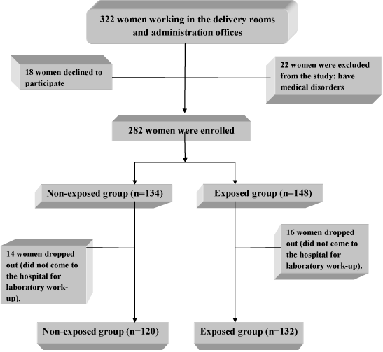
322 women working in the Radiology departments and administration offices of the Faculty of Medicine were invited to participate in the study, 18 women declined to participate and 22 women were excluded secondary to exclusion criteria. Out of 282 enrolled women, 30 women dropped out (did not come to the hospital for delivery). So, 252 women completed the study, 132 women working in the Radiology departments (exposed group) and 120 women from administration offices served as non-exposed group (Figure 1: The flow diagram).

Figure 1: Flow diagram of recruitment and retention of participants in the study.
Demographic, medical and occupational data were collected through direct interviews and a pre-designed questionnaire followed by thorough clinical examination, laboratory and imaging investigations.
Working at times other than normal daylight hours of approximately 7:00 AM to 6:00 PM was considered as shift work [3].
According to our local hospital policy, once pregnancy is confirmed, all females worked in the Radiology department were shifted to the rec
ord-keeping service at the same department till delivery to be away from further exposure to ionizing radiations. All enrolled nurses were wearing a radiation dosimeter during their work prior to pregnancy which is checked once pregnancy is confirmed and the readings were recorded.
Clinical examination entails general examination with recording of vital signs, weight, height, breast and thyroid gland examination followed by obstetric examination and ultrasonography to exclude women with medical disorders. Three dimentional obstetric ultrasound was done at 22-24 weeks after 2 D ultrasound to confirm the presence of congenital malformations.
Enrolled women were followed up from the start of pregnancy till the end of the puerperium and received the same management at the hospital which included regular antenatal care visits every 1-3 weeks in the outpatient clinic and delivery at the Menoufia University hospital.
Outcome measures
Maternal outcome: development of miscarriage defined as interruption (threatened miscarriage) or termination of pregnancy before the 20th week of pregnancy (either spontaneous or induced), venous thromboembolism (VTE), postpartum hemorrhage (PPH) and maternal mortality. Induced abortion means termination of pregnancy secondary to fetal demise. Anembryonic pregnancy means the presence of empty gestational sac with diameter of ≥ 20 mm with absent fetal pole while missed or silent miscarriage means absent fetal cardiac pulsations.
Fetal and neonatal outcome: congenital malformations, small for gestational age (SGA) defined as a birth weight <5th percentile, prematurity (delivery < 37 weeks), intrauterine fetal demise (IUFD), admission to neonatal intensive care unit (NICU) and neonatal death (defined as death during the first four weeks after delivery).
Statistical analysis
The data collected were tabulated & analyzed by IBM SPSS Statistics.
(Statistical package for the social science software) statistical package version 20. Quantitative data were expressed as mean & standard deviation and analyzed by applying student t- test for comparison of two groups of normally distributed variables while two groups of non normally distributed variables by applying Mann-Whitney Test.
Qualitative data were expressed as number and percentage and analyzed by applying Chi-square test and for 2×2 table and at least one cell has expected number less than 5 Fisher's exact test was applied. P value ≤ 0.05 was considered statistically significant. P-values in bold in the tables is statistically significant.
Results
The annual radiation exposure dose (mean ± standard deviation) of the exposed group in their radiation bandage was 3.2±1.7 mSv.
Table 1 depicts maternal characteristics. There was no significant difference between exposed and non-exposed group regarding age, parity, body mass index, duration of employment, shift work, heavy lifting (>10Kg) and prolonged standing (p>0.05).
Table 2 reveals maternal outcome. Female workers in the exposed group exhibit a higher rates of spontaneous miscarriage (25.7% versus 6.6%, p<0.001) with odd’s ratio (OR) of 4.86(2.1-10.9) and antepartum hemorrhage (10.6% versus 3.3%, p<0.05) with OR of 3.4(1.1-10.7) compared with non-exposed group. There was no significant difference between both groups regarding occurrence of venous thromboembolism and postpartum hemorrhage (p>0.05).
Exposed group (n=132)


Non-exposed group (n=120)


Student t-test


P-value


Age (in years)


31.6±4.2


30.9±4.5


1.02


>0.05


Parity


1.4±2.3


1.2±2.2


0.49*


> 0.05


Body mass index (Kg/m²)


25.2±3.4


24.8±3.7


0.62


> 0.05


Duration of employment (year)


10.6±4.2


10.2±4.9


0.73


>0.05


Shift work


104


88


0.75†


>0.05


Heavy lifting (>10 Kg)


56


48


0.07†


>0.05


Standing duration (>3 h)


112


96


0.72†


>0.05
Table 1: Maternal characteristics.
Exposed group (n=132)


Non-exposed group (n=120)


Chi square


P-value


Odd’s ratio (lower & upper limit at 95% CI)


Risk ratio (lower & upper limit at 95% CI)


Miscarriage Spontaneous                     -Induced  -Threatened


54(40.9%)                           34(25.7%)  8(6.1%)  12(9.1%)


22(18.3%)     8(6.6%)  8(6.6%) 6(5%)


14.16   15.15 0.001  1.03


<0.001      <0.001    >0.05 >0.05


3.08(1.7-5.5) 4.86(2.1-10.9) 0.9(0.3-2.4) 1.9(0.6-5.2)


1.6(1.2-2) 1.7(1.4-2.1) 0.95(0.5-1.5)  1.3(0.9-1.8)


Antepartum hemorrhage


14(10.6%)


4(3.3%)


3.98*


<0.05


3.4(1.1-10.7)


1.5(1.1-2.04)


Venous thromboembolism


4(0.7%)


1(0.8%)


0.63*


>0.05


3.7(0.4-33.7)


1.5(0.9-2.4)


Postpartum hemorrhage


10(7.5%)


6(5%)


0.34


>0.05


1.5(0.5-4.4)


1.2(0.8-1.8)


Maternal mortality


0


0


-


-


-


-
*Fischer's exact test
Table 2: Maternal outcome.
Exposed group (n=132)


Non-exposed group (n=120)


Fischer’s exact test


P-value


Odd’s ratio (lower & upper limit at 95% CI)


Risk ratio (lower & upper limit at 95% CI)


Congenital malformations


12(9.1%)


2(1.6%)


5.26


<0.05


5.9(1.2-26.9)


1.7(1.3-2.2)


Small for gestational age


26(19.6%)


12(10%)


3.89*


<0.05


2.2(1.06-4.6)


1.3(1.07-1.7)


Prematurity


10(7.5%)


6(5%)


0.34*


>0.05


1.5(0.5-4.4)


1.2(0.8-1.8)


Intrauterine fetal demise


6(4.5%)


2(1.6%)


0.89


>0.05


2.8(0.5-14.2)


1.4(0.9-2.2)


Admission to NICU


15(11.3%)


3(2.5%)


6.17


<0.05


5(1.4-17.7)


1.6(1.3-2.1)


Neonatal mortality


0


0


-


-


-


-
*Chi square test, NICU=Neonatal intensive care unit.
Table 3: Fetal and neonatal outcome.
Table 3 shows fetal and neonatal outcome. There was a higher rates of congenital malformations (9.1 % versus 1.6%, p<0.05) with OR of 5.9(1.2-26.9), small for gestational age (19.6% versus 10%, p<0.05) with OR of 2.2(1.06-4.6) and admission to NICU (11.3% versus 2.5%, p<0.05) with OR of 5(1.4-17.7). There was no significant difference between both groups regarding occurrence of prematurity and intrauterine fetal demise (p>0.05).
Discussion
The effects of radiation exposure on the fetus depend on the amount of radiation received and the gestational age of the fetus at the time of exposure [6,7].
Accidental exposure to ionizing radiations of less than 5 rads (50mSv or 50mGy) is not harmful to the fetus as stated by the American College of Radiology Resolution and the ACOG Committee Opinion No. 299 [7]. These guidelines could be applied when counseling pregnant woman or health care professional after accidental exposure to radiation in the workplace.
In this study, the rate of spontaneous miscarriage was 25.7% with odd’s ratio (OR) of 4.86(2.1-10.9) with the rate of induced abortion 6.1% for anembryonic pregnancy and missed abortion.
This could be attributed to occupational exposure to ionizing radiation during the pre-implantation period (blastogenesis) as exposure to a radiation dose greater than 0.1 Gy (10 rad) during this period is associated with a risk of failure to implant, representing an “all or none” phenomenon of early embryonic development [7].
Also, teratogenic effects are highly significant during the period of organogenesis (weeks 3 to 7 of gestation) which can be explained by serious damage to DNA or cell death [5,8].
Earlier studies showed an increased risk for spontaneous miscarriage with self-reported first trimester exposure to ionizing radiations with ORs ranging from 1.5–2.3 in samples that included from 18 to 223 exposed cases [6,9,10].
However, other studies did not find statistically significant associations between occupational exposure and spontaneous miscarriage [11-13].
Congenital fetal malformations affect 12(9.1%) fetuses in the exposed group in the current study with OR of 5.9(1.2-26.9) and risk ratio of 1.7(1.3-2.2) which occur in the nervous, cardiovascular and urinary systems.
Maternal occupational exposure to ionizing radiation within the first trimester of pregnancy is associated with a higher rate of birth defects with relative risk of 3.2 (1.2-8.7) in a previous retrospective study [14].
Prenatal exposure to ionizing radiation can result in intrauterine lethality and malformation of organs chiefly the central nervous system. At the preimplantation period (weeks 0 to 2 of gestation), exposure to a radiation dose of 100 to 150 mGy (the equivalent of more than three pelvic CT scans) may have lethal effects [15].
In this study, prolonged periods of standing (>3 hours) and heavy lifting (>10 Kg) were not associated with increased rate of preterm labour and delivery but, elevates the rate of small for gestational age in the exposed group.
Prolonged standing (>8 hours per day) and long working hours (>40 hours per week) may increase the risk of preterm birth and may negatively influence intrauterine growth [16,17].
The highest annual dose recorded in female workers in this study (wearing a radiation dosimeter) was 5 mSv, which represented only 25% of the of the annual dose limit of 20 mSv or 2 rads [18].
Inability to estimate the actual radiation dosage exposure during the period from conception till diagnosis of pregnancy (about 3 weeks) constitutes unintended limitation of this study.
Female workers in Radiology departments seeking for pregnancy should abstain from exposure to ionizing radiations 1-3 months before getting pregnant to improve their obstetric outcome as the fetal exposure dosimeter badge is not readily available at our institution.
Conclusion
Although female working in the Radiation departments in this study were shifted to another duty upon occurrence of pregnancy to withdraw them from further exposure to ionizing radiations, they still suffer from poor obstetric outcome in terms of higher odds of spontaneous miscarriage, congenital fetal malformations, small for gestational age, antepartum hemorrhage and neonatal admission to ICU.
References
- Lawson CC, Rocheleau CM, Whelan EA, Lividoti Hibert EN, Grajewski B, Spiegelman D, et al. Occupational exposures among nurses and risk of spontaneous abortion. 2012; 206: 1-8.
- Zhu JL, Hjollund NH, Olsen J. Shift work, duration of pregnancy, and birth weight: the National Birth Cohort in Denmark. 2004; 191: 285-291.
- Downes J, Rauk PN, Vanheest AE. Occupational hazards for pregnant or lactating women in the orthopaedic operating room. 2014; 22: 326-332.
- International Commission on Radiological Protection. Pregnancy and medical radiation. Ann ICRP. 2000; 30: 1-43.
- Brent RL. Saving lives and changing family histories: appropriate counseling of pregnant women and men and women of reproductive age, concerning the risk of diagnostic radiation exposures during and before pregnancy. Am J Obstet Gynecol. 2009; 200: 4-24.
- Selevan SG, Lindbohm ML, Hornung RW, Hemminki K. A study of occupational exposure to antineoplastic drugs and fetal loss in nurses. 1985; 313: 1173–1178.
- ACOG Committee on Obstetric Practice: ACOG Committee Opinion. Number 299, September 2004 (replaces No. 158, September 1995). Guidelines for diagnostic imaging during pregnancy. 2004; 104: 647-651.
- Uzoigwe CE, Middleton RG. Occupational radiation exposure and pregnancy in orthopaedics. J Bone Joint Surg Br. 2012; 94: 23-27.
- Stucker I, Caillard JF, Collin R, Gout M, Poyen D, Hemon D. Risk of spontaneous abortion among nurses handling antineoplastic drugs. 1990; 16: 102–107.
- Valanis B, Vollmer WM, Steele P. Occupational exposure to antineoplastic agents: self-reported miscarriages and stillbirths among nurses and pharmacists. 1999; 41: 632–638.
- Hemminki K, Kyyronen P, Lindbohm ML. Spontaneous abortions and malformations in the offspring of nurses exposed to anaesthetic gases, cytostatic drugs, and other potential hazards in hospitals, based on registered information of outcome. 1985; 39: 41–147.
- Skov T, Maarup B, Olsen J, Rorth M, Winthereik H, Lynge E. Leukaemia and reproductive outcome among nurses handling antineoplastic drugs. 1992; 49: 855–861.
- Fransman W, Roeleveld N, Peelen S, de Kort W, Kromhout H, Heederik D. Nurses with dermal exposure to antineoplastic drugs: reproductive outcomes. Epidemiology. 2007; 18: 112–119.
- Wiesel A, Spix C, Mergenthaler A, Queisser-Luft A. Maternal occupational exposure to ionizing radiation and birth defects. Radiat Environ Biophys. 2011; 50: 325-328.
- Fattibene P, Mazzei F, Nuccetelli C, Risica S. Prenatal exposure to ionizing radiation: Sources, effects and regulatory aspects. 1999; 88: 693-702.
- Domingues MR, Matijasevich A, Barros AJ. Physical activity and preterm birth: A literature review. 2009; 39: 961-975.
- Snijder CA, Brand T, Jaddoe V, Hofman A, Mackenbach JP, Steegers EA, et al. Physically demanding work, fetal growth and the risk of adverse birth outcomes: The Generation R Study. 2012; 69: 543-550.
- Domanska AA, Bienkiewicz M, Olszewski J. Evaluation of exposure to ionizing radiation among gamma camera operators. 2013; 64: 503-506.