Case Report
J Fam Med. 2014;1(1): 2.
Micro Abscess Formation in Adenomyotic Focus, a Rare Case
Sahin B1, Demirtas O1, Karabulut A1* and Akbulut M2
1Department of Obstetrics and Gynecology, University of Pamukkale, Turkey
2Department of Pathology
*Corresponding author: Aysun Karabulut, Pamukkale University Medical School, Department of Obstetrics and Gynecology, Çamlaralti Mah.20070 Denizli/TURKEY
Received: July 23, 2014; Accepted: Aug 20, 2014; Published: Aug 23, 2014
Abstract
Adenomyosis was defined as ectopic endometrial tissue in myometrium. A few cases of abscess formation in adenomyotic foci were reported previously. Herein, we presented a 53-year-old perimenopausal woman admitted with an abnormal vaginal bleeding and pelvic pain and hysterectomy material revealed micro abscess formation in adenomyotic foci. Micro abscess formation in adenomyotic foci may be further complicating the clinical picture by leading sepsis in postoperative period. Intensive antibiotic therapy in postoperative period has a crucial importance in treatment of suspected cases.
Keywords: Adenomyosis; Micro abscess formation; Myometrium
Case Presentation
A 53-year-old, multiparous, perimenopausal woman with complaint of vaginal discharge, and abnormal vaginal bleeding for last 5 days was admitted to outpatient clinic. In past medical history, she had chronic pelvic pain and dysmenorrhea which was resistant to non steroidal anti-inflammatory drugs for past 5 years. Menstrual history revealed normal cycles except heavy menses time to time.
Painful cervical movements and adnexal tenderness with globally enlarged uterus were detected on pelvic examination. Tran’s vaginal sonography revealed 130 x 103 x 96 mm uterus with 7 mm endometrial thickness, and 36x38 mm intramural myoma and minimal free fluid in the uterine cavity. Histopathologic examination of endometrial biopsy revealed secretary Endometrium.
Laboratory examination was remarkable for leukocytosis (14.000/ mm3) and elevated CA 125 (126.2 U/ml) level. Antimicrobial therapy composed of Gentamicin (Genta®, I.E. Ulagay, Istanbul, Turkey) and Clindamycin (Cleosin®, Pfizer, Istanbul, Turkey) were administered for a week followed by total abdominal hysterectomy and bilateral alpingo-oophorectomy. The patient was discharged without any complication three days after the surgery. Ca 125 levels returned to normal two weeks following the operation.
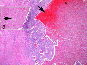
On histopathologic examination, adenomyotic foci characterized by endometrial glands embedded in their own stroma and extending deep into the myometrium were detected. Polymorpho nuclearleukocytes infiltrating the glandular epithelium and the stroma and micro abscess formation in adenomyotic focus was seen on pathologic exam (Figure 1).

    Figure 1:  Micro abscess formation in the foci of Adenomyosis (hematoxylin
and eosin; original magnification x 100).
(a) Ectopic endometrial islands containing polymorph nuclear leukocytes
infiltrating the glandular epithelium and stroma.
Discussion
Adenomyosis is a commonly seen gynecological pathology characterized by histologically benign invasion of Endometrium into the myometrium. It results in a diffusely enlarged uterus which microscopically exhibits ectopic non-neo plastic, endometrial glands and stroma surrounded by the hypertrophic and hyper plastic myometrium [1]. Growth and invagination of endometrial glands into the myometrium is the most popular theory accused for the Adenomyosis formation [2]. According to current literature, the prevalence of Adenomyosis changes between 5 to 70 %, and it was reported as approximately 20 to 30 % in the hysterectomy materials due to benign causes [3,4]. About 70 % of the women with a diagnosis of Adenomyosis were in premenopausal period [3]. Although it’s high prevalence in histopathologic specimens, preoperative diagnosis is difficult and tricky. Ultra sonography is very valuable in diagnosis; however magnetic resonance imaging forms the gold standard [5]. In this report, we presented a case with abnormal vaginal bleeding and pelvic pain resistant to medical therapy undergoing hysterectomy, and micro abscess formation in adenomyotic focus was diagnosed histopathologically.
Presenting symptoms of Adenomyosis are mostly non-specific; therefore preoperative diagnosis of Adenomyosis is usually difficult and depends on the clinical suspicion. Patients may present with pelvic pain resistant to conservative treatment and slight to moderate elevation of Ca 125 levels may be seen [6]. Chronic pelvic pain and abnormal vaginal bleeding are the prominent symptoms in our case and moderately elevated Ca 125 level was also noted on laboratory exam.
Abscess formation in endometriomas was reported previously, but only one case of abscess formation and one case of micro abscess formation in adenomyotic foci were detected in our literature search [7,8]. Erguvan, et al reported a 54-year-old, postmenopausal woman suffering from inguinal pain, night sweats, and hot flashes. A 95x85 mm leimyoma like lesion with a 53 mm x 43 mm cystic space in uterus mimicking malignancy was detected on sonography exam. Postoperative pathologic exam revealed abscess formation in adenomyotic focus. Although the patient had fewer in early postoperative period, later follow-up was uneventful. It was the first case of abscess formation in Adenomyosis reported in literature. However they could not isolate any microorganism from the lesion [9]. On the other hand, our patient did not have fewer despite elevated leukocyte count in postoperative period. Preoperative use of wide spectrum antibiotics probably played a role in this scene.
Shu-Fen Weng, et al described another case of Adenomyosis with micro abscess formation in a 50-year-old nulliparous women presenting with persistent vaginal bleeding, lower abdominal pain and fever, and sepsis was developed in postoperative period [10]. Different from the previous one, micro abscess formation looked like multiple separate islands, and polymorph nuclear leukocyte infiltration in ectopic endometrial foci were detected in this case. Similarly, we detected polymorpho nuclear leukocyte infiltration and micro abscess formation on pathologic exam, but probably due to early antibiotic therapy postoperative period was uneventful in ours.
Although Adenomyosis is known as estrogen-dependent lesion, interestingly both cases reported previously [9,10] were in perimenopausal period. The same was true for our case. Under the light of current literature, it is hard to interpret, whether Adenomyosis is aggravated in perimenopausal period due to unbalanced estrogen and they become vulnerable to abscess formation. In background history of all cases chronic pain resistant to medical treatment was the common finding. In previous two reports sepsis was developed postoperatively [9,10]. Luckily, it was not the situation in our case, probably due to antibiotic therapy (Gentamicine and Clindamycin) given preoperatively for suspected endometritis. Only elevated white blood cell count and CRP levels were noted, and they were resolved with continuation of antibiotic therapy.
Conclusion
In conclusion, Adenomyosis may present with nonspecific signs and symptoms, and micro abscess formation in adenomyotic foci may further complicate situation and carry risk of postoperative sepsis. Surgery is the main treatment modality in cases refractory to medical treatment, and intensive antibiotic therapy is required to minimize the risk of sepsis.
References
- Benagiano G, Brosens I. History of adenomyosis. Best Pract Res Clin Obstet Gynaecol. 2006; 20: 449-463.
- Ferenczy A. Pathophysiology of adenomyosis. Hum Reprod Update. 1998; 4: 312-322.
- Wallwiener M, Taran FA, Rothmund R, Kasperkowiak A, Auwärter G, Ganz A, et al. Laparoscopic supracervical hysterectomy (LSH) versus total laparoscopic hysterectomy (TLH): an implementation study in, 952 patients with an analysis of risk factors for conversion to laparotomy and complications, and of procedure-specific re-operations. Arch Gynecol Obstet. 2013; 288: 1329-1339.
- Hendrickson MR, Kempson RL. Non-neoplastic conditions of the myometrium and uterine serosa. 4th edn. Fox H, editor. In: Obstetrical and Gynaecological Pathology. Churchill-Livingstone. 1995: 511–518.
- Taran FA, Stewart EA, Brucker S. Adenomyosis: Epidemiology, Risk Factors, Clinical Phenotype and Surgical and Interventional Alternatives to Hysterectomy. Geburtshilfe Frauenheilkd. 2013; 73: 924-931.
- Babacan A, Kizilaslan C, Gun I, Muhcu M, Mungen E, Atay V. CA 125 and other tumor markers in uterine leiomyomas and their association with lesion characteristics. Int J Clin Exp Med. 2014; 7: 1078-1083.
- Martino CR, Haaga JR, Bryan PJ. Secondary infection of an endometrioma following fine-needle aspiration. Radiology. 1984; 151: 53-54.
- Lipscomb GH, Ling FW, Photopulos GJ. Ovarian abscess arising within an endometrioma. Obstet Gynecol. 1991; 78: 951-954.
- Erguvan R, Meydanli MM, Alkan A, Edali MN, Gokce H, Kafkasli A. Abscess in adenomyosis mimicking a malignancy in a 54-year-old woman. Infect Dis Obstet Gynecol. 2003; 11: 59-64.
- Weng SF, Yang SF, Wu CH, Chan TF. Microabscess within adenomyosis combined with sepsis. Kaohsiung J Med Sci. 2013; 29: 400-401.