Abstract
Developmental Hip Dysplasia (DDH) is a prevalent hip condition characterized by acetabular irregularities at birth, progressively deteriorating during growth, creating an unfavourable biomechanical environment. Consequently, resulting clinical manifestations range from subluxation of the head of the femur to severe osteoarthritis, femur head necrosis, and weight-bearing area cartilage deterioration. DDH is a complex musculoskeletal disorder with diverse pathophysiological presentations, ranging from asymptomatic cases with minor radiographic anomalies to those involving slight joint instability, irreversible hip dislocation, acetabular dysplasia, and subluxation. If left unnoticed, DDH can lead to additional femoral damage, degeneration of joint cartilage, and ultimately, profound mobility impairment, irrespective of age. Clinical symptoms include slight hip instability, restricted abduction movement in babies, very less mobility or flexibility on one side, limping, toe walking, and adult-onset osteoarthritis. Physiotherapy is extremely important in correcting posture, muscle weakness, joint awareness, and tendon irritation. Hip extensor and external rotator strength training, locomotor therapy, and increased body awareness can improve these elements. Large muscles act as stabilizers, providing additional support to the hip. Weight loss and therapeutic exercises are beneficial in managing DDH. Hippotherapy, a tailored therapeutic technique employing horse movement to engage patients, has shown promise in encouraging participation, maintaining motivation, and fostering a pain-free, playful environment while facilitating movement. This study includes a case of 6- year- old girl with hip developmental dysplasia This study emphasizes how crucial it is to act quickly to improve outcomes and encourage early progress in DDH patients, therefore improving their overall health.
Keywords: Gait training; Physical rehabilitation; Hippotherapy; Early identification and rehabilitation; Developmental dysplasia of hip
Introduction
One of the most frequent hip problems is Developmental Hip Dysplasia (DDH), which occurs when acetabular abnormalities at birth worsen gradually during growth and generate an inappropriate biomechanical environment. As a result, secondary clinical alterations vary from femoral head subluxation to cartilage deterioration in severe osteoarthritis, the weight-bearing area, and femoral head necrosis [1]. Developmental Dysplasia of the Hip (DDH) is an intricate musculoskeletal illness with an expansive pathophysiology that varies from asymptomatic with only moderate radiographic aberrations to slight joint instabilities, irreversible hip dislocation, acetabular dysplasia, subluxation [2]. Hip dysplasia can happen on its own or in combination with other conditions such club foot, cardiac malformations, and renal issues. Untreated DDH, regardless of age, can result in further femoral damage, joint cartilage deterioration, and severe mobility limitation [3,4]. Many candidate (susceptible) genes, have been identified through association studies to have a significant role in the pathophysiology of DDH [5]. Developmental Dysplasia of the Hip (DDH) is when the hip's "ball and socket" joint fails to develop normally in infants and early children. Hip dysplasia is another term for congenital hip dislocation. Hip developmental dysplasia affects 1-3% of neonates and accounts for 29% of all initial hip replacements in persons under 60. 95-98% of DDH instances may be reversible. Teratologic dislocation occurs in 2% of DDH patients and is often irreversible. After one month, 60% will return to normal with no therapy [6].The ever-changing relationship between the acetabulum and femur creates the hip joint. DDH is caused by any interruption with normal interaction between these two during infancy or in utero. Maligned exchange for an extended time causes long-term alterations such as thickening of the capsule ligament teres and creating a thicker acetabular edge (neolimbus), further hindering contact and precluding femoral head movement [7]. Risk factors for DDH include firstborn baby, female gender, breech presentation, family history, oligohydramnios, metatarsus adductus, and spina bifida. Clinical symptoms include slight hip instability, restricted abduction movement in babies, very less mobility or flexibility on one side, limping, toe walking, and adult-onset osteoarthritis. Physiotherapy is essential for correcting incorrect posture, muscle weakness, joint awareness, and tendon irritation. Therapy can improve these characteristics, including hip extensor and external rotator strength, locomotor rehabilitation, and enhanced understanding of one's body. Large muscles act as neutralizers, providing additional stability for the hip [8]. This customized therapy treatment technique employs a horse's movement to impact the client or patient in various approaches [9]. In hippotherapy, the patient mounts and physically acclimates to the three- dimensional motions of the horse's stride instead of receiving technical riding instruction. Research has shown that hippocampal stimulation stimulates children to engage in treatment, maintains their motivation to engage, and creates a fun atmosphere while promoting pain-free mobility [10].
Patient Information
A girl of 6 year visited AVBRH (Acharya Vinoba Bhave Rural Hospital) with a complaint of difficulty walking since birth. The informant was the patient's mother. The informant complained of weakness in the right lower limb and ill development. Patient is primi gravida. The patient was delivered full term via the c-section. Investigations such as an x-ray and MRI were done, which revealed right side hip developmental dysplasia. The patient was advised for surgery, and She went through open reduction internal fixation with plate osteosynthesis on 8/04/2023. After the surgery, skeletal traction was applied for 20 days to the patient under Total Intravenous Anesthesia [TIVA] on 24/04/2023. Derotation osteotomy of the right femur with pelvis osteotomy for DDH's right side under general anesthesia was done on 15/05/2023, and for 19 days, the patient was immobilized in a cast. The patient was referred to musculoskeletal physiotherapy for preoperative and postoperative rehabilitation.
Clinical Findings
The oral consent of patient was taken before the examination, and the patient was conscious and well aware of person, place and time. On observation, the patient presented a waddling gait, limited hip abduction, reduced mobility and flexibility, and postural control was affected; on palpation, the Ortolani and Galeazzi signs are positive. On examination, reduced range of motion of knee and ankle joint, strength of affected lower limb (right side) was reduced, tightness of hip adductors, weakness of hip abductors and pelvic muscles and impaired dynamic balance were seen. Table 1 depicts preoperative range of motion of bilateral lower limbs and table (2) demonstrates manual muscle testing of bilateral lower limb.
    Joint

    Right (Affected Extremity)

    Left


    Hip joint


    Flexion

    Not assessable

    0-120°


    Axtension

    Not assessable

    0-30°


    abduction

    Not assessable

    0-45°


    Adduction

    Not assessable

    0-30°


    External rotation

    Not assessable

    0-45°


    Internal rotation

    Not assessable

    0-45°


    Knee joint


    Flexion

    0-90°

    0-135°


    Extension

    90o-0°

    135°-0


    Ankle joint


    Dorsiflexion

    0-10°

    0-20°


    Plantarflexion

    0-40°

    0-50°


    Inversion

    0-20°

    0-35°


    Eversion

    0-10°

    0-15°
Table 1: demonstrates ROM (Range of Motion) for bilateral lower limb.
    Muscles

    Right (Affected Extremity)

    Left


    HIP


    Flexors

    Not assessable

    4/5


    Extensors

    Not assessable

    4/5


    Abductors

    Not assessable

    4/5


    Adductors

    Not assessable

    4/5


    Internal    rotators

    Not assessable

    4/5


    External    rotators

    Not assessable

    4/5


    KNEE


    Flexors

    3/5

    4/5


    Extensors

    3/5

    4/5


    ANKLE


    Dorsiflexors

    3/5

    4/5


    Plantar    flexors

    3/5

    4/5


    Invertors

    3/5

    4/5


    Evertors

    3/5

    4/5


    "4": (Grade 4) Movement against    gravity and resistance


      "3": (Grade 3)    Movement against gravity over (almost) the full range
Table 2: demonstrates manual muscle testing for bilateral lower limb.
Clinical Diagnosis
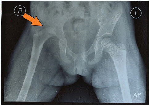
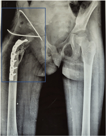
Diagnostic procedures such as an x-ray have been done, which revealed developmental dysplasia of the hip's right side, which can be seen in Figure 1 and Figure 2. Which are mentioned at end of manuscript.

Figure 1: X-ray from November 2022. Alpha angle is reduced; Tonnis angle is increased; Acetabular index is increased; Femoral neck shaft angle is increased.

    Figure 2: X-ray from September 2023.

Showing screw plate fixation for stabilization of femoral head in acetabulum and K-wire fixation is done for maintaining joint alignment and congruency.
Physiotherapy Intervention
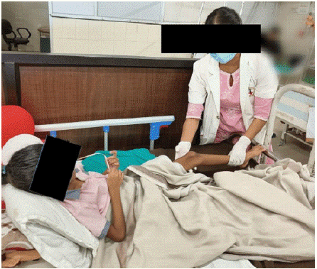
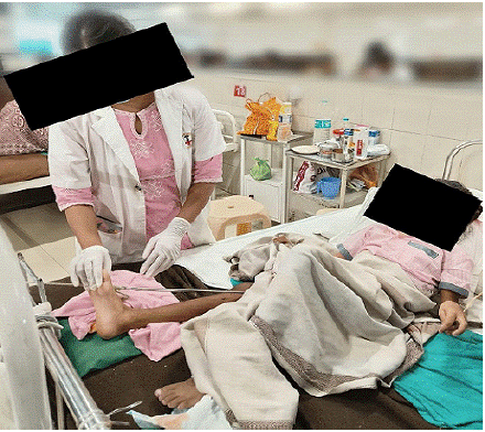
The patient was provided with preoperative physiotherapy intervention after diagnosing the patient with right side developmental dysplasia of the hip for which physiotherapy intervention was given for proper and early recovery post-operatively; hence, strengthening exercises for the left lower limb were shown, and ankle toe movements to prevent from deep vein thrombosis and diaphragmatic breathing exercises were given which were proven for better rehabilitation and early recovery of the patient. Table 4 represents physiotherapy intervention. Figure 3 shows active assisted range of motion exercise for left knee joint and Figure 4 shows passive range of motion exercise for affected extremity.
    Resisted    Isometrics

    Grading


    Ankle plantar flexors

    Weak painless


    Ankle dorsiflexors

    Weak painfull


    Ankle invertors

    Weak painfull


    Ankle evertors

    Weak painless
Table 3: Resisted isometric grading for ankle (right lower limb).
    Problem identified

    Physiotherapeutic    goals

    Intervention


    Lack    of awareness regarding physiotherapy treatment

    Patient and caregiver education.

    Educating    patients about recuperation processes, physiological and psychological agony    processes, methods of relaxation,    and cognitive strategies for coping and managing pain.


    Tightness    of hip adductors

    Improve flexibility of hip adductors

    Hip adductor    stretching: 1. Butterfly stretch: Set both of your feet firmly over the floor.    keep the soles of    the feet close to ensure both knees are pointing out. Draw your heels in as close to your groin as you can. Gently push your knees closer to the floor with your hands. Hold the position for 30 seconds to 1 minute. Relax and repeat 2–3 times more. 2.    lateral lunge stretch: Place your    legs approximately hip width apart. Step out to the side and maintain your    other foot on the flat surface. While maintaining the opposite knee straight, bend your "stepping" knee. Compared to backward and forwardward lunges, your torso will    tilt forward somewhat, and your shoulders will be slightly ahead of your knee. To return to the    beginning position, forcefully push off with your foot.


    Decreased mobility of hip joint

    To improve    mobility and range of motion of the affected limb

    Active    assisted ROM exercises for hip movements [flexion, extension, abduction,    external and internal rotation]. 10    reps, 1 set Dynamic assisted ROM exercises for the knee joint. 10 reps, 1 set Active assisted ROM exercises for the    ankle joint. 10 reps, 1 set


      Gait impairment

    To    improve gait and to make the patient    independent

    1.    Vojta therapy 2. Hippotherapy also helps to normalize gait patterns as the    horses' three- dimensional movement    replicates the typical movements of the human pelvis when walking. The horse's rhythmic, repeated, and    multidirectional movements stimulate the patient's anterior and posterior swinging movements [11].


    Dynamic balance impairement

    To    improve dynamic balance

    Tandem    walking, grapevine walking, high knee jumps, lunges [forward, backwards,    lateral], treadmill training for 15    minutes, step-ups.


    Pelvic muscle    weakness

    To    strengthen pelvic muscles

    Kegel exercise    Pelvic bridging


    Weakness    of hip abductors

    To strengthen hip abductors

    1.    Isometric hip abduction against the wall • Position yourself sideways and    slightly away from a wall, with the    affected side closest to the wall. • Keep the knees in line by bending the    knee of the leg closest to the wall    and pressing it against the wall. •    Maintain this position with approximately 70% effort - you should feel this working around the top, outer    part of the buttock.


    Reduced muscle to normalize


      Tone and achieve


      Postural control


      Tone and postural


      control Hippotherapy:    A walking horse's    movement generates 100 rhythmic impulses


      each minute.


      Repeatedly    challenging postural reflexes and keeping the patient seated on the


      horse enables patient to achieve and maintain balance and upright posture. As a


      result, balance, muscle control, postural control and paraspinal muscle growth


      are improved.
Table 4: Demonstrates physiotherapy intervention.

Figure 3: Active assisted range of motion exercise for left knee joint.

Figure 4: Demonstrates passive range of motion exercise for affected extremity.
Outcome Measures
Table 5 shows range of motion of bilateral lower limb (pre and post rehabilitation) and Table 6 shows manual muscle testing of bilateral lower limb (pre and post rehabilitation).
    Joint

    Degree of    freedom

    Pre-Rehabilitation

    Post    Rehabilitation


    Right

    Left

    Right

    Left


    Hip joint

    Flexion

    Not    assessable

    00-1200

    00-800

    00-1200


    Extension

    Not    assessable

    00-300

    00-150

    00-300


    Abduction

    Not    assessable

    00-450

    00-400

    00-450


    Adduction

    Not    assessable

    00-300

    00-150

    00-300


    External rotation

    Not    assessable

    00-450

    00-300

    00-450


    Internal rotation

    Not    assessable

    00-450

    00-300

    00-450


    Knee joint

    Flexion

    00-900

    00-1350

    00-1200

    00-1350


    Extension

    900-00

    1350-00

    1200-00

    1300-00


    Ankle joint

    Plantar flexion

    00-400

    00-500

    00-450

    00-500


    Dorsiflexion

    00-100

    00-200

    00-150

    00-200


    Inversion

    00-200

    00-300

    00-300

    00-350


    Eversion

    00-100

    00-150

    00-150

    00-150


    "4": (Grade 4) Movement against    gravity and resistance


      "3": (Grade 3)    Movement against gravity over (almost) the full range
Table 5: Shows range of motion of bilateral lower limb (pre and post rehabilitation) and Table 6: Shows manual muscle testing of bilateral lower limb (pre and post rehabilitation).
Discussion
This study provides information on the significance of early DDH diagnosis in physiotherapy practice [12]. We have noticed that stretching and strengthening has benefited the patient in a study conducted By Abhishek Sharma et al. 2022 study revealed that the Initial execution of an organized and individualized rehabilitation plan is beneficial for enhancing functional deficits. After an overview of the improvements and outcomes recorded in the investigation's findings, it is apparent that the impact of physical therapy portrayed the array of beneficial contributing variables to overall skeletal muscle equilibrium, as demonstrated by kids optimized overall development as their chronological age is approached [13]. Wojciech Kiebzak et al. As stated by Vojta, addressing a neuromotor hip joint formation problem should entail the application of universal motifs. Children with congenital hip dysplasia should begin rehabilitation as soon as possible [14]. Hippotherapy can help improve gait, balance, and postural control while increasing functional performance [11]. The systematic and tailored physiotherapy regimen that is performed early serves an indisputable effect in improving children's functional impairments. functional limitations in children. Based on an assessment of the children's development, it is evident that the benefits of physical therapy, including Vojta procedures, were not only local but also represented a variety of positive factors that supported overall muscular balance. This is demonstrated by the children's improved overall development relative to their chronological age. Because of the objective evaluation and investigation methods used, the documented results are concrete, emphasizing the usefulness and significance of physical therapy for newborns with hip dysplasia [15].
Benedetti et al. 2021 studied that DDH patients who went through THR, a strengthening exercise programme for the gluteal muscles resulted in an improvement in muscle strength, which enhanced efficiency and satisfaction with treatment [16]. Piechocka et al. 2018 mentioned that Physiotherapy is employed in surgical, conservative, and preventive treatments, and it plays a significant part in all of them. Physiotherapy is used to treat hip joints. It takes the form of massage, kinesiotherapy, and the selection of the right orthopedic equipment to correct the problem and preserve the therapeutic impact obtained following surgical treatment. It also allows and aids in the maintenance of normal limb function [17]. In patients with intra-articular hip pathology, conservative treatments, such as exercise modification and physical therapy, may be employed as the treatment of choice [18]. The application of massage, physiotherapy, gymnastics, and chromotherapy without using orthopedic structures to treat lower limbs with established hip dislocations or subluxations, which inevitably caused the joint's structural modifications to advance [19]. Aykurt, B et al. 1987 studied those 22 hips from 17 individuals who had been diagnosed with congenital dislocation of hip received postoperative therapy. The Physical Therapy and Rehabilitation Department carried out the rehabilitation interventions after the Department of Orthopedic Surgery, Ataturk University, performed surgery using the Radical Deduction Technique. The patients were evaluated by carefully taking into account a number of factors, including pain severity, range of motion, muscular strength, walking ability, and compliance with the extensive rehabilitation regimen. The patients' ability to regain a functional hip joint was finally made possible by the rehabilitation program's effectiveness, which was demonstrated by the improvement of normal hip range of motion and the growth of muscular strength [20].
Conclusions
The study revealed the efficacy of a tailored therapy strategy that focuses on gait and balance training to improve postural control and mobility and flexibility. Furthermore, patient awareness of the necessity of care following discharge was emphasized, with positive results. The study revealed positive effects of specialized therapies named hippotherapy and Vojta treatment that played a very important role in improving patients'gait, balance and postural control issues. Indeed, notwithstanding the observed amelioration in the patient's condition, it is imperative to underscore the significance of diligent and consistent follow-up as an integral component of the rehabilitation process. This case report serves as a beacon of hope for individuals facing DDH, healthcare professionals, and caregivers, demonstrating that with early intervention, a holistic approach, patient engagement, and a collaborative effort, successful rehabilitation, and improved quality of life are attainable goals in managing Developmental dysplasia of hip.
Abstract
- Zhang C, Jin D, Feng Y. Developmental dysplasia of the hip. Changqing Zhang (ed: Springer. 2020.
- Basit S, Hannan MA, Khoshhal KI. Developmental dysplasia of the hip: usefulness of next generation genomic tools for characterizing the underlying genes - a mini review. Clin Genet. 2016; 90.
- Kremli MK, Alshahid AH, Khoshhal KI, Zamzam MM. The pattern of developmental dysplasia of the hip. Saudi Med J. 2003; 24: 1118–20.
- Klisic PJ. Congenital dislocation of the hip-a misleading term: brief report. J Bone Joint Surg Br. 1989; 71.
- Zamborsky R, Kokavec M, Harsanyi S, Attia D, Danisovic L. Developmental dysplasia of hip: perspectives in genetic screening. Med Sci. 2019; 7.
- Agarwal A, Gupta N. Risk factors and diagnosis of developmental dysplasia of hip in children. Orthop Trauma. 2012; 3: 10–4.
- Nandhagopal T. De Cicco FL: Developmental dysplasia of the hip. StatPearls Publishing, Treasure Island; 2023.
- de Hundy M, Velmmix F, Bais JMJ, Hutton EK, de Groot CJ, Mol BWJ, et al. Risk factors for developmental dysplasia of the hip: a meta-analysis. Eur J Obstet Gynecol Reprod Biol. 2012; 165: 8–17.
- Debuse D, Chandler C, Gibb C. An exploration of German and British physiotherapists’ views on the effects of hippotherapy and their measurement. Int J Sports Phys Ther. 2005; 21: 219–42.
- Park ES, Rha DW, Shin JS, Kim S, Jung S. Effects of hippotherapy on gross motor function and functional performance of children with cerebral palsy. Yonsei Med J. 2014;55:1736–42.
- Koca TT, Ataseven H. What is hippotherapy? the indications and effectiveness of hippotherapy. North Clin Istanb. 2016; 2: 247–52.
- Vasilcova V, AlHarthi M, AlAmri N, Sagat P, Bartik P, Jawadi AH, et al. Developmental dysplasia of the hip: prevalence and correlation with other diagnoses in physiotherapy practice—a 5-year retrospective review. Children. 2022; 9.
- Sharma A, Vats S, Gupta R. Effectiveness of physiotherapy intervention in managing patient’s developmental dysplasia of the hip: a scoping review. SN Compr Clin Med. 2022: 4.
- Kiebzak W, Zurawski A, Dwornik M. Vojta method in the treatment of developmental hip dysplasia - a case report. Ther Clin Risk Manag. 2016; 12: 1271–1276.
- Marinela R. Early physical therapy intervention in infant hip dysplasia. Procedia Soc Behav Sci. 2013; 76.
- Benedetti MG, Cavazzuti L, Amabile M. Abductor muscle strengthening in THA patients operated with minimally-invasive anterolateral approach for developmental hip dysplasia. HIP Int. 2021; 31: 66–74.
- Piechocka E, Wrzesinski B, Wojtczak P, Ziólkowska A, Ciecierska D. Physiothreapeutic treatments in infants with congenital hip dysplasia. J Educ Health Sport. 2018; 8: 37-44.
- Kraeutler MJ, Garabekyan T, Pascual-Garrido C, Mei-Dan O. Hip instability: a review of hip dysplasia and other contributing factors. Muscles Ligaments Tendons J. 2016; 6: 343–53.
- Pozdnikin IY,Baskov VE: Errors of diagnosis and the initiation of conservative treatment in children with congenital hip dislocation. Pediatric Traumatol Orthop Reconstr Surg. 2017; 5: 42–51.
- Aykurt B, Aykurt M. Postoperative rehabilitation of the patients with congenital hip dislocation jere operated by radical reduction technique. Acta Orthop Traumatol Turc. 2006; 21: 178–82.
