Abstract
Limbus vertebra is defined as the presence of an ossicle or an adjacent bone affecting the margin angle of the vertebral bodies. The characteristic appearance on plain films is a detached fragment with triangular morphology and sclerotic margins. It represents a marginal herniation of the nucleus pulposus during childhood or adolescence through the vertebral end-plate and beneath the apophyseal ring.
Usually it is considered an incidental finding and an asymptomatic entity. However, several studies have suggested a significant relationship between the limbus vertebra and the low back pain (LBP), possibly due to an underlying intervertebral disc degeneration (IDD).
In order to contribute to the debate, we present the case of an adult male who has a double limbus vertebra and suffers from chronic LBP.
Keywords: Limbus vertebra; Radiological abnormalities; Low back pain
Abbreviations
LBP: Low Back Pain; NSAIDs: Nonsteroidal Anti-Inflammatory Drugs; CT: Computerized Tomography; MRI: Magnetic Resonance Imaging; ALV: Anterior Limbus Vertebra; IDD: Intervertebral Disc Degeneration
Case Presentation
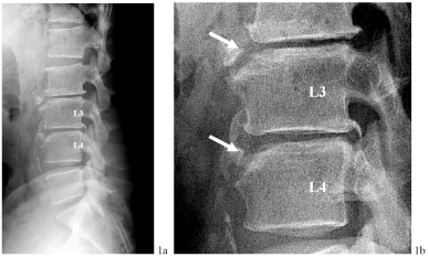
Male, aged 53 years. He worked as a truck driver, with activities that involved lifting heavy loads. He lived in a cattle area of Argentina during eight years. Chronic psoriasis on elbows and knees and an acute coronary syndrome in 2008 are the most relevant personal antecedents. He suffers from a chronic LBP that began at the age of 30 and the symptoms are almost daily, with few variations along these years. Occasionally the pain radiates to the back of the left thigh, without paresthesias. He denies the existence of “red flags” symptoms including fever, constitutional syndrome or inflammatory pain. The pain improves with rest and nonsteroidal anti-inflammatory drugs, but does not disappear completely. He describes morning stiffness that lasts about an hour. In the physical examination, neither arthritis nor enthesitis were noted and the muscular strength was normal, as well as the Achilles and patellar tendon reflexes. Radiculopathy and sacroiliac maneuvers were negative. Lumbar spine radiography showed rectification of the lumbar lordosis, avulsion of the anterior superior angle of L3 and L4 vertebral bodies, narrowing of the L2- L3 and L3-L4 spaces, posterior osteophytes and inter-apophyseal osteoarthritis (Figure 1). The patient underwent a blood analysis -including Rose Bengal test for brucellosis and acute phase reactants like erythrocyte sedimentation rate and C-reactive protein- and all results turned out normal or negative. Computerized tomography (CT) and magnetic resonance imaging (MRI) of the lumbar spine showed vertebral epiphysitis in L3 and L4, degenerative changes in disc spaces L2-L3 and L3-L4, a bulging disc between L2-L3, a central herniation in L3-L4 and a left side herniation in L5-S1, none of them with apparent compressional effect. MRI of sacroiliac joints did not show relevant abnormalities.

    Figure 1:  Anterior limbus vertebra affecting L3 and L4.
Lateral radiography of the lumbar spine of the patient (1a): Double limbus vertebra affecting L3 and L4.
Bone fragments on the anterior and superior end-plates of L3 and L4 vertebral bodies (Arrows, 1b). Furthermore, radiological signs of degeneration of the adjacent
discs: subchondral sclerosis, osteophytes and narrowing of the disc spaces L2-L3 and L3-L4.
Our differential diagnosis was directed to three entities: A psoriatic arthritis with axial involvement, a brucellar spondylitis with epiphysitis -Pedro Pons´ sign- [1] and finally, an anterior limbus vertebra (ALV) affecting two vertebral bodies with added radiological features of spinal osteoarthritis. The complementary tests discarded reasonably the first two entities and pointed out the ALV as the most likely diagnosis.
Discussion
The limbus vertebra was first described by C.G. Schmörl in 1927. It is a bone deformity produced as a consequence of an injury in an immature skeleton, at childhood or adolescence, caused by an intrabody marginal herniation of the nucleus pulposus. This results in the separation of a triangular bone fragment which apparently represents the ring apophysis and may ossify separately [2]. Its pathogenesis is related to Scheuermann's disease [3], an entity with which it usually coexists [4]. Plain lateral radiographs reveal a triangular piece with sclerotic margins next to a bone defect in the vertebral body. The superior anterior corner of a vertebral body in the middle lumbar spine is most frequently affected, whereas the inferior and posterior margin and other regions are less frequently found [2,5].
Recently, a TT genotype of COL11A1 polymorphism, the gene encoding the a1 chain of type XI collagen, has been associated with a significant risk for presenting the limbus vertebra [6]. Genetics and cumulative mechanical stress on an immature skeleton may play a role in the development of the ALV. Perhaps due to that, the ALV is more prevalent among athletes [7], specially those whose sport involves loading of the back in flexion such as gymnastics or weight lifting [3].
It is of general consensus that posterior limbus vertebra has potential clinical consequences, since the bone fragment may precipitate a narrowing in the spinal canal, causing pain and symptoms of nerve compression [8]. On the contrary, there is an increasing debate about the clinical significance of the ALV. Some authors consider ALV as an incidental finding, typically asymptomatic and with no relationship with LBP [9-12]. Interestingly, the great majority of reported cases of ALV refer to symptomatic patients, whereas there is a noticeable lack of reports in asymptomatic population. In fact, it has been pointed out [13] that the case described by Mupparapu et al of a 14-year-old male with a VLA in the fourth cervical vertebra discovered during a radiographic assessment for orthodontic treatment [4], may be at present the sole reported case of a true incidental finding of an asymptomatic ALV.
Decades ago, some MRI studies performed on adolescents demonstrated that the discs adjacent to ALV presented a certain degree of degeneration. Swischuk et al [14] analyzed retrospectively the MRI findings in twelve patients, aged 12-27, with various disc problems including Scheuermann´s disease, Schmörl nodes and ALV (four of them). All patients shared loss of disc height, altered disc hydration and variable herniation of nuclear material. Henales et al [15] revised 15 cases (children aged 10-15) with back pain and limbus vertebra. 5 patients reported vigorous sporting activity and 2 had had a previous trauma. In the 13 cases with ALV, besides the disc herniation, 8 presented subchondral sclerosis and in 6 cases, the intervertebral space was reduced. After a 12 years follow-up, 3 patients with initial ALV showed the radiological lesion of Schmörl´s hernia, and in the remaining cases the ALV and symptoms of LBP persisted.
More recently, Koyama et al analyzed 104 collegiate gymnasts (70 men and 34 women, aged 19.7±1 years) in a case-control study, to evaluate ALV and IDD using MRI. The authors observed that the ALV, after adjustment for sex and weight, was a strong predictor of IDD, especially in the upper half of the lumbar spine [16].
It has been suggested that the appearance of ALV may occur at an early age, prior to the IDD [16], and that the trigger of the changes that precipitate the IDD might be the end-plate lesion, produced by the intravertebral herniation of disc material [17]. Furthermore, the IDD seems to be accelerated by the accumulation of bending loads on an extruded disc [18].
Whereas there is evidence that ALV may be related to IDD, the relationship between IDD and back pain remains controversial. The intradiscal pathology seems to play a role in the back pain, both nonspecific and chronic [19]. Specifically, the LBP has been associated with changes in the annulus, with disc protrusions and with the decrease of signal intensity of the disc [19]. Different theories have been invoked, such as the pressure on the roots of spinal nerves caused by the disc bulging or by the extrusion of disc material. Moreover, it has been suggested that the narrowing of the intervertebral space could cause a disturbance in spine biomechanics and an increased pressure on adjacent structures, including disc spaces, facet joints and spinal ligaments, with associated pain arising from pressure on the affected nociceptors [20]. The presence of IDD does seem to increase the probability of LBP, even more when the number of affected discs is greater [19]. However, it is noteworthy that up to 30% of asymptomatic subjects have degenerative changes in the intervertebral discs [19]. In this same line, a systematic review, designed to assess how confidently LBP can be attributed to abnormalities on MRI, showed that MRI findings of disc protrusion and disc degeneration, among others, are associated with LBP, but individually none of them provides a strong indication that LBP is attributable to underlying pathology [21].
Regarding to our patient, with a double ALV that probably dates back from his adolescence, it is plausible that the occupational activity, with cumulative stress on the two previously extruded discs, L2-L3 and L3-L4, could have accelerated the disc degeneration and precipitated a chronic LBP at 30 years old. Plain radiography with a double ALV and radiographic findings of IDD [20], such as the narrowing of disc spaces L2-L3/L3-L4 and marginal osteophytes, as well as the CT/MRI findings of degenerative changes in disc spaces L2-L3 and L3-L4, a bulging disc L2-L3 and a central herniation in L3- L4, are both consistent with that clinical presentation.
Conclusion
This case could support the possible association between the ALV and the degeneration of the intervertebral disc, and in this line, as some authors have stated before [13], this entity should not be considered as an incidental finding. In our opinion, discovering an ALV in the context of a subacute/chronic LBP justifies the recommendation of a CT or MRI study, directed to evaluate the adjacent intervertebral discs as well as to make a correct diagnosis.
References
- Tuna N, Ogutlu A, Gozdas HT, Karabay O. Pedro Pons' sign as a Brucellosis complication. Indian J Pathol Microbiol. 2011; 54: 183-184.
- Ghelman B, Freiberger RH. The limbus vertebra: an anterior disc herniation demonstrated by discography. AJR Am J Roentgenol. 1976; 127: 854-855.
- Hollingworth P. Back pain in children. Br J Rheumatol. 1996; 35: 1022-1028.
- Mupparapu M, Vuppalapati A, Mozaffari E. Radiographic diagnosis of Limbus vertebra on a lateral cephalometric film: report of a case. Dentomaxillofac Radiol. 2002; 31: 328-330.
- Yagan R. CT diagnosis of limbus vertebra. J Comput Assist Tomogr. 1984; 8: 149-151.
- Koyama K, Nakazato K, Min S, Gushiken K, Hatakeda Y, Seo K, et al. COL11A1 gene is associated with limbus vertebra in gymnasts. Int J Sports Med. 2012; 33: 586-590.
- Bennett DL, Nassar L, DeLano MC. Lumbar spine MRI in the elite-level female gymnast with low back pain. Skeletal Radiol. 2006; 35: 503-509.
- Goldman AB, Ghelman B, Doherty J. Posterior limbus vertebrae: a cause of radiating back pain in adolescents and young adults. Skeletal Radiol. 1990; 19: 501-507.
- Sanal HT, Yilmaz S, Simsek I. Limbus vertebra. Arthritis Rheum. 2012; 64: 4011.
- Yen Y1, Wu FZ1 . Giant limbus vertebra mimicking a vertebral fracture. QJM. 2014; 107: 937-938.
- Learning Radiology: Limbus vertebra. [Accessed on November 15 2015].
- Quintana I, Mellado JM, Yanguas N, Martin J, Ibáñez D, Solanas S et al. Lumbar spine radiography in the emergency room: pearls and pitfalls for the radiologist on call. European Congress on Radiology, 2014 [Accessed on November 16 2015].
- Martínez-Carpio PA, Bedoya del Campillo A, Leal MJ, Lleopart N. Lumbalgia mecánica crónica en pacientes con vértebra limbus anterior: revisión de la literatura y presentación de tres casos clínicos. Rev Arg Reumatol. 2013; 24: 36-42.
- Swischuk LE, John SD, Allbery S. Disk degenerative disease in childhood: Scheuermann's disease, Schmorl's nodes, and the limbus vertebra: MRI findings in 12 patients. Pediatr Radiol. 1998; 28: 334-338.
- Henales V, Hervás JA, López P, Martínez JM, Ramos R, Herrera M. Intervertebral disc herniations (limbus vertebrae) in pediatric patients: report of 15 cases. Pediatr Radiol. 1993; 23: 608-610.
- Koyama K, Nakazato K, Min SK, Gushiken K, Hatakeda Y, Seo K, et al. Anterior limbus vertebra and intervertebral disk degeneration in japanese collegiate gymnasts. Orthopaedic Journal of Sports Medicine. 2013; 1: 2325967113500222.
- Wang Y, Videman T, Battié MC. Lumbar vertebral endplate lesions: prevalence, classification, and association with age. Spine (Phila Pa 1976). 2012; 37: 1432-1439.
- Hsu K, Zucherman J, Shea W, Kaiser J, White A, Schofferman J, et al. High lumbar disc degeneration. Incidence and etiology. Spine (Phila Pa 1976). 1990; 15: 679-682.
- Luoma K, Riihimäki H, Luukkonen R, Raininko R, Viikari-Juntura E, Lamminen A. Low back pain in relation to lumbar disc degeneration. Spine (Phila Pa 1976). 2000; 25: 487-492.
- Pye SR, Reid DM, Smith R, Adams JE, Nelson K, Silman AJ, et al. Radiographic features of lumbar disc degeneration and self-reported back pain. J Rheumatol. 2004; 31: 753-758.
- Endean A, Palmer KT, Coggon D. Potential of magnetic resonance imaging findings to refine case definition for mechanical low back pain in epidemiological studies: a systematic review. Spine (Phila Pa 1976). 2011; 36: 160-169.
