
Abstract
Background: Pregnancy with arteriovenous malformations can lead to life threatening complications if not properly recognized.
Case: A 22 year old white female 32 weeks pregnant with history of Hereditary Hemorrhagic Telangiectasias (HHT) and previous coiling 8 years prior presents with syncope and hypoxia. The patient is found with blood in her nasal and oral passage. She is intubated and transferred for further evaluation.
Computerized tomography of the chest and pulmonary angiogram reveal the presence of arteriovenous malformations, previous coils and distal pulmonary hemorrhage. Following balloon embolization and recoiling, there is no evidence of arteriovenous malformations. The patient’s platypnea (dyspnea upright) and orthodeoxia (hypoxia upright) resolve and she was discharged home.
Conclusion: Screening for arteriovenous malformations with prior history of hereditary hemorrhagic telangiectasia is essential to avoid complications in pregnancy. As pregnancy progresses at 12, 24 and 36 weeks respectively, blood volume increases leading to potential for life threatening hemoptysis in presence arteriovenous malformations. Close monitoring in patients with HHT for playpnea and orthodeoxia is essential to avoid catastrophic bleeding.
Keywords: Osler Weber Rendu; Syncope; Pregnancy; Orthodeoxia; Arteriovenous malformations
Teaching Points
1) Osler Weber Rendu in pregnancy can lead to massive hemoptysis due to increased vascular volume.
2) Pregnancy with hypoxia should be evaluated for platypnea and orthodeoxia.
3) Patients with appropriate history and physical exam findings should undergo coiling of arterio-venous malformations to prevent catastrophic hemoptysis.
Introduction
Pregnancy with arteriovenous malformations can lead to life threatening complications.
Hereditary Hemorrhagic Telangiectasia (HHT) is a rare disorder with a nonspecific presentation.
We present a case of HHT in pregnancy with catastrophic hemoptysis. Treatment options as well as clinical sequelae are presented.
Case Presentation
A 22-year-old white woman at 32 weeks gestation was found unconscious with hypoxia and blood in her nasal and oral passages. The patient was intubated and transferred to the emergency department. The patient’s mother gives a history of similar presentation 8 years prior.
The patient was extubated and gave a history of fatigue and platypnea for several days. She denied hemoptysis prior to this event.
Physical examination revealed sinus tachycardia (pulse of 112). She was afebrile and blood pressure was 112/60. She had a grade 2/6 systolic murmur with lungs clear to auscultation and trace peripheral edema.
Arterial blood gas demonstrated sever hypoxia (7.46/31/69/22/95% oxygen saturation on room air) in upright position on 100% fio2. Serum chemistries and liver function tests were within normal limits.
Her clinical and radiologic presentation was consistent with HHT. The patient had no evidence of pulmonary embolism. She underwent repeat vascular coiling resulting in resolution of the arteriovenous malformations. Arterial blood gas following the coiling procedure showed resolution of the hypoxia (7.49/33/83/24/97% on room air). The patient’s platypnea and orthodeoxia resolved and she was discharged home. She had an uneventful vaginal delivery 5 weeks after the coiling procedure.
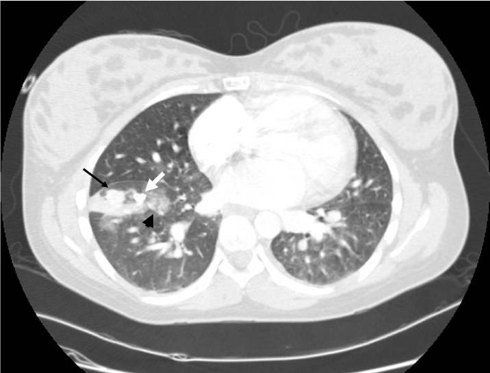
    Figure 1:  Computerized tomography reveals presence of arteriovenous
malformations (light black arrow) distal to the coils (heavy white arrow) and
pulmonary hemorrhage (black arrowhead).
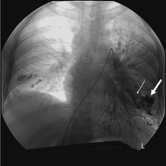
    Figure 2:  Pulmonary angiogram reveals the previous coils (light white arrow)
and significant distal arteriovenous malformations (heavy white arrow).
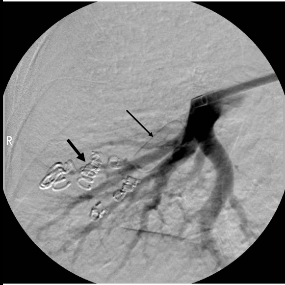
    Figure 3:  Following embolization with balloon (light black arrow) and recoiling
(heavy black arrow), there is no evidence of arteriovenous malformations.
Comment
Pregnancy poses a definite risk in HHT. With an overall incidence of 1 in 5000, HHT is inherited as an autosomal dominant trait. Offspring have a 50% risk of inheriting HHT [1].
Arteriovenous malformations (AVM) of the pulmonary, hepatic and cerebral circulations have been increasingly described. Pulmonary arteriovenous malformations will enlarge during pregnancy with
increased blood flow and alterations in vascular tone specifically at 12, 24 and 36 weeks gestation. Hypoxia during pregnancy results from shunting or vascular rupture [2].
Patients typically present with characteristic telangiectasias of the oral mucosa, fingertips, nosebleeds, gastrointestinal bleeding, and iron deficiency anemia and hemoptysis. While the differential for hemoptysis and syncope is broad including acute pulmonary embolism with infarct, mitral stenosis, bronchial artery rupture in setting of congenital heart disease with pulmonary artery hypertension and massive hematemesis with aspiration, our patient had a known history of arteriovenous malformations.
Diagnosis involves a high index of suspicion coupled with clinical sequelae of arteriovenous malformations.
Women with HHT should be screened for pulmonary and spinal AVM’s as at time of delivery; epidurals can pose a risk [2]. Pulmonary AVM’s can be detected via CT chest, contrast echocardiography or pulmonary angiogram [3].
Treatment during pregnancy consists of embolisation in an experienced clinical center. Even patients with silent AVM’s are at risk of hemorrhage and paradoxical embolism consisting of cerebral abscess and embolic strokes.
HHT type 1 and type 2 occur as autosomal dominant traits with mutation in endoglin and ALK-1 genes respectively [3,4]. HHT patients demonstrate decreased endoglin and ALK-1 levels.
Conclusion
Pregnancy coupled with Hereditary Hemorrhagic Telangiectasia needs careful pre and perinatal care. Arteriovenous malformations have the potential for causing serious complications throughout pregnancy in the mother and fetus therefore screening is essential.
References
- Begbie M, Wallace G, Shovlin C. Hereditary Haemorrhagic Telangiectasia (Osler-Weber-Rendu syndrome): a view from the 21st century. Postgrad Med J. 2003; 79: 18-24.
- Shovlin C, Winstock A, Peters A, et al. Medical complications of pregnancy in hereditary haemorrhagic telangiectasia. QJM. 1995; 88: 879-887.
- Azuma H. Genetic and molecular pathogenesis of hereditary hemorrhagic telangiectasia. J Med Invest. 2000; 47: 81-90.
- Dagdeviren A, Muftuolu SF, Cakar AN, et al. Endoglin (CD 105) expression in human lymphoid organs and placenta. Anat Anz. 1998; 180: 461-469.