
Abstract
Tuberous sclerosis complex (TSC) is a genetic neurocutaneous syndrome characterized by the classical triad of intellectual disability, seizures, and skin findings. Cutaneous findings of hypopigmented macules and facial angiofibromas are usually the initial presenting symptoms in early childhood. However, a minority of patients do not develop such features early in life; instead they may present with alternative skin findings later in life causing a delay in diagnosis. Herein, we present the case of a 58-year old female with a history of seizures and renal disease who developed a Shagreen patch and ungual fibromas later in life. She was diagnosed with TSC in our dermatology clinic based on the presence of ungual fibromas. We report this case to highlight the necessity of recognizing late onset skin features of TSC, particularly in patients with seizures, to prevent delays in tumor screening and management.
Keywords: Tuberous sclerosis complex; Periungual fibromas; Shagreen patches; Collagenoma
Abbreviations
TSC: Tuberous Sclerosis Complex
Introduction
Tuberous sclerosis complex (TSC) is an inherited neurocutaneous syndrome characterized by the development of multi-organ hamartomas, usually of the brain, skin, kidneys, and eyes [1]. Estimated birth incidence of TSC is 1 in 6000 to 1 in 10,000 live births with a population prevalence of 1 in 20,000 [2,3]. The syndrome is inherited in an autosomal dominant pattern or presents as new spontaneous mutations in TSC1 and TSC2, encoding tumor suppressive genes hamartin and tuberin, respectively [1]. Dysfunctional hamartin and tuberin lead to constitutive activation of mammalian target of rapamycin (mTOR), promoting cell growth that result in the hamartomas of TSC.
Diagnosis of TSC relies on identifying key clinical features. The “classic triad” includes the presence of cutaneous angiofibromas, intellectual disability, and early onset seizures [4]. Angiofibromas, also known as fibrous papules of the face or adenoma sebaceum, present as multiple dome-shaped papules distributed symmetrically on the cheeks, nose, and chin. Although the full triad occurs in less than a third of the patients, nearly 100% have characteristic skin findings, which include, in addition to facial angiofibromas, hypopigmented macules, Shagreen patches, and ungual fibromas.
The 2012 International Tuberous Sclerosis Complex Consensus Group listed 11 major and 6 minor clinical features; either two major or one major and two minor criteria are sufficient for a definitive diagnosis [5]. Major cutaneous features include Shagreen patches (a type of connective tissue hamartoma or collagenoma presenting as a pink, yellow plaque with an orange peel like texture), =3 facial angiofibromas, =3 hypopigmented macules at least 5-mm in diameter, and =2 ungual fibromas, also known as Koenen tumors (skin colored nodules appearing on the proximal nail folds). More severe cases may present with multi-organ complications including pulmonary lymphangioleiomyomatosis, renal disease, cardiac rhabdomyomas, and subependymal giant cell astrocytomas of the brain [6].
Although TSC most commonly manifests and is diagnosed in infancy or childhood, cases have been reported of adult diagnoses [7,8]. We report a 58-year old female who initially presented with asymptomatic dermatologic findings and was subsequently diagnosed with TSC.
Case Presentation
A 58-year-old Caucasian female presented to our dermatology clinic for a nonspecific complaint. Incidentally, visible growths were noted along her nail folds, which led to a more thorough skin exam. Physical examination revealed discrete, smooth, skin-colored to pink firm papules along nail folds of multiple fingers (Figure 1) and toes (Figure 2); irregular, skin-colored, rubbery plaques on lower back (Figure 3), and stippled hypopigmented macules on the bilateral lower extremities. Skin findings on fingers and toes were previously diagnosed as warts. Upon further questioning, the patient endorsed a history of seizures of unknown etiology since infancy despite a detailed neurologic workup. She had been following with a nephrologist for chronic kidney disease in setting of known multiple bilateral renal cysts. Family history was positive for seizures, renal disease and similar nail fold findings in her daughter.
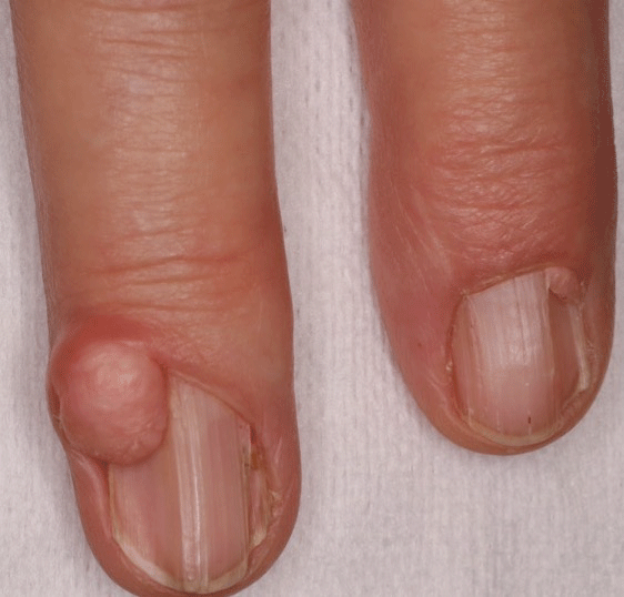
    Figure 1A:  Large discrete, smooth, skin-colored to pink papules (ungual
fibromas) along nail folds of fingers.
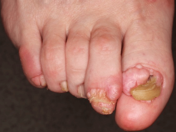
Figure 2: Multiple ungual fibromas along nail folds of multiple toes.
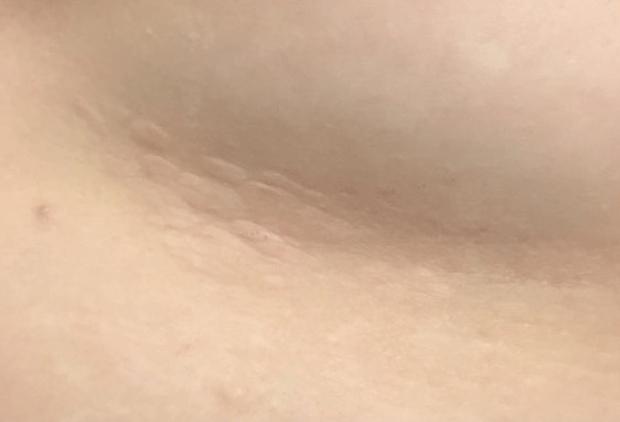
Figure 3: Irregular, skin-colored, rubbery plaque on right lower back.
Histopathology of the lesion on the right third digit revealed compact collagen with an increased number of spindle shaped fibroblasts as seen in periungual fibromas (Figure 4). Lesion on the right lower back showed thickened bundles of collagen arranged in a slight haphazard array in the mid-dermis with an increase in the number of stellate fibroblasts. A methyl trichrome stain demonstrated thick collagenous stroma almost entirely replacing dermis and adnexal structures including hairs, eccrine glands and coils (Figure 5), findings consistent with collagenoma (Shagreen patch). Overall, our patient fulfilled at least 2 major diagnostic criteria (Shagreen patch and =2 ungual fibromas) for TSC. Biopsy findings, in conjunction with clinical history and physical exam, led to a cohesive diagnosis of TSC.
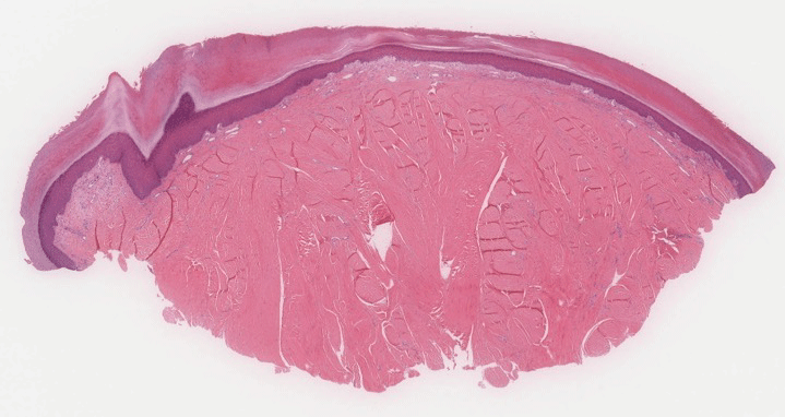
    Figure 4:  Biopsy of third finger digit showing collagen with an increased
number of spindle shaped fibroblasts as seen in ungual fibromas (H&E, 40x).
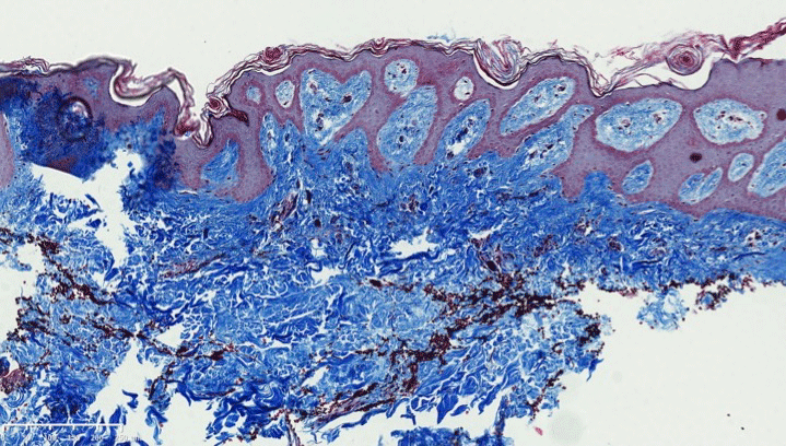
    Figure 5:  Shagreen patch with thick collagenous stroma almost entirely
replacing dermis and adnexal structures (Methyl trichome stain, 40x).
Discussion
Although TSC is typically an early childhood diagnosis, several retrospective reviews demonstrate that a late diagnosis may be more prevalent than initially perceived [2,9]. One reason for diagnostic delay is that sufficient criteria may not manifest until adulthood [9]. Furthermore, up to 25% of patients may never develop the characteristic facial angiofibromas that typically appear earlier in childhood [10]. Presence of ungual fibromas that commonly appear after puberty may be the key to diagnosis in this group of patients [11]. Ungual fibromas are usually noticed while the patient is evaluated for a different complaint or if the lesions present a cosmetic concern [12]. In our case, the diagnosis of TSC was made late in adulthood based on the incidental discovery of adult onset ungual fibromas.
Staley and colleagues compared presenting symptoms of early versus late TSC diagnosis in 243 patients; 81% of patients were diagnosed prior to the age of 10 whereas only 12% were diagnosed after 21 years of age [2]. Patients diagnosed in adulthood were frequently prompted to seek evaluation only after a family member had received a diagnosis of TSC. More importantly, 76% of patients with adulthood diagnoses were receiving treatments for either renal or dermatologic complaints and 48% reported a prior history of seizures. Similarly, our patient had existing renal cysts and a longstanding seizure disorder.
Seibert et al. observed clinical features of TSC in adult women [9]. Patients with TSC presenting in adulthood reported the late appearance of skin manifestations when compared with patients with a childhood TSC diagnosis. Furthermore, while children were more likely to initially present with seizures, adults with a later diagnosis were more likely to present with pulmonary and renal complications.
Above retrospective reviews demonstrate the challenges of diagnosing TSC in adults and the necessity of recognizing late presenting cutaneous manifestations. While our patient presented with nonspecific complaints, the presence of ungual fibromas led to a thorough skin and medical history examination. Hypopigmented macules and facial angiofibromas, which are more prominently observed in TSC, were not striking in this patient. Nonetheless, a clinical diagnosis of TSC was achieved [13].
Patients diagnosed with TSC during adulthood should undergo a detailed evaluation of organ involvement. It is crucial to establish a baseline for organs that could be affected with disease progression [14]. Most patients receive initial brain computed tomography (CT) or magnetic resonance imaging to evaluate for neurologic concerns or assess the presence of cranial lesions. Older patients are more likely to have renal involvement and therefore a renal ultrasound should be considered. Pulmonary disease is more prevalent in adult women and is rarely seen in children and men. Therefore, management guidelines recommend a baseline chest CT for adult women with additional testing needed if pulmonary symptoms present [14]. Our patient's neurologist and nephrologist were contacted regarding her new diagnosis. Her pulmonologist who was treating her asthma was also notified. Her daughter was scheduled for a dermatologic evaluation. Most importantly, the patient felt a sense of reprieve for finally receiving a diagnosis after all these years.
In summary, although the classic characteristics of TSC are well known, some patients experience delay in diagnosis. Subtle diagnostic features may be missed by the pediatricians; other patients may not develop typical clinical findings early in life. Multiple ungual fibromas presenting after puberty can provide important clues for this subset of patients. It is important for physicians to conduct careful examinations when they encounter ungual fibromas. An earlier TSC diagnosis allows for better management and treatment of multiorgan involvement. In cases of a late diagnosis, a careful assessment for complications that may already be present is imperative.
References
- Curatolo P, Bombardieri R, Jozwiak S. Tuberous sclerosis. Lancet. 2008; 372: 657-668.
- Staley BA, Vail EA, Thiele EA. Tuberous sclerosis complex: diagnostic challenges, presenting symptoms, and commonly missed signs. Pediatrics. 2011; 127: e117-125.
- Rosset C, Netto CBO, Ashton-Prolla P. TSC1 and TSC2 gene mutations and their implications for treatment in Tuberous Sclerosis Complex: a review. Genet Mol Biol. 2017; 40: 69-79.
- Schwartz RA, Fernandez G, Kotulska K, Jozwiak S. Tuberous sclerosis complex: advances in diagnosis, genetics, and management. J Am Acad Dermatol. 2007; 57: 189-202.
- Roach ES, Gomez MR, Northrup H. Tuberous sclerosis complex consensus conference: revised clinical diagnostic criteria. J Child Neurol. 1998; 13: 624-628.
- Franz DN, Bissler JJ, McCormack FX. Tuberous sclerosis complex: neurological, renal and pulmonary manifestations. Neuropediatrics. 2010; 41: 199-208.
- Zarei M, Collins VP, Chandran S, Valler D, Higgins JN, Compston DA, et al. Tuberous sclerosis presenting in late adult life. J Neurol Neurosurg Psychiatry. 2002; 73: 436-438.
- Afriansyah A, Yusuf AM, Nusaly H. Bilateral Giant Renal Angiomyolipoma in a Patient with Tuberous Sclerosis Complex: A Case Report. Acta Med Indones. 2018; 50: 61-65.
- Seibert D, Hong CH, Takeuchi F, Olsen C, Hathaway O, Moss J, et al. Recognition of tuberous sclerosis in adult women: delayed presentation with life-threatening consequences. Ann Intern Med. 2011; 154: 806-813, W-294.
- Jozwiak S, Schwartz RA, Janniger CK, Michalowicz R, Chmielik J. Skin lesions in children with tuberous sclerosis complex: their prevalence, natural course, and diagnostic significance. Int J Dermatol. 1998; 37: 911-917.
- Liebman JJ, Nigro LC, Matthews MS. Koenen tumors in tuberous sclerosis: a review and clinical considerations for treatment. Ann Plast Surg. 2014; 73: 721-722.
- Unlu E, Balta I, Unlu S. Multiple ungual fibromas as an only cutaneous manifestation of tuberous sclerosis complex. Indian J Dermatol Venereol Leprol. 2014; 80: 464-465.
- Cardis MA, DeKlotz CMC. Cutaneous manifestations of tuberous sclerosis complex and the paediatrician's role. Arch Dis Child. 2017; 102: 858-863.
- Roach ES, DiMario FJ, Kandt RS, Northrup H. Tuberous Sclerosis Consensus Conference: recommendations for diagnostic evaluation. National Tuberous Sclerosis Association. J Child Neurol. 1999; 14: 401-407.