Abstract
We report a case of a 9-year-old boy who presented with a non-tender, progressively growing mass on his right flank that was not present at birth. After imaging studies were conducted the patient was diagnosed and treated for an extracranial arteriovenous malformation (AVM). The patient was referred to the pediatric and plastic surgical services for surgical removal of the lesion. A literature review was conducted to look at the diversity of vascular lesions and resultant clinical sequelae in children. Findings suggest that magnetic resonance imaging along with other imaging modalities is critical for unique presentations of AVMs, in order to dictate subsequent management, future prognosis, and prevent potential life-threatening sequelae.
Keywords: Hemangioma; Arteriovenous malformations; Blood vessels; Neoplasms
Introduction
Vascular lesions of infancy are common and are divided into two categories – hemangiomas and malformations [1,2]. Infantile hemangiomas (IHs) are considered to be the most common vascular tumors of infancy [1-4]. IHs typically present within the first few days to months of life, and experience a rapid growth phase with 70% of the lesions undergoing complete involution by the age of seven [2,5]. Malformations, more so arteriovenous malformations (AVMs), are products of morphogenesis errors and subsequent vascular channel abnormalities [1]. AVMs are typically present at birth, undergo a rapid proliferative phase, and demonstrate no tendency for spontaneous involution, thus raising concern for clinical sequelae [1,6]. Due to the far less prevalent nature of AVMs, they are frequently misdiagnosed in early life [6]. We report a case of a 9-yearold boy with a progressively enlarging right flank mass presenting with clinical features of both an AVM and IH. To our knowledge, there have not been previously reported cases of undefined vascular lesions in the post-infancy period.
Case Presentation
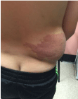
A 9-year-old male with no significant past medical history presented to the primary care clinic for a well-child visit. The patient had emigrated from Ecuador and denied any acute complaints or concerns. Physical examination was unremarkable apart from the examination of the flank, which demonstrated a large mass on the right posterolateral aspect (Figure A and Figure B). The mass was warm, erythematous and non-tender to palpation.
According to the parents, there was no presence of a mass or vascular lesion at birth. The presenting mass developed when the patient was a few months of age and continued to grow without any regression. Imaging, nor a biopsy was ever performed, and the parents were informed that the mass would likely continue to grow.
At our facility, the patient underwent imaging studies to further evaluate the lesion. Magnetic resonance imaging (MRI) with and without contrast was performed and demonstrated a 6.6 x 10.5 x 11.2 mass centered within the subcutaneous tissues along the lateral aspect of the right lower rib cage (Figure C). This mass consisted of a large area of fatty proliferation with numerous blood vessels extending throughout the lesion. On contrasted imaging, there were several large arterial blood vessels seen supplying this lesion. There were also multiple venous structures seen draining the lesions and extending along the course of the dilated arterial structures; findings consistent with a high flow vascular malformation. He was referred to the pediatric and plastic surgical services for further evaluation.

    Figure 1:  A posterior view on gross examination of the patient during the
initial visit revealing a large flank mass.

    Figure 2:  A lateral view on gross examination of the patient during the initial
visit revealing a large flank mass.

    Figure 3:  A T1-weighted MRI revealing a subcutaneous mass with vascular
invasion.
A complete resection of the AVM was performed without incident (Figure D). The patient returned to the clinic several weeks for a post-operative follow-up and showed no signs of acute distress, complaints, or concerns.

Figure 4: A post-operative photo revealing resection of the vascular lesion.
Discussion
Vascular lesions in the pediatric population are a common presentation, and are classically divided into two categories – hemangiomas and malformations [1,2]. IHs are benign tumors of vascular tissue that exhibit abnormal endothelial cell proliferation [2,3]. IHs are not present at birth, undergo a course of growth and subsequent involution with a regression onset by one year of life [2,5]. IHs can be located anywhere on the body, and can affect various organs thus raising the concern for significant consequences [2]. Vascular malformations are thought to be a result of an interruption at a particular stage of vessel development [1,6]. Malformations are products of morphogenesis errors and subsequent vascular channel abnormalities [1]. Distinctively, malformations are characteristically present at birth, grow along with the child and regression typically does not occur [1,6]. The lesion found on examination of the presented patient was not present at birth, similar to IHs, yet continued to grow showing no signs of regression which are characteristic findings in vascular malformations. Both hemangiomas and malformations have overlapping similarities yet, characteristically unique differences.
Infantile hemangiomas can be clinically classified as localized, segmental, or indeterminate lesions [3,5]. Localized lesions are more common, they arise from a single focus, and are clearly well-defined [3,5]. Segmental lesions present as a patch or plaque-like lesion that is distributed over a cutaneous territory [3,5]. Segmental lesions carry a higher complication rate and are most often seen in the Hispanic population [3,5]. Chiller et al., have reported that the anterior cheek is the most commonly involved anatomical segment in the population they studied, followed by the forehead of the infant [3]. Grossly, IHs can be described as superficial, deep, or combined. Superficial IHs are most common, and hence the name they appear as a plaque like nodule or bright red papule lying above normal skin [5]. These lesions take on names such as “capillary” or “strawberry” hemangiomas [7,8].
There are several complications associated with His [3,5]. Ischemic ulceration secondary to venous hypertension is the most common complication [3,5]. Other notable complications include pain, infection, airway compromise and hemorrhage that can lead to high-output cardiac failure [3,5]. The presentation of more than five cutaneous “strawberry” hemangiomas can be associated with a hepatic hemangioma, which warrants further workup to prevent potential life threatening high-output cardiac failure [8]. A segmental IH with periorbital involvement, or one which presents on the head and neck are closely associated with syndromes such as PHACE [3,5,9]. The associated abnormalities include (posterior fossa brain malformations, hemangiomas, arterial anomalies, coarctation of the aorta, other cardiac defects, and eye abnormalities) [3,5,9]. When the abnormalities of this syndrome include urogenital abnormalities or spinal involvement the collection of symptoms is referred to as LUMBAR syndrome [5,9]. There are several other cutaneous vascular anomalies that can undergo a benign, locally aggressive, or malignant course [5,9]. Congenital hemangiomas, tufted angioma, pyogenic granuloma are generally rare and benign. More notably there is Kaposi sarcoma, a locally aggressive vascular tumor, and angiosarcoma, a malignant tumor of endothelial cells [5,9]. The patient presented in this case did not present with other notable abnormalities suggestive of these syndromes.
AVMs are classified based on the primary aberrant vessel type: arterial, venous, capillary or venulocapillary, lymphatic or mixed [5,8]. The most common examples of capillary, or venulocapillary malformations include “nevus flammeus” and “port wine stain.” Malformations of venous etiology are loosely termed “cavernous hemangioma” and “venous hemangioma” however, cavernous hemangioma has also been used to refer to deep, or subcutaneous hemangiomas, and thus clinicians typically avoid its usage [5,8]. Vascular flow characteristics (i.e., slow-flow or fast-flow) are also used to classify vascular malformations [6]. Capillary, lymphatic, and venous malformations are characteristically slow-flow in nature, while arterial and arteriovenous malformations make up fast-flow malformations [6].
Almost always present and detected initially in infancy, AVMs typically remain quiescent during childhood [1]. The Schobinger classification describes the different stages of arteriovenous malformations from the less severe stage I (quiescence) to the more severe stage IV (decompensation) (Table 1) [6]. Malformations have the tendency of compressing surrounding tissues with ensuing pain, and functional impairment [6]. Clinical findings in AVMs vary from warm, pink-bluish stains with shunting on Doppler to ulceration, bleeding, tissue necrosis and most severely cardiac failure [6]. Cardiac failure is a result of hemodynamic dysfunction that leads to a “steal phenomenon” and subsequent life-threatening hemorrahages [6].
    Stage
    Clinical Finding


    I (quiescence)
    Warmth, pink-bluish stains, shunting on Doppler. Arteriovenous    malformation can mimic capillary or involuting hemangioma


    II (expansion)
    Enlargement, pulsation, thrill, bruit, tortuous tense veins


    III (destruction)
    Dystrophic skin changes, ulceration, bleeding, pain or tissue    necrosis. Bony lytic lesion can occur


    IV (decompensation)
    Cardiac failure
Table 1: The Schobinger Staging of AV Malformations [6].
The assessment of IH is clinical in nature. The diagnosis of IH is based primarily on the clinical features of the vascular lesion. Initially the diagnosis of AVM was clinical with the aid of symptoms and Doppler ultrasound results [6]. However, the gold standard for a definitive AVM diagnosis, like many other vascular pathologies, is angiography [6]. Angiography provides information on the structure of the lesion and presence of shunting; both are critical data points that assist in the planning of further intervention [6]. Recent studies have found utility in transarterial lung perfusion scintigraphy (TLPS); a nuclear medicine method that assesses the shunting status of the AVM and offers an improved diagnosis [6]. Clinicians use the Schobinger staging criteria based on clinical findings to determine the severity of symptoms [6]. The Schobinger staging is important in choosing the most appropriate treatment, and most authors recommend that treatment be offered for stages II, III, IV lesions [6]. Paltiel et al., [1] reported that the only significant multivariate predictor in distinguishing a hemangioma from a vascular malformation was the presence of a solid-tissue mass favoring the diagnosis of hemangioma [1].
It is critical to distinguish between IH and AVMs as the management course between the two are different [1]. Despite the characteristic regression, and subsequent involution of most IHs, some lesions require the need for intervention [2]. Corticosteroids were initially used in the management of problematic IHs however, due to the side-effect profile therapy has shifted away from its usage [2]. Randomized controlled trials have shown the effectiveness and safety of propranolol, a beta-blocker, in the treatment of IHs; which necessitated fewer surgical interventions and fewer side effects when used [2]. Treatment of arteriovenous malformations is based on symptoms and risk of complications [6]. The preferred treatment for AVMs is arterial embolization with or without surgical exicision [1]. Vaisnyte et al., reported an 83%, 93% and 10% success rate when AVMs were treated by endovascular, surgical, or combined means respectively [6].
Additionally, hemangiomas and vascular malformations alike are manifestations of a variety of different genetic syndromes hence a variety of inheritance patterns. Most notably AVMs and IHs are associated with Osler-Weber-Rendu syndrome, Sturge-Weber syndrome, and Klippel-Trenaunay-Weber syndrome.
This case highlights that it is difficult to rely on clinical judgement when assessing vascular lesions in the post-infancy period, especially when they demonstrate rapid growth, no evidence of prior involution, and were not present at birth.
In conclusion, vascular malformations are present at birth, and unlike infantile hemangiomas they typically do not undergo regression but grow commensurately with the patient. Approximately 30% of the malformations are found in the head and neck region [1]. Infantile hemangiomas become evident several months into life, and tend to regress and involute. There are rare cases when AVMs may initially present as an IH resulting in a false diagnosis. Angiography and MRI are among the best studies to assess the severity of AVMs. Appropriate assessment and diagnostic imaging are critical in the evaluation of AVMs as treatment with arterial embolization and/or surgical resection lessens the chances of subsequent clinical sequelae.
References
- Paltiel HJ, Burrows PE, Kozakewich HP, Zurakowski D, Mulliken JB. Soft-tissue vascular anomalies: utility of US for diagnosis. Radiology. 2000; 214: 747-754.
- Chen TS, Eichenfield LF, Friedlander SF. Infantile Hemangiomas: An Update on Pathogenesis and Therapy. Pediatrics. 2013; 131: 99-108.
- Chiller KG, Passaro D, Frieden IJ. Hemangiomas of infancy: clinical characteristics, morphologic subtypes, and their relationship to race, ethnicity, and sex. Arch Dermatol. 2002; 138: 1567-1576.
- Niazi TN, Klimo P, Anderson RCE, Raffel C. Diagnosis and Management of Arteriovenous Malformations in Children. Neurosurg Clin N Am. 2010; 21: 443-456.
- Darrow DH, Greene AK, Mancini AJ, Nopper AJ, SECTION ON DERMATOLOGY, SECTION ON OTOLARYNGOLOGY–HEAD AND NECK SURGERY and SECTION ON PLASTIC SURGERY. Diagnosis and Management of Infantile Hemangioma. Pediatrics. 2015; 136: e1060-e1104.
- Vaišnyte B, Vajauskas D, Palionis D, et al. Diagnostic methods, treatment modalities, and follow-up of extracranial arteriovenous malformations. Medicina (Kaunas). 2012; 48: 388-398.
- Fishman SJ, Mulliken JB. Hemangiomas and vascular malformations of infancy and childhood. Pediatr Clin North Am. 1993; 40: 1177-1200.
- Garzon MC, Huang JT, Enjolras O, Frieden IJ. Vascular malformations: Part I. J Am Acad Dermatol. 2007; 56: 371-374.
- Eivazi B, Werner JA. Extracranial vascular malformations (hemangiomas and vascular malformations) in children and adolescents - diagnosis, clinic, and therapy. GMS Curr Top Otorhinolaryngol Head Neck Surg. 2014; 13: Doc02.
