Abstract
Introduction: Pancytopenia is a clinical problem which is a common and wide differential diagnostic spectrum. and may occur with various mechanisms. In this study we aimed to determine the most common etiologic causes in patients with pancytopenia.
Materials and Methods: The records of patients aged 18 years and older, who applied to the Health Sciences University Bakirkoy Dr. Sadi Konuk Training and Research Hospital between 2012 and 2017 and who were diagnosed with pancytopenia according to WHO criteria were retrospectively reviewed.
Statistical Method: Mann-Whitney-U test was used for 2 groups and Kruskal-Wallis test was applied for 3 and more groups. Since no normal distribution was provided as a descriptive statistic, median and change interval values were given for continuous data.
Results: A total of 766 patients, 475 (62%) women and 291 (38%) men, were included in the study. In these patients, non-hematologic causes were found in 77.7% and hematologic causes in 22.3% of patients with pancytopenia. Hematological etiologies were 72.2% benign and 27.8% malignant. Nonhematological causes were divided into groups as renal diseases (6.1%), rheumatological diseases (2.4%), infective diseases (10.8%), endocrinological diseases (3.9), hypersplenism (14.5%), immunosuppressive drug use (17.5%), solid organ cancers (10.8%) and unidentified reasons (34.3%).
Conclusion: Pancytopenia should be evaluated carefully and the etiology should be detected quickly and corrected by appropriate treatment. It is an appropriate approach to exclude, first the non-hematological causes (especially immunosuppressive drug use, hypersplenism, infection and solid organ cancers, respectively) and the benign causes of hematological reasons.
Keywords: Pancytopenia; Anemia; Bisitopenia
Introduction
The definition of pancytopenia adopted by the World Health Organization (WHO) includes the combination of all three parameters: Hemoglobin (Hb) - for non-pregnant women - ‹12g/ dl and ‹13g/dl for men, absolute neutrophil count ‹ 1800 /microl, platelet count ‹150000 /mm3 [1]. In healthy adults, hematopoiesis occurs in the marrow where mature blood cells migrate to other regions with the circulatory system. The balance between blood cell production, distribution in other organs, and ongoing cellular destruction determines the levels of circulating blood cells [2-5]. Pancytopenia may occur with various mechanisms. The etiologic classification consists of bone marrow infiltration (hematological malignancies, metastatic cancers, myelofibrosis and infectious diseases, tuberculosis, fungal infections, etc.), bone marrow aplasia (vitamin B12 or folate deficiency, aplastic anemia, infectious diseases such as HIV, viral hepatitis, parvovirus B19 and drugs) and blood cell destruction or sequestration (disseminated intravascular coagulation, thrombotic thrombocytopenic purpura, ineffective erythropoiesis - myelodysplastic syndrome, megaloblasitis disorders, hypersplenism). Although pancytopenia is a common clinical problem with a wide differential spectrum, there is not enough information about the incidence of causes except for a few studies [6-8]. In our study, the aim was to determine the most common etiologies in patients with pancytopenia and to contribute to the shortening of the transition period for appropriate treatment by making a rapid diagnosis.
Materials and Methods
The records of patients 18 years and older, who applied to the Health Sciences University Bakirkoy Dr. Sadi Konuk Training and Research Hospital Internal Medicine outpatient clinics from, 2012 to 2017 and who were diagnosed with pancytopenia according to WHO criteria were retrospectively analyzed. Gender, age, Hemoglobine (Hb), hematocrit (Hct), white blood cell count, platelet count, mean corpuscular volume (MCV), reticulocyte count, lactate dehydrogenase (LDH), vitamin B12, folic acid, serum iron, ferritin levels, TSH, fT4, fT3, drug (immunosuppressive) use, presence of hepatomegaly and/or splenomegaly, bone marrow aspiration and biopsy results and diagnoses leading to pancytopenia after the analyzes were recorded. The patients were divided into 2 groups according to hematological and non-hematological etiologies, which primarily led to pancytopenia. Hematologic etiology group was further divided into two groups as benign and malignant causes. The non-hematologic etiological group was further divided into subgroups as; infectious, rheumatologic, endocrinological, renal diseases, hypersplenism, immunosuppressive drug use, solid organ cancers and others (undetectable).
Statistical method
The normality tests were performed for each variable in the study and Kolaporov-Smirnov and Shapiro-Wilk tests were performed. Since the variables were not normally distributed due to p ‹0.05, non-parametric methods were preferred in the analyzes. Mann- Whitney-U test was used for 2 groups and Kruskal-Wallis test was applied for 3 and more groups. Since no normal distribution was provided as a descriptive statistic, median and change interval (maxmin) values were given for continuous data. Frequency (frequency) distribution tables for categorical data were interpreted. Data are presented as percentage and number. The analyzes were performed with SPSS 22.0 statistical analysis program and significance level was considered as p ‹0.05.
Results
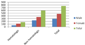
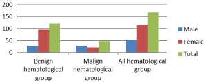
A total of 766 patients, 475 (62%) women and 291 (38%) men, were included. The mean age of men was 60.6 years, the mean age of women was 55.5 years, and the average age of all patients was 57.5 years. Non-hematological causes were found in 77.7% and hematological causes in 22.3% of patients with pancytopenia. Gender distribution among both groups is shown in Graphic 1. Hematological etiologies were 72.2% benign and 27.8% malignant. Gender distribution in hematological subgroups is shown in Graphic 2. Non-hematological causes were divided into groups as renal diseases (6.1%), rheumatological diseases (2.4%), infective diseases (10.8%), endocrinological diseases (3.9), hypersplenism (14.5%), immunosuppressive drug use (17.5%), solid organ cancers (10.8%) and unidentified reasons (34.3%). Gender and etiology distribution of non-hematological group is shown in Table 1. Differences between the hematological and non-hematological groups (Table 2) and benign and malignant groups from the hematological subgroups (Table 3) were shown in the tables below. Age (p=0.024), LDH (p=0.000), serum iron (p=0.032), ferritin (p=0.000) and vitamin B12 (p=0.000) levels were significantly higher in the non-hematological group. According to the comparison between hematological groups; Hb (p=0.000), Hct (p=0.000), white blood cell count (p=0.000) and platelet count (p=0.002) were significantly higher in benign hematological group. Serum iron (p=0.001), ferritin (p=0.000) and vitamin B12 (p=0.004) levels were significantly higher in the malignant hematological group. Fifty-five (17.2%) out of 319 patients with abdominal ultrasonography had hepatomegaly and 92 (28.8%) had splenomegaly. Bone marrow aspiration and biopsy was performed in only 33 (4.3%) of all patients. Because in other patients there was a non-haematological cause and no need to performe bone marrow biopsy.

Graphic 1:: Gender distribution among groups.

Graphic 2: Gender distribution between hematological subgroups.
    Etiology
    Male
    Female
    Total / %


    Infectious causes
    22
    42
    64 / 10,7


    Rheumatological
    0
    14
    14 / 2,3


    Hypersplenism
    33
    53
    86 / 14,4


    Endocrinological causes
    5
    18
    23 / 3,8


    Immunosuppressive drug use
    49
    55
    104 / 17,4


    Renal causes
    18
    18
    36 / 6,05


    Solid organ cancers
    38
    26
    64 / 10,7


    Other reasons
    71
    133
    204 / 34,2


    Total
    236
    359
    595 / 100
Table 1: Nonhematological group sex - etiology distribution.
    Group
    N
    Average row
    Mann-Whitney-U statistics
    p


    Age
    hematologic
    171
    352.41
    45556
    0.024*


    non-hematologic
    595
    392.44


    Total
    766


    Hb
    hematologic
    171
    373.93
    49236
    0.521


    non-hematologic
    595
    386.25


    Total
    766


    Hct
    hematologic
    171
    389.82
    49791
    0.671


    non-hematologic
    595
    381.68


    Total
    766


    White Blood Cell Count
    hematologic
    171
    372.79
    49040
    0.472


    non-hematologic
    595
    386.58


    Total
    766


    Platelet Count
    hematologic
    171
    384.25
    50745
    0.96


    non-hematologic
    595
    383.29


    Total
    766


    LDH
    hematologic
    171
    302.59
    37036
    0.000*


    non-hematologic
    595
    406.75


    Total
    766


    MCV
    hematologic
    171
    369.5
    48478
    0.346


    non-hematologic
    595
    387.52


    Total
    766


    TSH
    hematologic
    171
    144.42
    8183.5
    0.319


    non-hematologic
    595
    155.95


    Total
    766


    Ft4
    hematologic
    171
    124.73
    5179.5
    0.975


    non-hematologic
    595
    125.07


    Total
    766


    Serum iron
    hematologic
    171
    155.59
    10917.5
    0.032*


    non-hematologic
    595
    176.03


    Total
    766


    Ferritin
    hematologic
    171
    137.38
    9006.4
    0.000*


    non-hematologic
    595
    181.61


    Total
    766


    Folate
    hematologic
    171
    119.68
    5863.5
    0.853


    non-hematologic
    595
    117.97


    Total
    766


    Vitamin B12
    hematologic
    171
    140.6
    9447.1
    0.000*


    non-hematologic
    595
    194.14


    Total
    766
Table 2: Differences between hematological and non-hematological groups of variables, Mann-Whitney-U test results.
    Group
    N
    Average row
    Mann-Whitney-U statistics
    p


    Age
    benign
    122
    79.88
    2242.5
    0.023*


    malign
    47
    98.29


    Total
    169


    Hb
    benign
    122
    93.73
    1802
    0.000*


    malign
    47
    62.34


    Total
    169


    Hct
    benign
    122
    93.43
    1839
    0.000*


    malign
    47
    63.13


    Total
    169


    White Blood Cell Count
    benign
    122
    93.23
    1862.5
    0.000*


    malign
    47
    63.63


    Total
    169


    Platelet Count
    benign
    122
    92.3
    1977
    0.002*


    malign
    47
    66.06


    Total
    169


    LDH
    benign
    122
    81.65
    2458.5
    0.151


    malign
    47
    93.69


    Total
    169


    MCV
    benign
    122
    81.36
    2422.5
    0.113


    malign
    47
    94.46


    Total
    169


    TSH
    benign
    122
    39.47
    1567.8
    0.584


    malign
    47
    36.52


    Total
    169


    Ft4
    benign
    122
    24.29
    1254.9
    0.304


    malign
    47
    28.65


    Total
    169


    Serum iron
    benign
    122
    46.73
    1796.3
    0.001*


    malign
    47
    68.47


    Total
    169


    Ferritin
    benign
    122
    46.7
    1952.2
    0.000*


    malign
    47
    72.23


    Total
    169


    Folate
    benign
    122
    34.58
    1162.1
    0.378


    malign
    47
    39.38


    Total
    169


    Vitamin B12
    benign
    122
    50.52
    1836.3
    0.004*


    malign
    47
    70.15


    Total
    169
Table 3: Differences between benign and malign hematological groups, Mann-Whitney-U test results.
Discussion
Pancytopenia can be fatal if it cannot be diagnosed early [9]. Therefore, rapid detection of the underlying cause is extremely important in terms of coping with the disease and prognosis. It is important to investigate the the most common pancytopenia etiologies and which may be less frequent but more serious, in the differential diagnosis. Gayathri BN et al. reported a mean age of 41 years and male gender as a dominant in a prospective study of 104 pancytopenia patients aged between 2 and 80 years in India. Also, splenomegaly was more common than hepatomegaly in their study [10]. M. Premkumar et al. found that the mean age was 32.8 / year and male gender was dominant in their study which evaluating the hematological etiology with 140 pancytopenia patients. As the etiological frequency; Megaloblastic anemia (60.7%), infectious causes (16.4%), aplastic anemia (7.8%) and leukemia (9.2%) were detected [11]. In a study conducted by Imbert et al. with 213 adult pancytopenia patients in France, it was observed that malign hematological causes were more frequent that was not compatible with our study. According to this study, malignant myeloid disorders (acute myeloid leukemia, MDS and myelofibrosis) 42% and malignant lymphoid disorders 18% accounted for 60% of all hematological etiologies. The group containing the benign etiologies such as megaloblastic anemia was found to be 17% [8]. It was thought that this difference could be related with adequate nutrition and sociocultural level of the patient population. Dr. Atif Sitwat Hayat et al. Found that 72.94% of the patients were male and 27.05% were female. In the etiological evaluation, they found that non-cancerous causes were more frequent with a rate of 63.52% [12]. Bhagwan Singh Yadav et al., found the mean age of 35.15 ± 12.6 years and an equal female / male ratio in gender distribution, in their study with 58 pancytopenia patients above the age of 18 [13]. In the study of T. N. Dubey et al., which included 70 patients over 13 years of age, the male / female ratio was 1.4/1. In the etiological evaluation, megaloblastic anemia was in the first place with a rate of 41.4%. Aplastic anemia with the ratio of 22.9%, hypersplenism 15.7% and leukemic diseases 14.2% were also found in the etiology [14].
In our study, the mean age was 57.5 year and different from the literature the female gender was dominant. The difference in mean age was considered to be related only to the inclusion of the adult population in our study. We also showed that non hematological causes more common than hematological ones and similar to literature, we showed that benign causes (72.8%) were more frequently in the hematological etiology.
Conclusion
Pancytopenia should be evaluated carefully and the etiology should be detected quickly and corrected by appropriate treatment. In studies conducted, gender dominance is different for each study, so it is not true to say that pancytopenia is more common in male or female sex. According to our study, it is an appropriate approach to exclude, first the non-hematological causes (especially immunosuppressive drug use, hypersplenism, infection and solid organ cancers, respectively) and the benign causes of hematological reasons. When family physicians encounter patient with pancytopenia, they should be calm and after diagnosis treat the benign causes. If there is no benign cause than they should refer the patients to advanced center immediately.
References
- Valent P. Low blood counts: immune mediated, idiopathic, or myelodysplasia. Hematology Am Soc Hematol Educ Program. 2012; 2012: 485.
- Young NS, Abkowitz JL, Luzzatto L. New Insights into the Pathophysiology of Acquired Cytopenias. Hematology Am Soc Hematol Educ Program. 2000: 18-38.
- Pascutti MF, Erkelens MN, Nolte MA. Impact of Viral Infections on Hematopoiesis: From Beneficial to Detrimental Effects on Bone Marrow Output. Front Immunol. 2016; 7: 364.
- Marks PW. Hematologic manifestations of liver disease. Semin Hematol. 2013; 50: 216-221.
- Risitano AM, Maciejewski JP, Selleri C, Rotoli B. Function and malfunction of hematopoietic stem cells in primary bone marrow failure syndromes. Curr Stem Cell Res Ther. 2007; 2: 39.
- Savage DG, Allen RH, Gangaidzo IT, et al. Pancytopenia in Zimbabwe. Am J Med Sci. 1999; 317: 22-32.
- Tilak V, Jain R. Pancytopenia-a Clinico-Hematologic Analysis of 77 Cases. Indian J Pathol Microbiol. 1999; 42: 399-404.
- Imbert M, Scoazec J-Y, Mary J-Y, Jouzult H, Rochant H, Sultan C. Adult Patients Presenting with Pancytopenia: A Reappraisal of Underlying Pathology and Diagnostic Procedures in 213 Cases. Hematologic Pathology. 1989; 3: 159-167.
- Khodke K, Marwah S, Buxi G, Yadav RB, Chaturvedi NK. Bone marrow examination in cases of pancytopenia. J Indian Acad Clin Med. 2001; 2: 55–59.
- Gayathri B N, Rao KS. Pancytopenia: A clinico hematological study. J Lab Physicians. 2011; 3: 15-20.
- M Premkumar. Cobalamin and Folic Acid Status in Relation to the Etiopathogenesis of Pancytopenia in Adults at a Tertiary Care Centre in North India. Anemia. 2012: 707402.
- Atif Sitwat Hayat. Pancytopenia; Study For Clinical Features And Etiological Pattern At Tertiary Care Settings In Abbottabad 1 2 3 4 Dr. The Professional Medical Journal.
- Bhagwan Singh Yadav, Amit Varma and Priyanka Kiyawat. Clinical profile of pancytopenia: a tertiary care experience. International Journal of Bioassays ISSN: 2278-778X CODEN: IJBNHY.
- TN Dubey. The Common Causes Leading to Pancytopenia in Patients Presenting in Hospital of Central India. International Journal of Contemporary Medical Research. 2016; 3.
