Introduction
Varicose veins and chronic venous insufficiency are common disorders of the venous system in the lower extremities that have long been regarded as not worthy of treatment, because procedures to remove them were once perceived as worse than the condition itself. All too frequently, patients are forced to learn to live with them, or find "creative" ways to hide their legs. The treatment of varicose veins is most successful when the point of superficial reflux in the leg, particularly at the saphenofemoral junction is eliminated. The endo-venous ablation procedure decreases the elevated venous pressure even in secondary varicosities leading to their resolution. The relatively simple office-based procedure allows patients to return to work early with minimal morbidity and has long-lasting results. Remaining varicosities are then eliminated with phlebectomy or chemical ablation.
Yet, it remains widely unrecognized that 15-20% of patients who present with superficial venous disease have a more proximal source of reflux in the pelvis and may require evaluation and treatment of their “pelvic reflux disease” as well [1,2]. More importantly, unidentified and untreated PVR can be a major cause of recurrent leg varicose veins in up to 30% of patients [3], and in 35% of patients who present with non-saphenous venous reflux [4]. Our lack of understanding of the common occurrence of pelvic venous reflux as well as the close interplay between chronic pelvic pain, pelvic varices, with chronic superficial venous insufficiency in lower extremities and varicose veins has hindered our ability to treat many patients effectively. Patients are often told that their vulvar and thigh varicose veins (particularly if occurring following a pregnancy) will get better by themselves, and patients are never informed that these varicosities may well be secondary to pelvic venous reflux [5,6]. Noteworthy is the fact that many patients with unusual varicosities in the gluteal, flank, and posterior thighs may have a normal venous duplex examination with no traditional truncal or saphenous reflux. Therefore, it is not surprising that many patients are not referred and are not able to get appropriate treatment of their pelvic venous reflux disease. Lastly, even doctors who have some knowledge of pelvic congestion syndrome often think that the condition is restricted to females and do not diagnose pelvic reflux disease in males. Of particular significance is the presentation of a young male patient with a painful left varicocele in whom further evaluation is mandatory to rule out left renal vein thrombosis or compression by a renal tumor.
Anatomic Considerations
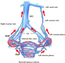
Each ovary is drained by a plexus forming one major vein measuring normally 5mm in size. The left ovarian plexus drains into left ovarian vein, which empties into left renal vein; the right ovarian plexus drains into the right ovarian vein, which drains into the anterolateral wall of the inferior vena cava (IVC) just below the right renal vein. An interconnecting plexus of veins drains the ovaries, uterus, vagina, bladder, and rectum (Figure 1).

    Figure 1: Pelvic venous drainage. Rich network of connecting veins between
the gonadal veins, uterus, rectum, and pelvic veins.
The lower uterus and vagina drain into the uterine veins and then into branches of the internal iliac veins; the fundus of the uterus drains to either the uterine or the ovarian plexus (utero-ovarian and salpingo ovarian veins) within the broad ligament. Vulvoperineal veins drain into the internal pudendal vein, then into the inferior gluteal vein, then the external pudendal vein, then into the saphenous vein, or into the circumflex femoral vein, and then into the femoral vein. The ovarian veins are duplicate in 30-40% of patients while the internal iliac vein consists of two trunks in 35% of female and 23% of male specimens (27% of all specimens) [1,2].
Pathophysiology
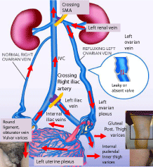
The physiologic abnormality that underlies pelvic venous reflux disease (whether primary or secondary) is a significant increase in the pelvic venous pressure and overfilling of the various venous reservoirs in the abdomen and pelvis. When the capacitance of the renal reservoir is exceeded due to ovarian vein reflux or venous compression, the pelvic reservoir will start filling. It may reach full capacity, at which point the pelvic escape veins will then fill the leg reservoir resulting in either unusual varices in the vulvar, gluteal or inner thig areas, or truncal reflux with varicosities associated with the saphenous systems in the leg (Figure 2 and 3).

Figure 2:

Figure 3:
In Pelvic congestion syndrome, the ovarian vein is dilated and the significant reflux into the pelvis with the resulting hypertension leads to distension of the draining veins around the ovary, with the pressure eventually reaching the various veins connecting the ovaries with the uterus. The uterine veins will act as an escape point and crossing vein around the uterus will enlarge to accommodate the increased venous volume, which then drains into the contralateral ovarian vein back into the IVC, a pattern easily identified by venography or during laparoscopy (Figure 3). The pressure can also be transmitted to the pelvis and escape veins develop to the lower extremities thru vulvoperineal, posterior thigh, and gluteal veins (Figure 2 and 3). Venous reflux in the ovarian vein leads to congestion and sluggish drainage from engorged pelvic veins and pelvic varices. The stretching of the vein wall, compression of surrounding nerves, and release of various neurotransmitters all play a role in causing the pain [7].
Causes of Venous Reflux
In pelvic reflux diseases, the development of “congestion” or sluggish drainage of the pelvic veins may occur as a result of primary vein valvular insufficiency from either congenital or acquired vein incompetence such as absent or floppy valves, or may be due to secondary pelvic venous insufficiency from venous compression.
In primary pelvic venous reflux, venous insufficiency occurs as a result of the absence or dysfunction of venous valves in the ovarian and iliac veins, variant venous anatomy with atresia or hypoplasia of the main veins, venous kinking from uterine malposition, as well as structural and hormonal changes of parity. Unique anatomical factors render pelvic veins particularly liable to become dilated, even without pregnancy. Many pelvic veins are devoid of valves and have weak attachments between the adventitia and supporting connective tissue. Anatomical studies demonstrate that ovarian venous valves are absent congenitally in 15% of the female patients on the left and 6% on the right, with valves being incompetent in 41% of the female patients on the left and 46% on the right [8]. Using duplex ultrasound, venous reflux in the left ovarian and iliac veins was identified in 58% patients presenting with PCS, and only in 4% of right ovarian veins [9]. In addition, estrogens and hormones play an important role as evidenced by the fact that incompetent valves and increased venous diameter are found more frequently in multiparous women, and ovarian varicosities are seen more frequently after pregnancy. Indeed, the capacity of pelvic veins may increase 60-fold over the nonpregnant state, leading to venous dilatation and hence valvular incompetence. A heightened estrogen state is further evidenced by the fact that treatment with progestins or Gonadotropin releasing hormone receptor to suppress ovarian function by blocking estrogens may provide temporary relief.
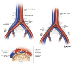
In secondary pelvic reflux, retroperitoneal and pelvic veins are compressed along their anatomical paths. When present, the venous compression results in functional obstruction and significantly interferes with the venous return from the left ovarian and/or iliac veins into the Inferior Vena Cava. There are three sites of compression that are commonly described but the most common anatomical compression consists of the May-Thurner Syndrome (a compression of the iliac veins by the crossing iliac arteries and which can occur at various points as shown in Figure 4). Other sites of compression include the Nutcracker Syndrome (compression of the left renal vein by the superior mesenteric artery (Figure 2), and retro-aortic left renal vein (an anatomical variant where the left renal vein passes behind the aorta and the L4-L5 vertebrae).

Figure 4: Various points of compression of iliac veins by crossing arteries.
As we gain more understanding of when and how pelvic venous reflux may cause chronic pelvic pain, it has become increasingly clear that the term pelvic congestion syndrome falls short of describing the various etiologies underlying the development of pelvic venous reflux disease. Although left ovarian venous reflux in the setting of pelvic congestion syndrome may often be the source of pelvic venous reflux, a number of patients present with pelvic reflux disease without the presence of ovarian vein reflux. When studied, these patients turn out to have iliac vein compression or primary internal iliac venous reflux with no ovarian reflux. Therefore, the term pelvic venous reflux diseases is been advanced to describe two separate but related syndromes with concomitant anatomic and physiologic abnormalities originating in venous reflux either in the ovarian veins, or in tributaries of the internal iliac veins; it encompasses both the condition of pelvic congestion syndrome and pelvic varices not associated with ovarian vein reflux.
Clinical Presentation
Chronic pelvic pain is a common symptom in women and accounts for at least 10% of outpatients visits to gynecologists [9]. While endometriosis represents the most common cause of chronic pelvic pain (40% patients), pelvic congestion syndrome (PCS) is the second most common cause accounting for 30% of visits in the US [10]. A recent review of 2384 abdomino-pelvic computed tomography scans performed on women at one institution reported that 8% of all premenopausal women had documented chronic pelvic pain of unclear etiology and dilated ovarian and pelvic veins on crosssectional imaging studies but not all had evidence of dilated ovarian veins suggesting another etiology for the venous hypertension and pelvic varices [11].
A detailed history and physical examination may often help differentiate whether the patient is presenting primarily with pelvic congestion symptoms or with pelvic varices suggesting iliac venous compression (Table 1).
TABLECREATED
Table 1: Symptoms and signs of pelvic venous reflux diseases.
When pelvic reflux is secondary due to venous compression and outflow obstruction, additional symptoms and signs may also develop (Table 1). In the Nutcracker Syndrome, patients may experience hematuria, flank pain, and recurrent UTI”s due to venous engorgement of the left renal vein (the right renal vein is not normally subject to compression as it drains directly in the inferior vena Cava and rarely drains in the right renal vein). In May-Thurner Syndrome, patients will often present with chronic leg swelling, sometimes starting in their twenties and thirties. The venous compression compromises venous return resulting in sluggish or stagnating blood flow, and may lead to acute deep venous thrombosis of the iliac vein. When extensive iliofemoral thrombosis occurs, a limb threatening condition known as phlegmasia cerulea dolens may then develop, and if tis unrecognized and untreated may lead to venous gangrene and limb loss. In fact, at least 30-40% of patients who present with iliac vein thrombosis are commonly found to have iliac venous compression and should be emergently referred to a vascular surgeon for further evaluation and treatment of the thrombosis13. With proximal outflow obstruction suprapubic, crossing veins from one femoral vein to the other may develop and are often visible or identified by ultrasound. The presence of such crossing veins may warrant additional evaluation.
With the development of peri-colonic and peri-vesicular veins, patients may experience irritable bowel syndrome (IBS), irritable bladder or hemorrhoids some patients find relief of these symptoms after treatment of the pelvic venous reflux [14,15]. Dilated pelvic veins due to PVR has been suggested as a cause of impotence in males [16] with reports of selective embolization of the pelvic veins restoring erectile function [17].
Patient Profile
It is essential to start with a detailed history and physical, which may give valuable clues as to the likelihood that a particular patient is experiencing pelvic venous reflux disease looking for the following:
• Chronic pelvic pain of greater than 6 months duration in association with leg symptoms and varicose veins in the lower extremities in premenopausal females.
• Presence of varicosities in unusual areas including posterior gluteal, inner thighs, vulvar varices or varicoceles in males.
• Presence of leg symptoms in young females with h/o menorrhagia, dyspareunia or post coital pain.
• Recurrent varicose veins after saphenous ablation or stripping with evidence of pelvic varices on ultrasound.
• Persistent chronic leg pain and swelling in the absence of saphenous reflux.
• Chronic left leg swelling with a history of iliac DVT or recurrent DVT in the lower extremities.
• Chronic leg swelling, pelvic symptoms with symptoms of IBS, hemorrhoids.
Our Approach to Diagnosis and Treatment
Once relevant symptoms and signs are identified, non-invasive studies including trans-pelvic and transvaginal duplex ultrasound are ordered initially to confirm the diagnosis and demonstrate pelvic varicosities. CT or MR venography will confirm the diagnosis. Venography is then undertaken to perform treatment. In many cases is highly suspicious, we go directly to conventional venography.
• Duplex ultrasound: This technique has already been shown very useful investigation for PVR [18]. Published criteria for investigating PVR using external transcutaneous duplex ultrasound include:
i) An ovarian vein larger than 6mm with reflux greater than one second.
ii) Four or more pelvic varices larger than 4mm.
iii) Dilatation of peri-uterine veins with siphon effect to the contralateral side.
iv) Compression of iliac or renal veins with flow disturbance and elevated velocities by duplex.
• The addition of transvaginal venous duplex ultrasonography, with the patient, elevated in the head-up position at 45° allows physiological reflux to be seen in the lower pelvic veins, as well as the presence of pelvic varicosities and any exit points from the pelvis. The addition of maneuvers such as the Valsalva maneuver and Kegal squeeze enhance this test further.
• CTV, MRV: However, these cross-sectional imaging techniques are usually performed with the patient lying flat, preventing any physiological reflux and pelvic varicosities may not be dilated.
Conventional Venography involves the introduction of radiopaque contrast injected under pressure. Venography can be performed at different angles, and to have any chance of seeing venous reflux, the patient needs to be tilted into a head-up angle.
Treatment
The medical treatment for pelvic congestion and pelvic reflux consists of both pharmacological and compressive treatments to minimize the hormonal effects on the veins and reduce venous hypertension. Pharmacologic treatments are used including simple analgesia, or the use of agents to suppress ovarian function or increase venous contraction. Medroxyprogesterone acetate (MPA) is a progesterone agonist and suppresses estrogen, while Goserelin is a gonadotropin releasing hormone agonist and suppresses sex hormones. They both may be a good option for short term treatment. Recently, a synthetic steroid consisting of a single-rod nonbiodegradable implant containing and releasing the desogestrel metabolite etonogestrel has been used in the treatment of pelvic pain. The etonogestrel implant seems to be a viable option for long-term medical treatment of PVI, but a long-term study with more subjects is necessary to evaluate the effectiveness and recurrence of PVI after removal.
Compression garments have also been tried to reduce the dilation of pelvic varicosities. While there have been published reports on the benefits of external compression (similar to the use of proven elastic stockings in the legs), results are not uniform [19].
In the past, the surgical treatment for the various pelvic reflux conditions consisted of rather extensive and radical open surgical procedures including hysterectomy with oophorectomy, varicocelectomy, or rerouting the vessels to relieve the compression. These open procedures are currently reserved for patients who require additional surgeries like excision of fibroids, or nephrectomy for cancer to be done at the same setting.
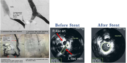
Currently, the gold standard treatment for pelvic truncal venous incompetence is embolization, usually using metal embolization coils or vascular plugs, sclerosing agents like glue, or a combination of both [20]. In order to be effective, the definitive treatment of PVR and/or pelvic varicosities has to include the ablation of incompetent pelvic truncal veins and the reduction of pelvic varicosities. That requires the identification of which trunks are incompetent, recognizing that the internal iliac veins are much more commonly involved than the gonadal veins [7]. In our own practice, we start with a transfemoral approach under local anesthetic as a walk-in walk-out office-based procedure in our angiographic suite. However, as both the gonadal veins and the internal iliac veins pass upward and therefore have origins superiorly, a trans-jugular route allows easier cannulation into the relevant vein, and we quickly move to the jugular approach if needed. Dilated veins in the pelvis can also be treated with coil embolization and or vascular plugs, the latter being quite expensive (Figure 5).

Figure 5: Treatment of iliac compression with stenting and ovarian vein reflux with coil embolization.
We have used vascular plugs on occasion but they remain quite expensive and do not seem to save time or reduce the overall cost of the procedure [21,22].
In cases of venous compression, angioplasty and stenting remains the gold standard. More recently, intravascular ultrasound has been an invaluable tool in both the direct and real time diagnosis of renal or iliac vein compression as well as the precise and accurate deployment of venous stents (Figure 5). In fact, IVUS has already had a significant impact on the management of pelvic venous reflux diseases [23].
Outcomes
Yet, despite improved imaging criteria and improving embolization techniques, one of the most frustrating problems with the treatment of PCS is that complete symptom relief after treatment remains variable at 75%–100% (Table 2).
TABLECREATED
Table 2:
In a guideline published by the SVS and the AVF in 2011, guideline authors suggest “treatment of pelvic congestion syndrome and pelvic varices with coil embolization, plugs, or transcatheter sclerotherapy, used alone or together (2B)”. The 2B recommendation indicates a “weak” recommendation based on moderate quality evidence, where the benefits of the technology are considered closely balanced with risks and burdens (Gloviczki et al., 2011).
As of to date, no guidance has been published from CMS for pelvic vein embolization.
Classification and Scoring of Pelvic Venous Reflux Diseases
There has been a push recently to develop a “CEAP” type scoring system that can be used for PCS, and many experts are presenting at venous meetings their alternative views as to how such a scoring system would work. However, most proposals remain cumbersome and impractical at present, and it is unlikely that there will be a simple “CEAP type” scoring system for PCS in the near future. A diseasespecific assessment tool for patients with PCS has been published and may provide a good basis for understanding the diverse number and nature of signs and symptoms of the pelvic venous disease [24].
In August 0f 2019, the International Union of Phlebology issued a consensus guideline for the treatment of pelvic reflux disease with similar recommendations to the ones discussed below [25].
Our Approach to Diagnosis and Treatment
It is essential to start with a detailed history and physical, which may give valuable clues as to the likelihood that a patient is experiencing pelvic venous reflux disease looking for the following:
• Chronic pelvic pain of greater than 6 months duration in association with leg symptoms and varicose veins in the lower extremities in premenstrual females.
• Presence of varicosities in unusual areas including posterior gluteal, inner thighs, vulvar varices or varicoceles in males.
• Recurrent varicose veins after saphenous ablation or stripping with evidence of pelvic varices on ultrasound.
• Persistent chronic leg pain and swelling in the absence of saphenous reflux.
• Chronic left leg swelling with a history of iliac DVT or recurrent DVT in the lower extremity.
• Chronic leg swelling, pelvic symptoms with symptoms of IBS, hemorrhoids.
Once we suspect the diagnosis, we obtain a standing venous reflux study as well as a duplex ultrasound of the IVS, iliac and pelvic veins and follow the protocol published by the vascular team at Stony Brook University [26]. If the pelvic varices need to be further studied, a transvaginal ultrasound is helpful. Assuming the non-invasive studies are consistent with venous compression or pelvic reflux, we will then discuss at length with the patient the pros and cons of additional evaluations and options for treatment. We then consult with both the referring physician and the patient’s gynecologist if they have one. We do believe that shared decision making provides the patient with the best tools for an informed decision. Patients are then offered venography at which time a decision is made to proceed with iliac venous stenting in the setting of a May-Thurner Syndrome, or ovarian and pelvic vein embolization for reflux disease. It is important to note that now, while the treatment of venous compression with iliac stenting is a covered benefit under most insurances, only a handful of commercial insurances will cover treatment for pelvic reflux disease. A national effort is underway among various societies and advocacy groups to change that trend and allow patients to receive treatment. Currently, we will do peer to peer review, document our findings extensively both from the visits and from venography if done. If still denied, the patients are then counseled on the costs of the embolization part of the procedure. It is not uncommon for patients to elect to pay for the treatment out of pocket, or even change their insurance so they are covered and are able to undergo the procedure.
Notwithstanding the lack of uniformity in the treatment outcomes, making the correct diagnosis and providing the appropriate treatment, particularly in young female patients has been one of the most gratifying times in my practice. A number of the young female patients that are referred to me have been chastised by their spouses and families for being an unwilling or unworthy spouse. Following an office- based intervention, their symptoms improve significantly, if not resolve, and they invariably express their deepest gratitude for the significant change it has caused in their lives. I can recount many of them telling me with tears of joy running down their face: “I will forever remain grateful, you have changed my life”.
References
- Marsh P, Holdstock J, Harrison C, Smith C, Price BA, Whiteley MS. Pelvic vein reflux in female patients with varicose veins: Comparison of incidence between a specialist private vein clinic and the vascular department of a National Health Service District General Hospital. Phlebology. 2009; 24: 108- 113.
- Dabbs EB, Dos Santos SJ, Shiangoli I, Holdstock JM, Beckett D, Whiteley MS, et al. Pelvic venous reflux in males with varicose veins and recurrent varicose veins. Phlebology. 2018; 33: 382-387.
- Whiteley AM, Taylor DC, Dos Santos SJ, Whiteley MS. Pelvic venous reflux is a major contributory cause of recurrent varicose veins in more than a quarter of women. J Vasc Surg Venous Lymphat Disord. 2014; 2: 411-415.
- Labropoulos N, et al. Nonsaphenous superficial vein reflux. Journal of Vascular Surgery. 2001; 3495: 872-877.
- The Impact of Pelvic Congestion Syndrome. 2018.
- Brown CL, Rizer M, Alexander R, Sharpe EE 3rd, Rochon PJ. Pelvic congestion syndrome: Systematic review of treatment success. Semin Intervent Radiol. 2018; 35: 35-40.
- Phillips D, Deipolyi AR, Hesketh RL, Midia M, Oklu R. Pelvic congestion syndrome: Etiology of pain, diagnosis, and clinical management. J Vasc Interv Radiol. 2014; 25: 725-733.
- Jurga-Karwacka A, Karwacki GM, Schoetzau A, Zech CJ, Heinzelmann- Schwarz V, Schwab FD. A forgotten disease: Pelvic congestion syndrome as a cause of chronic lower abdominal pain. PLoS ONE. 2019; 14: e0213834.
- Kim AS, Greyling LA, Davis LS. Vulvar varicosities: A Review. Dermatol Surg. 2017; 43: 351-356.
- Liddle AD, Davies AH. Pelvic congestion syndrome: Chronic pelvic pain caused by ovarian and internal iliac varices. Phlebology. 2007; 22: 100-104.
- Ahlberg NE, Bartley O, Chidekel N. Right and left gonadal veins. An anatomical and statistical study. Acta Radiol Diagn (Stockh). 1966; 4: 593–601.
- Ashour MA, Soliman HE, Khougeer GA. Role of descending venography and endovenous embolization in treatment of females with lower extremity varicose veins, vulvar and posterior thigh varices. Saudi Med J. 2007; 28: 206-212.
- Kasirajan K, Gray B, Ouriel K. Percutaneous AngioJet thrombectomy in the management of extensive deep venous thrombosis. J Vasc Interv Radiol. 2001; 12: 179-185.
- Holdstock JM, Dos Santos SJ, Harrison CC, Price BA, Whiteley MS. Haemorrhoids are associated with internal iliac vein reflux in up to one-third of women presenting with varicose veins associated with pelvic vein reflux. Phlebology. 2015; 30: 133-139.
- Farquhar CM, Hoghton GB, Beard RW. Pelvic pain – Pelvic congestion or the irritable bowel syndrome? Eur J Obstet Gynecol Reprod Biol. 1990; 37: 71-75.
- Hwang TI, Yang CR. Penile vein ligation for venogenic impotence. Eur Urol. 1994; 26: 46-51.
- Narayanan S. “Dramatic Results” of Cyanoacrylate Glue Ablation in Younger Patients with Pure Venous Erectile Dysfunction. Venous News. UK: BIBA Publishing. 2018; 6: 14.
- Whiteley MS, Dos Santos SJ, Harrison CC, Holdstock JM, Lopez AJ. Transvaginal duplex ultrasonography appears to be the gold standard investigation for the haemodynamic evaluation of pelvic venous reflux in the ovarian and internal iliac veins in women. Phlebology. 2015; 30: 706-713.
- Gavrilov SG, Karalkin AV, Turischeva OO. Compression treatment of pelvic congestion syndrome. Phlebology. 2018; 33: 418-424.
- Gloviczki P, Comerota AJ, Dalsing MC, Eklof BG, Gillespie DL, Gloviczki ML, et al. The care of patients with varicose veins and associated chronic venous diseases: Clinical practice guidelines of the society for vascular surgery and the American venous forum. J Vasc Surg. 2011; 53: 2S-48S.
- Whiteley MS, Lewis-Shiell C, Bishop SI, Davis EL, Fernandez-Hart TJ, Diwakar P, et al. Pelvic vein embolisation of gonadal and internal iliac veins can be performed safely and with good technical results in an ambulatory vein clinic, under local anaesthetic alone – Results from two years' experience. Phlebology. 2018; 33: 575-579.
- Whiteley MS. The current overview of pelvic congestion syndrome and pelvic vein reflux. Indian J Vasc Endovasc Surg. 2018; 5: 227-233.
- Gagne PJ, Tahara RW, Fastabend CP, Dzieciuchowicz L, Marston W, Vedantham S, et al. Venography versus intravascular ultrasound for diagnosing and treating iliofemoral vein obstruction. J Vasc Surg Venous Lymphat Disord. 2017; 5: 678-687.
- Gibson K, Minjarez R, Ferris B, Neradilek M, Wise M, Stoughton J, et al. Clinical presentation of women with pelvic source varicose veins in the perineum as a first step in the development of a disease-specific patient assessment tool. J Vasc Surg Venous Lymphat Disord. 2017; 5: 493-499.
- Antignani PL, et al. Diagnosis and treatment of pelvic congestion syndrome: UIP consensus document. International Angiology. 2019; 38: 265-283.
- Labropoulos N, Jasinski PT, Adrahtas D, Gasparis AP, Meissner MH. A standardized ultrasound approach to pelvic congestion syndrome. Phlebology. 2017; 32: 608-619.
