Abstract
46 year old male with acute onset of right upper extremity paralysis accompanied with slurred speech. Initial concerns for a thromboembolic stroke were ruled out by MRI. Characteristic MRI findings were consistent with a medium to high grade glioma. Craniotomy for lesion biopsy and cerebrospinal fluid analysis ruled out malignancy, but was consistent with Multiple Sclerosis. Teriflunomide was started with follow-up MRI 16 months after which demonstrated interval resolution a ring-enhancing lesion.
Keywords: Multiple sclerosis; Unilateral paralysis; High grade glioma; Teriflunomide; Parietal craniotomy; Vasogenic edema
Case Presentation
A 46-year-old male with a three-week history of sudden right upper extremity paralysis and slurred speech presented to our clinic to establish care with a new primary care physician. Patient was seen and evaluated in the emergency room for sudden paralysis and was informed that he had suffered a stroke. Initial CT identified a nonspecific white matter hypo-attenuation in the left frontal lobe, which was interpreted as a subcortical white matter infarct. Unfortunately, he left against medical advice due to unsatisfactory care.
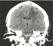
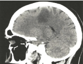
During the office visit, the patient was found to have continued paralysis of the right upper extremity, but no evidence of slurred speech. Patient was sent immediately to the emergency room for further evaluation. While in the ER, the patient underwent a repeat CT brain, which identified a focal area of subcortical hypo-density within the superior frontal/precentral gyrus (Figure 1 and 2). CT chest identified a 2mm soft tissue nodule in the right apex.

    Figure 1: Coronal view on CT with focal area of subcortical hypo-density in
the superior frontal/precentral gyrus.

    Figure 2: Sagittal view on CT with focal area of subcortical hypo-density in
the superior frontal/precentral gyrus.
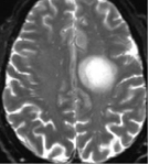
Patient was admitted to the family medicine service with subsequent consultation with neurology and neurosurgery. Brain MRI identified a 3.1cm irregularly rim-enhancing mass in the left mid centrum semi-ovale likely representing a high-grade glioma with surrounding vasogenic edema (Figure 3). Initial perception by both specialists included a medium to high grade lesion that would require biopsy for confirmation. Subsequently, patient underwent a left parietal craniotomy for brain parenchyma tissue sampling.

    Figure 3: MRI axial view with a 3.1cm irregularly rim-enhancing mass in
the left mid centrum semi-ovale likely representing a high-grade glioma with
surrounding vasogenic edema.
The morphological and immunohistochemical features of the tissue sample were consistent with an active inflammatory demyelinating disease with no evidence of malignancy or thromboembolic stroke.
During follow-up evaluation with neurology, it was determined that a lumbar puncture was necessary. Cerebrospinal fluid culture was negative, but greater than five well-defined gamma restriction bands were present. The CSF IgG synthesis rate was +0.4 with a CSF IgG Index of 0.65.
Based on pathology and cerebrospinal fluid results, this patient was diagnosed with Multiple Sclerosis and subsequently treated with Teriflunomide.
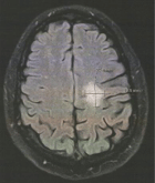
Repeat MRI 16 months after starting Teriflunomide demonstrated an interval resolution of a prior large intra-axial incomplete ringenhancing lesion with a large residual area of gliosis measuring 24.8x19.5 mm in AP and transverse diameters (Figure 4). This patient has gradually regained motor function with shoulder flexion and abduction but has not returned to full strength and function.

    Figure 4: MRI axial view with interval resolution of a prior large intraaxial
incomplete ring-enhancing lesion with a large residual area of gliosis
measuring 24.8 x 19.5 mm in AP and transverse diameters.
Discussion
Environmental and genetic factors have been implicated as risk factors for the development of Multiple Sclerosis. The prevalence is 50-300 per 100,000 people with approximately 2.3 million currently living with the diagnosis of MS. Vitamin D deficiency, obesity, exposure to Epstein-Barr virus and tobacco use have been shown to play a part in the development of Multiple Sclerosis. Chromosome 6 has been implicated in the development of numerous autoimmune diseases, but specifically serotype DR2 (HLA-DR15 and HLA-DR16) have been consistently replicated in MS [1].
The adaptive immune system consisting of B and T cells are causally related in the development of Multiple Sclerosis. Inflammation associated with MS is limited to the central nervous system, which theorizes that antigens are specifically expressed and limited to this region. Two hypothesis currently exist that may explain the immune response targeting the central nervous system. The CNS intrinsic model hypothesis believes that the initial event takes place in the CNS leading to the release of antigens in the periphery leading to an autoimmune response. The CNS extrinsic model hypothesis believes that the initial event occurs outside the central nervous system, but leads to an autoimmune response targeting the CNS [1].
Multiple sclerosis is the result of axonal and neuronal loss, demyelination, and astrocytic gliosis. Permanent clinical disability associated with MS is related to neurodegeneration with features including numbness, tingling, blurred vision, gait impairment, imbalance, poor coordination, and bladder dysfunction.
The revised 2017 McDonald criteria is currently the standard used for the diagnosis of Multiple Sclerosis. An individual experiencing a typical attack, defined as unilateral or bilateral optic neuritis, focal supratentorial syndrome, focal brainstem or cerebellar syndrome, partial or complete myelopathy, complete ophthalmoplegia, encephalopathy, headache, alteration of consciousness, meningismus, or fatigue may be diagnosed with MS based on one of four clinical criteria with additional supportive criteria.
Category one involves two or more attacks with clinical evidence of two or more lesions, or two or more attacks with clinical evidence of one lesion, and historical evidence of a prior attack that suggests a lesion in a different location.
Category two involves two or more attacks with clinical evidence of one lesion that is supported by disseminated in space as defined by a clinical attack implicating a different site, or one or more T2 lesions in two or more regions within the central nervous system.
Category three involves one attack and clinical evidence of two or more lesions with dissemination in time to include an additional clinical attack along with the presence of enhancing and non-enhancing MRI lesions, or new T2 or enhancing MRI lesion compared with the baseline scan. Oligoclonal bands must be present in the cerebrospinal fluid.
Category four includes one attack and clinical evidence of one lesion with dissemination in space to include an additional attack implicating a different CNS location defined by one or more T2 lesions in two or more areas and dissemination in time to include an additional attack with enhancing and non-enhancing MRI lesions, or new T2 or enhancing lesions when compared to baseline scans. Oligoclonal bands must be present in the cerebrospinal fluid [2].
In the acute setting, treatment includes methylprednisolone. Oral methylprednisolone at 500mg daily for five days has been shown to not be inferior to intravenous methylprednisolone at 1000mg daily for three days.
Disease modifying medication, which target neuroinflammation and neurodegeneration, are the main stay of treatment for Multiple sclerosis. Medication such as Teriflunomide exerts its effect on stimulated lymphocytes through non-competitive, reversible inhibition of the mitochondrial enzyme dihydro-orotate dehydrogenase. This effect results in disruption of the cell cycle in the S phase of the proliferating T and B cells thereby reducing the inflammatory process [3].
Summary
Multiple Sclerosis is the great mimicker! As illustrated in the case, MS can often resemble other disease processes such as stroke and malignancy. The initial presentation of extremity paralysis was concerning for a stroke and based on imaging, which demonstrated a ring-enhancing lesion with surrounding vasogenic edema, further supported the premature diagnosis, but also raised concerns for a possible high-grade glioma. Based on the clinical presentation and imaging, considering MS as a high likelihood was not entertained. It was not until a biopsy was performed which confirmed Multiple Sclerosis as the insidious culprit. The clinical presentation for Multiple Sclerosis will never be identical amongst patients with MS. It is important when evaluating a patient with neurological symptoms to include this within the differential.
References
- Thompson AJ, Baranzini SE, Geurts J, Hemmer B, Ciccarelli O. Multiple sclerosis. Lancet. 2018; 391: 1622-36.
- Thompson AJ, Banwell BL, Barkhof F, Carroll WM, Coetzee T, Comi G, et al. Diagnosis of multiple sclerosis: 2017 revisions of the McDonald criteria. Lancet Neurol. 2018; 17: 162-173.
- Bar-Or A, Pachner A, Menguy-Vacheron F, Kaplan J, Wiendl H. Teriflunomide and its mechanism of action in multiple sclerosis. Drugs. 2014; 74: 659-74.
