
Abstract
Hypertrophic Cardiomyopathy (HOCM) is a genetic cardiac disease. It is not commonly seen at the primary care clinic. HOCM is well tolerated in pregnancy. However, sudden death is a recognized complication during pregnancy especially in patients with severe outflow tract obstruction. This case illustrates the case of a patient who initially presented with history of being amenorrhoeic for the past four months. During the examination, a systolic murmur was identified incidentally, and the patient was further worked up. She was diagnosed with HOCM. Although she was seen by various doctors throughout her life, the diagnosis was never made. Its variable presentation and clinical course may cause a delayed or missed diagnosis of HOCM. It is hoped that this case study will illustrate the importance of diagnosing and managing patients with HOCM appropriately.
Case Report
Miss NF is a single, 30-year-old lady who was referred to the primary care clinic for the determination of the Period Of Amenorrhea (POA) and Estimated Date Of Delivery (EDD) of her pregnancy. Miss NF had amenorrhea for the last 5 months. She was unsure of her Last Menstrual Period (LMP) as her menstrual cycle always been irregular. She only began suspecting that something was amiss when she felt sudden movements in her abdomen for one week prior to the presentation to the clinic. She denied feeling any symptoms of pregnancy in the initial stages.
At the clinic, a Urine Pregnancy Test (UPT) confirmed her pregnancy. The patient had no known medical illness. She had never been hospitalized before nor had any surgical procedure in the past. This was my patient’s first pregnancy and it was unplanned. She had been sexually active since the last 1 year with her only partner (boyfriend). She had never used any form of contraception. There was no history of any medical illness in the family. She neither smokes nor drinks alcohol.
On examination, she was pink, and not in respiratory distress. Her BP was 100/60 mmHg and her pulse rate was 74 beats per minute, regular rhythm and good volume. There was no pedal edema. Her cardiovascular examination revealed a loud systolic murmur along the left sternal edge (Grade III). There was no radiation to the neck or axilla. Apex beat was located in the mid clavicular line of the 5th intercostal space. There was no thrill palpable. The air entry was equal on bilateral lung field. There were no crepitations. On palpation, the abdomen was soft and non-tender. The uterus was palpable at about 18 weeks and the fetal parts were palpable.
In view of the newly diagnosed heart murmur, an echocardiogram was done, and it was noted that the patient had apical Hypertrophic Obstructive Cardiomyopathy (HOCM). Her ejection fraction was noted to be 64%. Her left ventricle was noted to be normal in size and of normal systolic function. The left ventricular wall motion was normal and pulmonary artery pressure was noted to be 29 mmHg. She was started on metoprolol 25 mg bd and followed up closely at the antenatal clinic. Patient ‘s infective screening (HIV Ag/Ab Combo), fasting and2 hours post glucose readings, hemoglobin levels (12.8g/ dl) and her renal profile were within normal parameters. During the fetal ultrasound, fetal heart activity was detected, and other fetal parameters were corresponded with her REDD.
She had a Caesarean section at 38 weeks POA as there was poor progress of labor and CTG revealed fetal distress. The caesarean section was uneventful. The patient was in stable condition and her girl baby was healthy and weighed 3.53kg.
Discussion
Cardiovascular diseases are seen in 0.5-4% of pregnant women.1 The most common disorders are rheumatic valvular disease, congenital heart disease and cardiomyopathy [1].
Hypertrophic cardiomyopathy is a relatively common genetic cardiac disease with a ratio of 1:500 in general population [2]. It is an autosomal dominant disease but 50% of cases are sporadic [3]. it is due to mutations of the cardiac sarcomere proteins. HOCM may present at any age.
Clinical diagnosis is by electrocardiography whereby there is identification of otherwise unexplained left ventricular wall thickening of more than 15mm in the presence of non-dilated cavity [4]. Anteroseptal hypertrophy is mostly involved whereas patterns such as concentric hypertrophy or pure apical involvement are less common [4].
Left ventricular morphology in Hypertrophic Obstructive Cardiomyopathy (HOCM). Figure 1 cited from the study of Johan MB et .al.2018 (Figure 1).
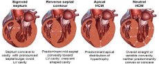
Figure 1: Left ventricular morphology in Hypertrophic Obstructive Cardiomyopathy (HOCM). Figure 1 cited from the study of Johan MB et .al.2018.
Annual mortality rates are 1%2 Poor prognostic values for this condition are age less than 14 years at presentation, syncope at presentation and a family history of HOCM or sudden death [4].
The identification of patients with HOCM who are at risk of dying suddenly is imperative in the clinical management. A study by Perry et al identified several risk factors that were associated with sudden death. They include a family history of sudden death, unexplained syncopal attacks, abnormal blood pressure response (BPR) during upright exercise testing and maximal LVWT (left ventricular wall thickness) [5].
Treatment strategies must be indivualised. Beta blockers, verapamil or disopyramide is used for exertional dyspnea.2 Beneficial effects of beta blockers on symptoms of exercise tolerance are due to a decrease in heart rate and the consequent prolongation of diastole [6]. Septal myotomy or myectomy operation is the standard of care for patients presenting with severe refractory symptom associated with marked outflow obstruction [2].
Treatment algorithm for the management of hypertrophic cardiomyopathy patients (Figure 2). Figure 1 was cited from the study of Takashi M.et al.2018. Hypertrophic cardiomyopathy is well tolerated in pregnancy even in the presence of an outflow gradient [1]. Maternal mortality is increased in patients with HOCM compared with the general population. Sudden death is a recognized complication of HOCM during pregnancy especially in the group of patients with severe outflow tract obstruction.3 However the absolute maternal mortality is noted to be low and confined to high risk patients. Studies by Autore showed that one quarter to one third of pregnant patients were receiving beta blockers for HOCM. Beta blockers have been described as safe in this population with little effect on fetal outcome [7]. All pregnant women with HOCM should attempt a vaginal delivery unless if there is an obstetric contraindication [3]. Vaginal delivery is preferred in this group of patients because it poses less cardiac risk because Caesaerean section is accompanied by approximately twice as much blood loss [9]. Epidural anaesthesia should be avoided in patients with HOCM due to increased risk for hypotension [9]. The use of prostaglandin for induction of labour is also not advisable due to secondary inherent vasodilatory effects [10]. HOCM is a genetic cardiac disease. However some genetically prone individuals might not manifest phenotypically identifiable disease until late in adult life.3 It is important to counsel patients regarding family planning. This is because there is a 50% chance of producing an affected child.3 The counseling should also include the need for life long clinical and echocardiography follow up as HOCM can present at any age.
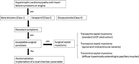
Figure 2: Treatment algorithm for the management of hypertrophic cardiomyopathy patients. Figure 1 was cited from the study of Takashi M.et al.2018.
All first degree relatives should be provided with adequate information to allow them to decide if they wish to be screened for HOCM.10 The children of affected parents like my patient’s baby should be screened every three years until puberty and then annually until they reach the age of 20 years .10Screening usually involves using the ECG and the echocardiogram. If there is no evidence of HOCM in early adulthood, it is unlikely that the condition will develop in later life [11].
Conclusion
Cardiovascular disease is one of the commonest non obstetric cause of maternal mortality worldwide. Hypertrophic cardiomyopathy is generally well tolerated in pregnancy. Beta blockers are safe in this population. HOCM is a genetic cardiac disease and therefore firstdegree family members must be counselled for screening.
Disclosure
The authors declared no conflicts of interest.
References
- Clinical Practice Guidelines Management of Heart Disease in Pregnancy
- Maron BJ. Hypertrophic cardiomyopathy, systematic review. JAMA 2002; 287: 1308-1320.
- Stergiopolus K, Shiang E, Bench T. Preganacy in patients with preexisting cardiomyopathies Journal of American College of Cardiology 2011; 58: 337- 350.
- Oxford Handbook of Clinical Medicine Hypertrophic cardiomyopathy 8th Edition; 146
- Perry ME, Jan P, Shaughan D, Sanjay S, Lorenzo M, Amanda V. Sudden Death in Hypertrophic Cardiomyopathy: Identification of High Risk Patient. Journal of American College of Cardiology 2010 ; 36: 2212-2218.
- Spirito P, Seidmann CE, William J, Mckenna. The management of hypertrophic cardiomyopathy 1997; 336: 775-785.
- Autore C, Bruzzi P, Ortolani P, Fragola P. Hypertrophic cardiomyopathy: patterns of progression Circulation 1995; 166-173.
- Rusleena T, Kasemsri STT. Short stature as an independent risk factor for cephalopelvic disproportion in a country of relatively small-sized mothers Arch Gynecol Obstet. 2012; 285: 1513-1516.
- Grewal JC, Siu Samuel, Ross H, e al. Pregnancy Outcomes in women with Dilated cardiomyopathy Circulation 2009; 120: 45-52.
- Perry E, Kenna WJ. Inherited heart conditions: hypertrophic cardiomyopathy. British Heart Foundation 2009.
- John Murtagh Part 1 Basis of General Practice Fourth Edition Mc Graw Hill 2009; 3-7.