Abstract
Acute respiratory distress can be life threatening if proper management is delayed. The cause of respiratory distress needs to be diagnosed quickly in order to administer appropriate and timely treatment. However, it is sometimes difficult to tease out various conditions that can present as acute respiratory distress. We present such a unique case of acute respiratory distress in a patient with anemia. We show how the ability to differentiate between cardiogenic and non-cardiogenic pulmonary edema can help in diagnosis and appropriate timely management of acute respiratory distress.
Keywords: Pulmonary edema; ARDS; TRALI
Abbreviations
TRALI: Transfusion Related Acute Lung Injury; RBC: Red Blood Cells; ABG: Arterial Blood Gas; BNP: Brain Natriuretic Peptide; TSH: Thyroid Stimulating Hormone; BiPAP: Bi-level Positive Airway Pressure; SARS-Cov-2: Severe Acute Respiratory Syndrome associated Corona Virus-2; COVID-19: Coronavirus Disease-19; ARDS: Acute Respiratory Distress Syndrome; RT-PCR: Reverse transcription polymerase chain reaction; PE: Pulmonary Embolism; HLA: Human Leukocyte Antigen
Case Presentation
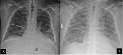
A 65-year-old man with past medical history of hypertension and persistent atrial fibrillation presents with weakness, exertional dyspnea, and black-tarry stools for last 3-4 days. He denied chest pain, cough, fever, nausea, vomiting, or abdominal pain. He denied exposure to any sick contact or any recent national or international travel. He has been a never smoker and doesn’t use alcohol. He is on Rivaroxaban for anticoagulation for atrial fibrillation. Physical examination was unremarkable with normal heart sounds, no murmurs, no rales, no jugular venous distention, clear lungs, and no hepatosplenomegaly. He was found to have hemoglobin of 6.7 mg/ dL and was transfused one unit of packed RBC, which improved his hemoglobin to 8.9 mg/dL. He developed acute respiratory distress and hypoxia immediately after transfusion. His oxygen saturation dropped to 70%. His ABGs were pH of 7.35, PCO2 of 36, PO2 of 71, and HCO3 of 19 on BiPAP with FiO2 of 100%. His BNP was 300. Previous echocardiogram one month ago was unremarkable with ejection fraction of 65%. TSH was with-in-normal limits. SARSCov- 2 testing was negative. Furosemide treatment was minimally effective. His initial Chest X-Ray (CXR) on arrival is shown in panel A and subsequent CXR is shown is Panel B (both CXRs are two hours apart) (Figure 1). Patient has provided informed consent.
What is the diagnosis?
• Flash Pulmonary Edema from Congestive Heart Failure
• Aspiration Pneumonia
• ARDS from Covid-19
• Transfusion Related Acute Lung injury (TRALI)
• Pulmonary Embolism
Discussion
• The correct answer is Transfusion-Related Acute Lung Injury (TRALI) is a non-cardiogenic pulmonary edema after a blood product transfusion without other explanation [1]. There is no other reasonable explanation for bilateral pulmonary infiltrates in our patient other than TRALI (see below). CXR in panel-A and panel-B are only 2 hours apart (Figure 1), TRALI manifests within 6 hours of receiving a blood product [2]. It occurs in about 1 in 5000 transfused blood products and allogeneic antibodies to leukocyte/HLA are found in about 80% cases as well as no antibodies are identified in 20% cases [3]. 10-20 % female blood donors and 1-5 % male blood donors have anti-leukocyte antibodies in their serum3. There is no specific treatment available, only supportive care. Our patient’s reaction was reported to blood blank and subsequent testing for antileukocyte antibodies was negative. Patient recovered with supportive care and discharged home after three days of hospital stay.

Figure 1: Patient’s Chest X-Ray (Two Hours Apart).
• Flash pulmonary edema occurs when there is rapid accumulation of fluid within the lung interstitium secondary to elevated cardiac filling pressures. This is a patient who does not have history of heart failure (Ejection Fraction 65%) and he had normal kidney functions. He did not have any signs or symptoms of fluid overload. He was given Furosemide 40mg intravenously with only minimal improvement clinically as well as in CXR findings. Table-1 shows the features differentiating cardiogenic from non-cardiogenic pulmonary edema.
    Condition
    Characteristics


    Cardiogenic
    Diffuse alveolar edema with a central distribution,    mediastinal widening, cardiomegaly, pleural effusion, peribronchial cuffing,    and Kerley B lines. It occurs due to increased capillary hydrostatic pressure    and responds to diuretic therapy.


    Noncardiogenic
    Diffuse “Batwing” pattern opacities with air    bronchogram due to altered pulmonary vascular permeability. Increased Lung    volumes. Absence of cardiomegaly, pleural effusion, and mediastinal widening.
Table 1: Differentiating features between cardiogenic and noncardiogenic diffuse
(bilateral) pulmonary infiltrates.
• Aspiration risk was low with no history of previous stroke, alcohol use, or dysphagia. The patient was alert and oriented x3. There were no typical features of aspiration pneumonia like fever or infiltrates in the right lung base. Therefore, aspiration pneumonia is unlikely in this patient.
• ARDS is an acute, diffuse, inflammatory form of lung injury and should be suspected in patients with progressive symptoms of dyspnea, increasing oxygen requirement, and bilateral alveolar infiltrates on imaging. The SARS-Cov-2 test is negative for COVID-19 in our patient. Despite some studies showing the false negative rate as high as 35% for the SARS-Cov-2 Reverse Transcription Polymerase Chain Reaction (RT-PCR) test [4], it is an unlikely diagnosis in him. He does not have any symptoms of cough or fever and no history of recent travel. Follow up testing could be considered. However, in this scenario, TRALI is more likely.
• Pulmonary Embolism (PE) cannot be diagnosed by CXR. There are also no significant risk factors for hypercoagulability; patient is a nonsmoker and no recent surgery or immobilization. He is also therapeutically anticoagulated with Rivaroxaban. Patient did not have typical findings to support diagnosis of PE such as tachycardia, electrocardiographic changes, or signs of acute right heart strain via biomarkers. CXR showed diffuse bilateral infiltrates, which is not usual in a PE.
References
- Toy P, Lowell C. TRALI--definition, mechanisms, incidence and clinical relevance. Best practice & research. Clin Anaesthesiol. 2007; 21: 183-193.
- Vlaar APJ, Toy P, Fung M, Looney MR, Juffermans NP, Bux J, et al. A consensus redefinition of transfusion-related acute lung injury. Transfusion. 2019; 59: 2465-2476.
- Kuldanek SA, Kelher M, Silliman CC. Risk factors, management and prevention of transfusion-related acute lung injury: a comprehensive update. Expert Rev Hematol. 2019; 12: 773-785.
- Xie C, Lu J, Wu D, Zhang L, Zhao H, Rao B, Yang Z. False negative rate of COVID-19 is eliminated by using nasal swab test. Travel Med Infect Dis. 2020: 101668.
