Introduction
In 2021, tuberculosis remains a world health problem. It is still the first cause of mortality by infectious disease in the world [1].
Fortunately, in developed countries, tuberculosis is a very rare pathology.
Genitourinary tuberculosis has been reported to account for 20% to 73% of all cases of extrapulmonary tuberculosis in the general population [2].
The epididymal involvement accounts for only about 20% of the genitourinary tuberculosis [3].
Isolated genitourinary tuberculosis (GUTB) is a rare and unusual presentation of tuberculosis that occurs in young male or female adults and that can cause infertility [4].
Most of the time, infertility is due to the inflammation and scarring that follow the infection, resulting in distortion of the normal anatomy and causing obstruction of the excretory tract [5].
The disease typically develops slowly. Early diagnosis is difficult, therefore delayed diagnosis and misdiagnosis are common [6].
Case Report
A 33 year old man, originary from India, without past medical history was sent to the urology clinic by his general practitioner for a suspicion of left epididymitis with no clinical improvement despite an empirical treatment by antibiotics.
The patient had a painful and indurated left testicle and an absence of ejaculation. Erection was normal and orgasm was present. The patient had no other urological symptoms and no general symptoms (no fever, no sweating, and no weight loss).
Clinical examination of the scrotum showed an enlarged and indurated left testicle and epididymis. The right testis was normal. No pathological lymph nodes were clinically palpable. Abdomen was tender. Digital rectal examination was normal.
The blood test was normal. Serum testicular markers were normal.
Urinary culture showed an abacterial pyuria. PCR analysis for Chlamydia Trachomatis and Neisseria Gonorrhea were negative.
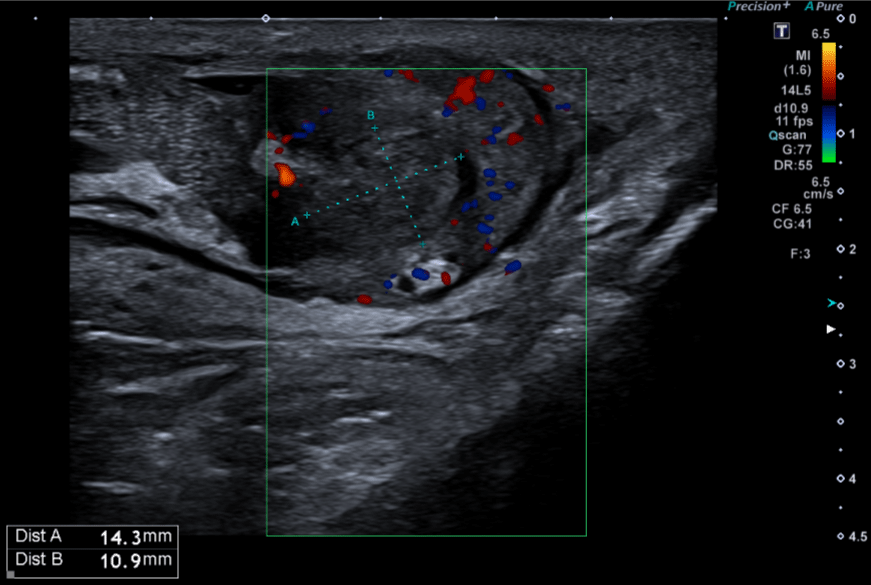
The scrotal ultrasound displayed a thickening of the body and especially of the tail of the left epididymis, with a non-vascularized heterogeneous hypoechoic 15 x 10 mm mass evoking a granuloma of sperm (Figure 1).

Figure 1: Echography of the left epididymitis evoking a granuloma of sperm.
The transrectal ultrasound of the prostate was normal. Seminal vesicles were non dilated.
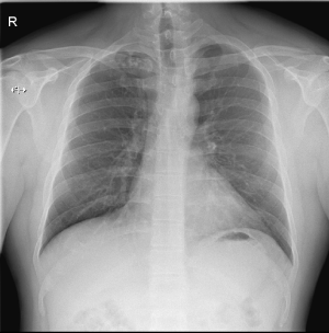
The chest X-Ray showed several small nodular opacities at the right apex (Figure 2).

    Figure 2: Chest X Ray showing several small nodular opacities at the apex

of the right lung.
The CT-Scan of the thorax showed post-infectious sequelae and some calcified mediastinal lymph nodes compatible with a residual lesion of tuberculosis.
The 18F-FDG PET/CT showed a pathological uptake in the epididymis and the testicular bursa on the left side with no other focus of suspected recurrence of tuberculosis.
The bacterial cultures in the broncho-alveolar samples were negative.
No intradermal injection of tuberculin was performed.
Specific urine culture for M. Tuberculosis revealed positive.
The wife of the patient had a negative screening.
Anti-tuberculous drugs were initiated. The first two months the treatment was Rifampicin 600mg/d, Isoniazid 300mg/d, Pyrazinamide 500mg 4/d, and Ethambutol 400mg 3/d). After initial two months, downgrading to (Rifampicin 300mg/d and isoniazid 300mg/d for six months.
During whole the treatment, the patient received also a supplementary in Vitamine B6 (pyridoxine), to prevent toxic neuropathy induced by isoniazid.
At the end of the treatment, the patient had a resolution of symptoms but no more ejaculation.
Discussion
The diagnosis and the management of the isolated tuberculous epididymitis is a challenge for physicians. Local symptoms can be insidious and the differential diagnosis with bacterial epididymitis, testicular tumor or azoospermia can be challenging.
The physiopathological mechanism is a hematologic or lymphatic spreading of the tuberculosis or a retrocanalicular pathway. A veneral transmission is very rare.
At first, tuberculosis lesions appear in the tail of the epididymis.
After that, the lesions gradually invade and spread throughout the body towards the head and finally whole the epididymis is infected. In most advanced cases, the testis can be involved.
It is important to notice that failure to respond to conventional antibiotic therapy, strongly suggest tubercular epidemiology.
Abacterial pyuria on microscopic urinalysis is considered as a typical manifestation of GUTB. However, according to some reviews, leukocytes in urine are present in 10 to 50% of the cases.
Hematuria is also a common symptom of GUTB but mainly caused by nefritis or cystitis.
In this case, hematuria and leucocyturia were absent.
According to the EAU Guidelines, a positive culture or histological analysis of biopsy specimens, possibly combined with PCR, is required for a definitive diagnosis of GUTB.
The intradermal injection of tuberculin only has a value if it is positive. People with a history of vaccination by BCG or BCG’s instillation for a superficial bladder cancer can be false positive.
Radiologically, it is difficult to make a difference between bacterial orchido-epididymitis and tuberculous epididymitis. Most of the time, the ultrasound shows an enlarged epididymis, predominantly the tail, and a marked heterogeneity.
The first line of treatment of GUTB consists of a combination of anti-tuberculosis drugs.
The first two months, a combination of three to four treatments on a daily (Rifampicin 600mg/d, Isoniazid 300mg/d, Pyrazinamide 500mg 4/d, and Ethambutol 400mg 3/d (or Streptomycin) basis is used.
Then, a second phase with only two drugs (Rifampicin 300mg/d and isoniazid 300mg/d) during four to seven months.
In case of bad response to the medical treatment, epididymectomy or orchiectomy should be performed. Preservation of fertility should be addressed and proposed at the same time.
There is a consensus to perform surgery in case or absence of evolution after two months of medical therapy or in case of unspecified diagnosis.
In case of delayed diagnosis, an abscess can involve the entire scrotum.
In that cases, the histology show diffuse necrotizing granulomas, with giant cell formation in the scrotum, testis, and epididymis.
At that point, the treatment would be performing an orchidectomy.
Conclusion
The diagnosis of GUTB is challenging because symptoms are insidious and unspecific. Therefore, the proper diagnosis is often delayed but should be mentioned in the differential diagnosis of epididymitis.
A positive BK urinary culture or a positive BK PCR on a urine sample or a histological analysis on biopsy is diagnostic.
The first line treatment is based on a combination of antituberculous drugs.
Epididymectomy or orchidectomy are performed in case of nonresponse to medical treatment or in case of unspecified diagnosis.
References
- https://www.who.int/fr/news-room/fact-sheets/detail/tuberculosis
- Chattopadhyay A, Bhatnagar V, Agarwala S, DK Mitra. Genitourinary tuberculosis in pediatric surgical practice. J Pediatr Surg 1997; 32: 1283-6.
- Papadopoulos A, Bartziokas K, Morphopoulos G, Anastasiadis, Makris D. A rare case of isolated tuberculous epididymitis in a young man presenting with a swollen testicle. OA Case Reports 2013; 2: 3.
- Surati, Suthar, Shah Surati KN, et al., Isolated tuberculous epididymoorchitis: a rare and instructive case report. Southeast Asian Journal of Case Report & Review. 2012; 1: 46–50.
- Kumar R. Reproductive tract tuberculosis and male infertility. Indian J Urol. 2008; 24: 392–5.
- Jiangwei Man, Lei Cao, Zhilong Dong, Junqiang Tian, Zhiping Wang, Li Yang. Diagnosis and treatment of epididymal tuberculosis: a review of 47 cases. 2020; 8: e82291.
- Viswaroop BS, Isolated tuberculous epididymitis: A review of forty cases. J Postgrad Med 2005; 51: 109-11.
- Chung JJ, Kim MJ, Lee T, Yoo HS, Lee JT, et al., Sonographic findings in tuberculous epididymitis and epididymo-orchitis. Journal of Clinical Ultrasound. 1997; 25: 390–394.
- Das P, Ahuja A, Datta Gupta S. Incidence, etiopathogenesis and pathological aspects of genitourinary tuberculosis in India: A journey revisited. Indian J Urol. 2008; 24: 356-361.
- Jiangwei Man, Lei Cao, Zhilong Dong, Junqiang Tian, Zhiping Wang, Li Yang. Diagnosis and treatment of epididymal tuberculosis: a review of 47 cases. 2020; 8: e82291.
- Mete C ek, Severin Lenk, Kurt G Naber, Michael C Bishop, Truls E Bjerklund Johansen, et al. Members of the Urinary Tract Infection (UTI) Working Group of the European Association of Urology (EAU) Guidelines Office. EAU Guidelines for the Management of Genitourinary Tuberculosis. European Urology. 2005; 48: 353-362.
- Figueiredo André A MD, PhD, Lucon Antônio M. Urogenital Tuberculosis: Update and Review of 8961 Cases from the World Literature. Rev Urol. 2008; 10: 207-217.
- Felix Kwizera, Stéphanie Hublet, Antoine Bufkens, et al., Tuberculose épididymaire révélée par des ganglions rétroperitonéaux.
- Mete C ek, Severin Lenk, Kurt G Naber, Michael C Bishop, Truls E Bjerklund Johansen, et al. Members of the Urinary Tract Infection (UTI) Working Group of the European Association of Urology (EAU) Guidelines Office. EAU Guidelines for the Management of Genitourinary Tuberculosis. European Urology. 2005; 48: 353-362.
- American Thoracic Society, CDC, Infectious Diseases Society of America Treatment of tuberculosis. Am J Respir Crit Care Med. 2003; 167: 603–662.
- Felix Kwizera, Stéphanie Hublet, Antoine Bufkens, et al., Tuberculose épididymaire révélée par des ganglions rétroperitonéaux.
- Michael U. Williams, Ashley Burris, Amy Zingalis, et al., Disseminated Tuberculosis Presenting as Chronic Orchiepididymitis in a Military Trainee: A Case Report and Review of the Literature. 2018.
