Abstract
Eccrine poroma is a rare, slow growing, benign tumor that originates from the eccrine sweat glands. It is reported that 47% of eccrine poroma cases occur on the foot, and it is commonly asymptomatic. Squamous cell carcinoma is a malignant neoplasm and it requires complete excision to prevent the chance of metastasis.
Squamous cell carcinoma of the foot is very rare, as only 2.4% of all cases are found on the foot. We report a patient presented with a lesion on the lateral aspect of the right foot at the mid calcaneal body. An initial biopsy of the lesion indicated that it was an eccrine poroma, and the lesion was surgically excised within three months. However, pathological examination of the excised tissue determined that the lesion had converted to a squamous cell carcinoma. The conversion of eccrine poroma to squamous cell carcinoma is rare and has previously not been reported in scientific literature.
Keywords: Eccrine poroma; Squamous cell carcinoma; Wound conversion.
Introduction
Eccrine poroma is a slow growing nodular tumor that grows from the acrosyngium of eccrine sweat glands and can be found on any cutaneous surface. Eccrine poromas are benign and account for less than 1% of primary cutaneous lesions [1]. They are commonly found in middle aged and elderly patients, and studies suggest that longterm radiation exposure and a past history of skin disease increase the likelihood of developing a poroma [1]. Although eccrine poromas have been diagnosed on unique regions of the body such as the face and neck, they are commonly found on the soles of feet [2]. Eccrine poromas are easily treated by excision of the lesion, and have a low probability of recurrence [1].
Unlike eccrine poromas, squamous cell carcinomas are malignant tumors that grow from the squamous epithelium of the dermis [3]. A squamous cell carcinoma is rarely seen in the foot [5]. The preferred method of treatment for squamous cell carcinoma is wide excision to effectively remove all carcinoma cells and prevent metastasis [3]. An estimated 60% of squamous cell carcinoma cases originate from actinic keratosis lesions, which are skin lesions caused by continuous exposure to ultraviolet radiation [4]. Burn wounds can also lead to the development of Marjolin’s ulcers, which have a 71% conversion rate to squamous cell carcinoma [5]. Squamous cell carcinoma can also develop from preexisting skin wounds such as ulcers, pressure sores, fistulas, and diabetic wounds [6,7]. Although there have been previous cases of squamous cell carcinoma development from wounds, there is no literature about the conversion of an eccrine poroma to a squamous cell carcinoma.
Case Report/Series
A seventy-one-year old female patient presented with a round granular lesion on the lateral aspect of the right foot at the mid calcaneal body. Patient has a past history of hypothyroidism and no known allergies. Lesion was nodular, limited to the skin and subcutaneous tissue, and measured 17x9x7 mm upon excision. A biopsy was performed and the lesion was diagnosed as an eccrine poroma. The lesion was completely excised three months later.
A pathological examination determined that the excised soft tissue mass was a squamous cell carcinoma. The squamous cell carcinoma was a grade one tumor with a maximum dimension of 11mm. Tumor was well differentiated and without vascular or perineural invasion. A pathology report showed no evidence of malignancy in the resection margins of the tumor.
Discussion
Eccrine poroma is a rare benign tumor that originates from the acrosyngium of eccrine sweat glands and is commonly presented in elderly patients. It is a nodular asymptomatic neoplasm that varies in color from pink to red, and has either a smooth or ulcerated surface [8]. Eccrine poromas typically grow on the extremities, and the majority of cases occur on the foot, which has a large amount of eccrine sweat glands. Dermoscopic studies indicate that eccrine poromas contain a variety of glomerular, hairpin, and linear irregular vessels, which can be used to differentiate these benign tumors from other skin lesions [8]. Radiation exposure, past history of skin disease, and the human papilloma virus are thought to cause the development of an eccrine poroma [1,9].
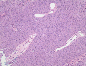
    Figure 1:  Histomicrograph of eccrine poroma (original magnification 200x,
hematoxylin and eosin).
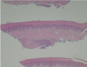
    Figure 2:  Histomicrograph of eccrine poroma (original magnification 40x,
hematoxylin and eosin).
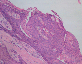
    Figure 3:  Histomicrograph of the excised squamous cell carcinoma (original
magnification 100x, hematoxylin and eosin).
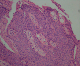
    Figure 4:  Histomicrograph of the excised squamous cell carcinoma (original
magnification 200x, hematoxylin and eosin).
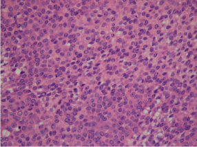
    Figure 5:  Histomicrograph of the excised squamous cell carcinoma (original
magnification 400x, hematoxylin and eosin).
Although eccrine poroma is benign, it does have the ability to mimicother skin cancers, and in rare cases eccrine poroma can develop malignant traits. Clinical case studies have reported that eccrine poromas can develop physical features of malignant skin tumors [9]. Kang et al. present a case of a vascular, bleeding eccrine poroma that was benign despite appearing malignant [9]. Furthermore, a study by Kassuga et al. presents a case of facial pigmented eccrine poroma that simulated a pigmented basal cell carcinoma [10]. Due to the possibility of an eccrine poroma developing malignant traits, the recommended treatment is complete excision followed by pathology testing.
The patient presented in this case is a unique instance of eccrine poroma conversion to squamous cell carcinoma. Our patient had a nodular lesion of eccrine poroma that was completely excised. However, from the date of the diagnosis to the date of the excision procedure, the eccrine poroma had rapidly converted to a squamous cell carcinoma. This result was confirmed by pathological examination of the excised tissue mass.
Squamous cell carcinoma is a malignant tumor that arises from the squamous epithelium. Unlike eccrine poromas, cases of squamous cell carcinoma in the foot are rare. Previously literature indicates that there are instances of squamous cell carcinoma developing from chronic skin wounds. Chronic scars such as burn wounds can convert to malignant tumors, which are called Marjolin’s ulcers [11]. The majority of Marjolin’s ulcers become squamous cell carcinoma, although the process of a malignant tumor developing from a wound can take several years to occur [11]. In a 2015 case study by Cho et al., squamous cell carcinoma developed quickly within one month from a chemical burn wound [11]. There have been additional reports of squamous cell carcinoma developing from ulcers, pressure sores, fistulas, and even diabetic foot wounds, although there is no literature about squamous cell carcinoma development from an eccrine poroma [6,7]. Since squamous cell carcinoma can originate from preexisting wounds, it is necessary to thoroughly test skin lesions for this malignancy.
In this case study we reported a rare eccrine poroma, located at the mid calcaneal body of the right foot, that converted to a squamous cell carcinoma. This rare case signifies the importance of prompt treatment, adequate size biopsy, and pathology testing of eccrine poroma as a precaution against the development of squamous cell carcinoma.
References
- Sawaya JL, Khachemoune A. Poroma: a review of eccrine, apocrine, and malignant forms. Int J Dermatol. 2014; 53: 1053-1061.
- Kong TH, Ha TH, Eom MS, Park SY. Eccrine poroma of the auricle: a case report. Korean J Audiol. 2014; 18: 151-152.
- Wani I. Metastatic squamous cell carcinoma of foot: case report. Oman Med J. 2009; 24: 49-50.
- Goldenberg G, Perl M. Actinic keratosis: update on field therapy. J Clin Aesthet Dermatol. 2014; 7: 28-31.
- Tobin C, Sanger JR. Marjolin’s ulcers: a case series and literature review. Wounds. 2014; 26: 248-254.
- Cocchetto V, Magrin P, Andrade de Paula R, Aidé M, Monte Razo L, Pantaleão L. Squamous cell carcinoma in chronic wound: Marjolin ulcer. Dermatol Online J. 2013; 19: 7.
- Chiao HY, Chang SC, Wang CH, Tzeng YS, Chen SG. Squamous cell carcinoma arising in a diabetic foot ulcer. Diabetes Res Clin Pract. 2014; 104: 54-56.
- Ferrari A, Buccini P, Vitaliano S, De Simone P, Mariani G, Marenda S, et al. Eccrine poroma: a clinical-dermoscopic study of seven cases. Acta Derm Venereol. 2009; 89: 160-164.
- Kang MC, Kim SA, Lee KS, Cho JW. A case of an unusual eccrine poroma on the left forearm area. Ann Dermatol. 2011; 23: 250-253.
- Kassuga LEBP, Jeunon T, Jenoun Sousa MA, Campos-do-Carmo G. Pigmented poroma with unusual location and dermatoscopic features. DermatolPract Concept. 2012; 2: 39-42.
- Cho HR, Kwon SS, Chung S, Kie JH. Rapidly developed squamous cell carcinoma after laser therapy used to treat chemical burn wound: a case report. World J Surg Oncol. 2015; 13: 28.
