Abstract
Peroneal tendon subluxation has typically been approached through an extensile technique. Reports of endoscopic technique to facilitate repair of the superior peroneal retinaculum are rare in the literature. In this technique paper we present our endoscopic approach to repair of a subluxing peroneal tendon complex.
Keywords: SPR repair; Peroneal tendon dislocation; Peroneal tendon subluxation; Tendoscopy
Introduction
Tendoscopy of the peroneal tendon complex has proven to be a useful tool to assist with diagnosis, debridement of synovitic tissue, release of adhesions, removal of distal muscle belly or peroneus quartus, groove deepening, superior peroneal retinaculum repair, debridement of tendon rupture and even tendon repair [1]. It is important to review the mainstay classification schemes because correctly characterizing pathology may assist with pre-operative planning, especially if deciding between an open or endoscopic procedure (Figures 1-4). Eckert and Davis characterized peroneal subluxation into three grades: Grade 1, Superior Peroneal Retinaculum (SPR) is elevated off the fibula; Grade 2, the fibrocartilagenous ridge avulses together with the SPR; Grade 3, avulsion of a small piece of cortical bone [2]. Raikin et al. characterized intra-sheath peroneal subluxation into two subtypes and likely what determines whether one approaches repair through an open technique or endoscopically: Type A, intra-sheath subluxation with an intact peroneus brevis tendon; Type B, intra-sheath subluxation with associated longitudinal tear of the peroneus brevis tendon [3]. Endoscopic repair is attractive for numerous reasons. Open surgical technique are not without morbidity including potential healing issues associated with a larger incision, infection and scar formation. It has been proposed that the mere act of dissection inherent to open surgery results in sectioning of the stabilizing structures that require repair
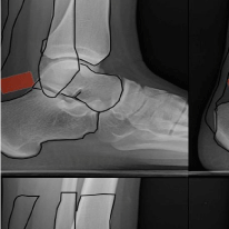
    Figure 1: Pertinent osseous anatomy is outlined in black overlaid on a
standard lateral radiographic projection of the ankle. The SPR is denoted by
a red rectangle.
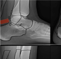
    Figure 2: Two small stab incisions are placed posterior to the fibula in
preparation for placement of two anchors into the distal fibula. Anchor
placement is performed under endoscopic guidance (see video for technique).
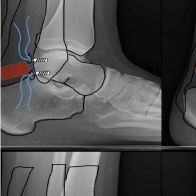
    Figure 3: Two anchors are placed into the distal fibula with each having two
sutures emanating from the stab incisions which facilitated entry. A gentle tug
on each anchor confirms sturdy placement.
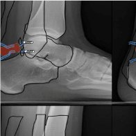
    Figure 4: A suture lasso is pierced through the skin being mindful of the likely
position of the sural nerve. The lasso is driven into deeper tissue to engage
the SPR. The lasso tip is then directed to exit the initial stab incision which
was placed for anchor delivery. Individually, suture is fed through the suture
lasso loop. The instrument is removed allowing for passage of suture through
the SPR and to then exit the skin. Each suture is fed individually. As such, this
requires performing this technique four times.
Surgical Technique
General anesthesia is induced and the patient is placed into the lateral decubitus position with special care to pad all osseous prominences to prevent neural injury (Figures 5-8). A high calf tourniquet is applied to the operative extremity. A standard ankle block is infiltrated with care to avoid the zone of operation. Relevant anatomical landmarks should be drawn with surgical ink and include the distal fibula, the peroneal tendons, the superior peroneal retinaculum, and likely position of the sural nerve (which has been shown to be located 14-mm posterior to the posterior edge of the fibula [4]. Two endoscopic portals are then established as originally described by van Dijk and Kort [5]. The distal portal is located 1.5 to 2 cm inferior to the edge of the fibular malleolus. Unlike small joint arthroscopy, the tendon sheath is not insufflated with fluid prior to insertion of the blunt obturator with associated cannula. An incision is placed into skin only. The blade should always be held parallel to the direction of underlying anatomy as to prevent iatrogenic injury. A spreading technique with a small hemostat allows for atraumatic dissection down to the level of the tendon sheath. The obturator with cannula is used to enter the peroneal tendon sheath. A 2.7-mm camera is used with 30-degrees of angulation. The proximal portal is established approximately 2 to 2.5-cm superior to the lower edge of the fibula in the retromalleolar zone. Fluid instillation is established by gravity. It is the preference of the authors to use normal saline for tendoscopic procedures. Evaluation of the peroneal tendon complex can be undertaken. Any intra-sheath synovitis can be carefully debrided with an arthroscopic shaver. Dynamic testing of the peroneal tendon complex can sometimes demonstrate subluxation/dislocation visualized from within the sheath. A guide wire is then inserted in the posterior to anterior direction into the fibula to assist with placement of the first anchor. Care should be taken to avoid injury to the peroneal tendons. A cannulated step drill prepares the bone for accepting a suture anchor. (Arthrex 3-mm Bio Composite SutureTak). The entire process is visualized and assisted by the endoscope. The second anchor is inserted in a similar fashion approximately 5-10-mm inferior to the first anchor. A curved suture retrieving instrument (Arthrex Micro Suture Lasso) is then inserted at the same level horizontally of each respective anchor slightly posterior. The suture retriever is advanced deeper to capture the deep fascia/peroneal tendon sheath/SPR and is directed to exit through the same area of penetrance through which the anchor was introduced. Suture emanating from each anchor is passed through the Nitinol loop associated with the suture retrieving instrument. The lasso allows for each suture emanating from the anchor to engage the deeper tissue to be grabbed to facilitate repair of the SPR. This is repeated for each strand of suture for both anchors. A small incision is placed between the sutures and a probe is then used to retrieve all suture through this central incision. With the ankle held in slight eversion and dorsiflexion, the sutures are tied. If appropriate deep fascia, peroneal retinaculum and tendon sheath has been cinched up to the posterior fibula, intra-operative provocative maneuvers should deem the tendon complex stable with absence of subluxation/dislocation. Small surgical wounds are closed with 4-0 Prolene. A well-padded Jones compression with posterior splint is applied to the extremity in neutral ankle position.
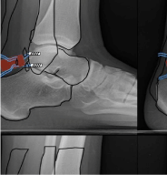
Figure 5: A small stab incision is placed lying between the two sets of suture.
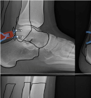
    Figure 6: A probe is passed into the central incision and all suture is grasped
and pulled through.
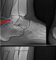
    Figure 7: With the ankle held in an everted and dorsiflexed position, suture
is tied.
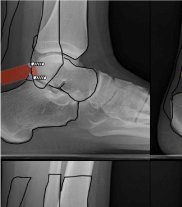
Figure 8: Once tied, suture ends are cut completing the repair.
Post operative management consists of 3 weeks immobilization in neutral position and strict non weight bearing status with crutch assistance. Patient is then placed in controlled ankle mobility boot and permitted to weight bear to tolerance. Physical medicine and rehabilitation is initiated at this time to begin gentle range of motion and strengthening. Full weight bearing with multi ligamentous ankle brace is permitted at six weeks. Return to full physical activity is permitted at 3 months.
Discussion
Lui described a technique of endoscopic SPR repair in two patients using three suture anchors [6]. Both patients demonstrated complete resolution of symptoms. Patients may complain of less subjective tightness at the peroneal tendons compared to patients who undergo an open repair (per Lui). He suggested that endoscopic repair should be limited to Eckert and Davis stages 1 and 2 and that stage 3 is not amenable to endoscopic repair. Guillo and Calder reported on their technique of endoscopic SPR repair [7]. Their technique involved using a large-gauge needle onto which a shuttle relay had been looped. This allowed for passage of suture through the SPR which ultimately become tied. Our technique is most similar to this approach; however, we believe that ours is slightly less technically demanding because our technique does not depend on grasping the looped shuttle relay within the tendon sheath. Our technique allows this to be more easily performed outside the skin and is performed without reliance on the endoscope. They utilized this technique in 7 patients, most of which were athletes. All patients were able to resume normal activities and sports to the same level. Only one patient presented with skin irritation due to underlying suture knots and this required repeat surgery to remove suture (Figures 4-7).
Endoscopic repair of longitudinal tendon tears has been described [1,5]. As such, this would not be a reason to preclude endoscopic repair of the SPR if this particular pathology coexists. With that said, this technique demands advanced endoscopic expertise and may be better approached through an open technique by the novice arthroscopist. Retromalleolar fibular groove deepening can also be endoscopically approached [1]. If more advanced pathology or difficulty in repair is encountered, the procedure can easily be converted to an open approach. We would also suggest that intrasheath peroneal subluxation may require a more involved procedure to include groove deepening. Raikin Type B lesions (intra-sheath subluxation with peroneus brevis tear) may not be amenable to endoscopic repair due to tendon repair requirements.
Awareness of the anatomical arrangement of the lateral ankle complex is pivotal especially with special regard to the sural nerve. It has been shown that the sural nerve is located on average 14-mm posterior and inferior to margin of the fibular malleolus [4] (Figures 8-10). This value can be used as a guideline as to prevent neural injury during the endoscopic procedure with use of the suture lasso instrument; however, it must be noted that anatomic variation does exist which can increase the risk of iatrogenic injury.
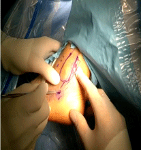
Figure 9: Creation of endoscopic portals for camera placement.
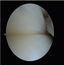
Figure 10: Peroneal tendons with brevis superior to longus.
Risks associated with open repair of dislocating/subluxating peroneal tendons has previously been reported. This includes longer rehabilitation period/need for immobilization, infection, wound healing issues, bone block fracture, permanent discomfort, recurrence of subluxation, and skin hypersensitivity [7]. We have found this technique applicable in our surgical practice. Although endoscopic approaches are not without risk and require greater arthroscopic expertise, we realize that further study is needed to gain a better understanding of this technique, especially in comparison to open procedures (Figures 11-13).
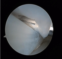
Figure 11: Guide pin insertion for placement of bone anchor with suture.
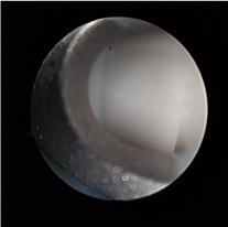
Figure 12: Placement of bone anchor with suture attached.
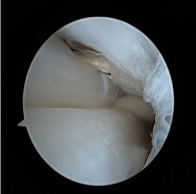
    Figure 13: Presenting sutures to be used for imbrication and tightening of
the SPR.
Conclusion
Peroneal subluxation syndrome is an uncommon but identified disorder that produces lateral ankle pain. Initial treatment can be conservative with taping , bracing, physical medicine and activity modification. Patients failing conservative management may require surgical repair of the disrupted superior peroneal retinaculum. In our efforts to minimize surgical trauma of open technique and restore anatomy with earlier return to activity an endoscopic approach has been presented. This technique is getting increasing favor amongst those surgeons inclined in performance of endoscopic/arthroscopic management of foot and ankle disorders.
References
- Vega J, Golano P, Batista J. Tendoscopic Procedure Associated with Peroneal Tendons. Techniques in Foot & Ankle Surgery. 2013; 12: 39-48.
- Eckert W, Davis EA. Acute Rupture of the peroneal retinaculum. The Journal of Bone and Jont Surgery. 1976; 58: 670-672.
- Raikin SM, Elias I, Nazarian LN. Intrasheath subluxation of the peroneal tendons. J Bone Joint Surg Am. 2008; 90: 992-999.
- Lawrence SJ, Botte MJ. The sural nerve in the foot and ankle: Anatomic study with clinical and surgical implications. Foot Ankle Int. 1994; 15: 490-494.
- Van Dijk CN, Kort N. Tendoscopy of the peroneal tendons. Arthroscopy. 1998; 14: 471-478.
- Lui TH. Endoscopic peroneal retinaculum reconstruction. Knee Surg Sports Traumatol Arthrosc. 2006; 14: 478-481.
- Guillo S, Calder JD. Treatment of recurring peroneal tendon subluxation in athletes: endoscopic repair of retinaculum. Foot Ankle Clin. 2013; 18; 293-300.
