Abstract
Background: Acute ankle fracture-dislocations require emergent reduction. Once the dislocation is successfully reduced, the ideal timing of operative fixation is not agreed upon, in part due to lack of study. At our institution, a protocol enables patients who have a successful closed reduction in the Emergency Department (ED) to go home and return to the clinic to schedule surgery. We sought to describe the rate at which initial reduction is lost between the ED and clinic visits, and to identify factors associated with loss of reduction.
Methods: We retrospectively reviewed all patients who were treated operatively for an ankle fracture from 2008-2013 at a single, Level 1 trauma center and identified 30 patients who had isolated, closed ankle fracture-dislocations that were successfully reduced and splinted in the ED. Adequate reduction was defined by achievement of congruent joint line with <5mm medial clear space. If reduction was maintained at the clinic visit, surgery was scheduled electively, defining a success. However, if reduction was lost in the interim between ED and clinic visits the patient was admitted from clinic for urgent surgical reduction and stabilization, defining a failure.
Results: Seventeen patients (57%) successfully maintained closed reduction and 13 (43%) experienced failure of closed reduction in the interim. Compared to the successful group, the failed group had significantly greater Posterior Malleolus (PM) fracture fragment size (5.1 mm vs. 3.0 mm, p = 0.029). When the ratio of PM fracture fragment size to complete articular surface was >0.1, rate of failure was 65% compared to 18% when the ratio was ≤0.1 (p = 0.016). Other assessments of radiographic and patient factors did not yield any significant difference between the failed and successful groups.
Conclusion: Greater PM fracture fragment size is associated with higher rates of interim failure of closed reduction of closed ankle fracture-dislocations. Injuries with a large PM fracture fragment may warrant consideration of operative intervention at the earliest available time.
Keywords: Ankle fracture-dislocation; Fracture management; Stability; Reduction; Radiographic assessment
Introduction
Closed ankle fracture-dislocations require emergent attention due to the threat of vascular compromise, progression to open fracture, or significant peri-articular soft tissue injury. In the isolated closed ankle fracture-dislocation without vascular compromise, urgent closed reduction is required to restore alignment, decrease soft tissue injury, and relieve pain. If adequate closed reduction cannot be achieved, urgent operative intervention may be warranted to optimize anatomic alignment, reduce further tissue damage and provide fixation [1-3].
In the event of a successful closed reduction, there is no widely accepted protocol for the management of these injuries. For definitive management, Open Reduction and Internal Fixation (ORIF) are well-supported in the literature for most patients [4-6] even if adequate closed reduction is achieved [6].
At our institution, a protocol was developed that enables patients who have a successful closed reduction in the Emergency Department (ED) to go home and return to the clinic in 5-7 days to evaluate soft tissues, obtain x-rays, and schedule elective ORIF. If the reduction is not maintained at this visit, the patient is admitted from clinic to have urgent surgical repair.
Previous studies have investigated factors associated with initial reducibility of closed ankle fracture-dislocations [2,3,7,8], however we are not aware of any study that describes failure to maintain closed reduction over an interval of a few days in these injuries, occurring between ED visit and clinic visit.
The first aim of this study is 1) to describe the rate at which initial reduction is lost in the interim between successful closed reduction in the ED and clinic follow-up. The second aim 2) is to identify patient and radiographic factors associated with loss of reduction. We hypothesized that, compared to successfully maintained reductions, those that that failed in the interim would have greater initial radiographic fracture displacement, greater radiographic evidence of syndesmotic disruption, and/or larger Posterior Malleolar (PM) fracture fragment size. We evaluated demographic and radiographic data in this consecutive series of closed ankle fracture-dislocations in order to support or refute the hypothesis.
Materials and Methods
Prior approval for this retrospective chart review was obtained through an institutional review board.
Criteria
A total of 243 patients underwent operative fixation of an ankle fracture between January 2008 and December 2012 at either the Level 1 trauma center, or the ambulatory surgery center in our health system. Of these patients, 56 had isolated, closed ankle fracture-dislocations that could be classified as bi- or tri-malleolar fractures or fracture-equivalents. Thirty of these patients had injuries that were successfully reduced in the ED and sent to follow-up in clinic. Exclusion criteria included open injuries, concomitant lower extremity fractures, pilon fractures, multisystem trauma requiring hospital admission, fracture-dislocations that failed closed reduction attempts, and fractures that did not require a reduction procedure.
Protocol
All patients presented initially through the ED of a single, Level 1 trauma center. Patients with ankle fracture-dislocations were managed according to a protocol. Closed reduction and plaster splinting were performed by Orthopaedic Surgery residents in the ED with fluoroscopic guidance utilizing an intra-articular block with or without conscious sedation, as described by White et al. [9]. A reduction was considered to be adequate if the post-reduction radiographs showed a congruent ankle joint line with medial clear space <5mm. Reduction was attempted up to 2 times in order to achieve radiographic alignment. If adequate reduction was maintained on standard 3-view post-reduction radiographs, patients were made non-weight bearing, discharged from the ED and scheduled for clinic visits in 5-7 days to arrange elective Open Reduction and Internal Fixation (ORIF). If reductions were unsatisfactory on post-reduction radiographs in the ED urgent operative reduction and ORIF vs. external fixation was performed based on the status of the soft tissues.
Data collection
Charts were reviewed to determine patient demographic data, chronology, and clinical plans. Radiographic data was collected digitally, and measurements were performed using Centricity PACS digital imaging software (GE Medical Systems, Little Chalfont, UK). A single researcher performed the initial measurements on all radiographs included in the study, and these were reviewed and verified by the senior author.
Injury mechanism was determined by the level of the fibula fracture and classified as supination or pronation based on the Lauge-Hansen system [10]. Talus displacement, in both coronal and sagittal plane, was measured and recorded as the percentage of talus uncovered by the tibial plafond. Radiographic length and displacement measurements were made digitally using landmarks described Leeds and Ehrlich [11].
    Success
    Failure
    P-value


    N
    17
    13


    Age (years)
    58.5±16.4
    56.4±21.5
    0.765


    Male (%)
    5 (29.4%)
    3 (23.1%)
    1.000


    BMI (kg/m2)
    32.2 (28.2,36.0)
    32.6 (30.3,33.3)
    0.926


    Right Side (%)
    15 (88.2%)
    8 (61.5%)
    0.345


    Surgical Delay (days)
    11.0 (7,15)
    9.5 (5.5,22)
    0.773
Table 1: Patient and treatment characteristics.
N


Success


Failure


P-value


17


13


Injury Mechanism


0.360


Supination


15 (86.7%)


9 (69.2%)


Pronation


2 (13.3%)


4 (30.8%)


Fibular Comminution


0.212


None


8 (47.1%)


2 (15.4%)


Mild-moderate


6 (35.3%)


7 (53.8%)


Severe


3 (17.6%)


4 (30.8%)


Posterior Malleolus


0.238


Fractured


10 (62.5%)


11 (84.6%)


Intact


6 (37.5%)


2 (15.4%)


Talus Displacement (coronal plane)


0.172


None


6 (37.5%)


1 (7.7%)


0-50%


7 (43.8%)


7 (53.8%)


50-100%


3 (18.8%)


2 (15.4%)


>100%


0 (0%)


3 (23.1%)


Talus Displacement (sagittal plane)


0.350


None


1 (5.9%)


0 (0%)


0-50%


8 (50.0%)


9 (69.2%)


50-100%


2 (12.5%)


3 (23.1%)


>100%


5 (31.2%)


1 (7.7%)


Medial Injury


0.663


Bony


12 (75.0%)


11 (84.6%)


Ligamentous


4 (25.0%)


2 (15.4%)
Table 2: Radiographic categorical measurements.
Data analysis
Data are presented using standard methods for continuous (n, mean, standard deviation, median, IQR, minimum and maximum) and categorical variables (counts and percentages). Continuous variables were tested for normality using the Kolmogorov-Smirnov test, and either a t-test or Wilcoxon rank sum test used to compare groups based on the distribution of each variable. Categorical variables were compared using either the chi-square test or Fisher’s exact test in the presence of small cell counts (<5). SAS version 9.2 (Cary, NC) was used for all analyses, and a p-value <0.05 was considered statistically significant. As the numerical radiographic variables are the primary measurements of interest, including posterior malleolus fracture fragment, a sample size of 56 subjects would allow us to have >80% power to observe an effect size (Cohen’s d) of 0.75 with a two-sided alpha level of ≤0.05.
Results
Patient variables
A total of 30 patients were included in the analysis after inclusion and exclusion criteria were applied, including 17 (57%) patients in the successful group and 13 (43%) patients in the failed group. There was 1 patient that had incomplete radiographic data due to inadequate initial injury films. Patient characteristics are shown in Table 1. There was no significant difference between the electively treated and urgently treated groups with regards to patient characteristics.
Categorical radiographic variables
Table 2 shows the relationship between interim reduction outcome and categorical variables. There was no statistically significant relationship identified between interim reduction outcome and amount of tibiotalar displacement, injury mechanism, fibular comminution, or bony vs. ligamentous medial injury.
Numerical radiographic variables
The associations between interim reduction outcome and numerical radiographic measurements are shown in Table 3. The median Posterior Malleolus (PM) fracture fragment size was 5.1 (4.2, 8.5) in the failed group compared to 3.0 (0.0, 5.1) in the successful group (p = 0.029). The trend for larger PM fracture fragment size in the failed vs. successful group persists when injuries without any PM fracture fragment are eliminated from analysis (6.8 ± 3.0 vs. 5.0 ± 1.7), however statistical significant is lost (p = 0.143).
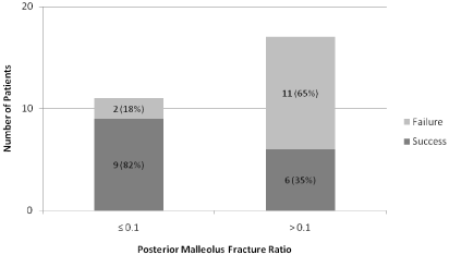
Figure 1 plots the proportion of patients treated urgently versus electively with regard to PM fracture ratio, defined as length of fractured fragment divided by length of total articular surface. When the ratio of PM fracture fragment size to complete articular surface was > 0.1, rate of failure was 65% compared to 18% when the ratio was ≤0.1 (p = 0.016).
N


Success(mm)


Failure(mm)


P-value


17


13


MM Fragment Size


9.0±7.5


9.4±5.5


0.853


MM Fragment Displacement


6.1±8.3


11.6±8.1


0.052


Medial Clear space


5.8 (4.1,7.2)


6.5 (4.8,8.4)


0.560


Fibular Shortening


0.6(0.0,7.9)


4.8 (3.7,5.5)


0.572


Fibular Sagittal Displacement


7.2±4.6


6.6±3.6


0.715


PM Fragment Size


3.0 (0.0,5.1)


5.1 (4.2,8.5)


0.029


PM Fragment Size (no zeros)


5.0 ±1.7


6.8 ±3.0


0.143


TF Clear Space


6.4 (4.9,7.3)


7.6 (6.0,13.6)


0.096


TF Overlap (AP view)


6.4 ±4.7


4.3±4.2


0.270


TF Overlap (mortise view)


3.4 (0.0,4.6)


0.0 (0.0,1.9)


0.089
Table 3: Radiographic continuous measurements.

Figure 1: Graph representing the distribution between failed and successful outcomes based on posterior malleolar fracture fragment ratio cutoff point of 0.1. P = 0.016 for chi-square test comparing proportions between the ≤ 0.1 and >0.1 groups.
There was a trend toward greater displacement of the medial malleolus fracture displacement in the failed vs. successful group (11.6 ± 8.1 vs. 6.1 ± 8.3), however this difference was not statistically significant (p = 0.052). Radiographic measurements of syndesmotic disruption (Tibiofibular clear space, tibiofibular overlap) also trended toward greater displacement in the failed group; however this was not statistically significant.
When the displacement measurements shown in Table 3 were made in the post-reduction radiographs instead of initial radiographs, no significant relationships were identified.
Discussion
The most appropriate protocol for early management of closed ankle fracture-dislocations is not well understood. Though ORIF is well supported in the literature for definitive management [4-6] the most appropriate timing of surgery following successful closed reduction remains unknown. At our institution, timing of surgery following successful closed reduction depends on maintenance of reduction at first clinic follow-up appointment. Loss of reduction requires urgent admission for surgery, whereas maintenance of reduction allows surgery to be scheduled electively. To our knowledge, there is no study that reports interim failure rate of closed reduction prior to definitive operative management. In the present study it was found that 43% successful closed reductions failed in the interim between ED visit and clinic visit, and that failure was associated with larger PM fracture fragment size.
This study has several limitations. First, patient compliance was not incorporated into the analysis in this study. Thus it is possible that patient non-compliance with immobilization and non-weight bearing restrictions may have contributed to interim loss of reduction. However, we did not find any documentation of noncompliance in the clinical records, and the similarities of patient characteristics between the two groups would support that compliance was similar between them. Second, this study is retrospective in nature and is thus susceptible to bias from limited available information. In order to minimize measurement bias or inconsistency, all radiographic measurements were performed systematically by a single investigator, and then confirmed by the senior author. Third, there was heterogeneity in the individual Orthopaedic Surgeons and Residents participating in this management protocol over the five year period of inclusion. While this may have led to some inconsistency with clinical techniques and decision-making, it may also render the data more generalizable for the larger population of ankle fracture-dislocations and more specifically to other providers at different acute treatment centers.
There are no previous studies that report rate of interim failure following successful closed reduction of ankle fracture-dislocations. Among all closed, isolated bi- or tri-malleolar fractures or fracture-equivalents in our analysis (including those that could not be reduced in the ED), the success rate for maintenance of closed reduction was 30.4% (17/56). This result is comparable to that of Federici and colleagues, who report that anatomic reduction is achieved in 32.4% (47/145) when closed reduction is attempted for definitive treatment [8]. Another study reports initial successful reduction in 71.8% (28/39), though this study was at a community hospital ED and did not include patients who failed interim immobilization at clinical follow-up [2].
There was no statistically significant association between reduction outcome and patient or categorical factors in this study. It has been previously shown that age, gender, and injury mechanism (supination vs. pronation) are not associated with ability to achieve reduction acutely [2]. However, it has also been reported that poor anatomic reduction is more likely with pronation injuries than with supination injuries [7]. Which was not the finding in the present study.
Prior studies that assess the predictive capability of radiographic findings in ankle fracture-dislocations have focused on implications long-term outcomes [5,10,11]. Additionally, it is known that initial dislocation is predictive of worse long-term outcomes among ankle fractures [1,7]. We present data to suggest that short term, interim reduction stability is associated with PM fracture fragment size. When PM fracture fragment ratio was >0.1, rate of failure was 65% compared to 18% when the ratio was ≤0.1 (p = 0.016). de Vries and colleagues report that relative PM fragment size is greater in fracture-dislocations than in nondisplaced fractures [7], which is consistent with our finding that PM fragment size is associated with interim injury instability.
It was hypothesized that radiographic measurements of more severe syndesmotic disruption, such as tibio-fibular clear space and tibio-fibular overlap, would be associated with higher rates of interim reduction failure. Though this study demonstrates a trend toward more radiographic syndesmotic disruption in the failure group, no statistically significant association was found. Previously, it has been reported that adequate reduction of the syndesmosis is paramount to achieving ankle stability in the long term [10]. Based on our findings it remains unclear whether initial syndesmotic measurements can predict reduction outcomes in the short term.
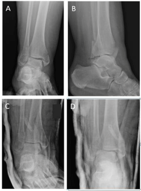
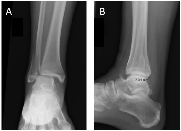
Figure 2 shows initial radiographs of an injury with a small PM fracture fragment size that was successfully reduced in the ED and maintained reduction at first clinic follow-up, allowing for elective surgery to be scheduled. Figures 3a and 3b show initial radiographs of an injury with a larger PM fracture fragment. Though this injury was successfully reduced initially (Figure 3c) that patient presented to clinic 4 days later and had lost reduction (Figure 3d).

Figure 2A and 2B: Initial AP and lateral radiographs of a closed right ankle fracture-dislocation that underwent successful closed reduction. PM fracture fragment length is 2.81mm, which gives relative PM ratio <0.1. At clinic presentation, reduction was maintained and patient underwent elective ORIF.
    Figure 3a and 3b: Initial AP and lateral radiographs of a closed right ankle fracture-dislocation that underwent successful closed reduction. PM fracture fragment length is 8.80mm, which gives a relative PM ratio >0.1. Despite initially successful appearance of reduction (3c), after an accident while riding his bicycle. Closed reduction and splinting in the ED was unsuccessful so he was taken to the OR urgently for external fixation. Initial injury films shown with a) AP view of the ankle demonstrating significant fibular shortening (28mm) and b) lateral view of the ankle demonstrating significant posterior malleolus fracture fragment size (20mm, PM ratio = >0.2).
Figure 3c and 3d: A 48-year-old male sustained a closed left ankle fracture-dislocation after tripping off a curb. He was successfully reduced and splinted in the ED and followed up in Orthopaedic trauma clinic, where he was scheduled for elective ORIF. Initial injury films shown with a) AP view of the ankle demonstrating mild fibular shortening (4mm) and b) lateral view of the ankle demonstrating small posterior malleolus fracture fragment size (3 mm, PM ratio = <0.1).
Value given is mean ± standard deviation, median (minimum, maximum) or as N (%).
Value given is N (%). P-value determined by chi-square test or Fisher’s exact test for small counts.
Value presented is mean ± standard deviation or as median (25th percentile, 75th percentile) in the case of irregular distribution, as determined by Kolmogorov-Smirnov test. P-value determined by student t-test or Wilcoxon rank sum for irregular distribution.
Knowledge about which injuries are likely to fail in the interim despite initial successful closed reduction may better inform providers when making decisions about acute surgical management. The results of this study suggest that injuries with larger PM fracture fragment size, specifically >0.1 relative to the articular surface, are likely to fail in the interim after closed reduction is achieved in the ED. Selection of the appropriate management pathway may help to better shape patient expectations and possibly promote healthcare savings as different ankle fracture management pathways vary significantly in cost [12].
Conclusion
Interim loss of reduction is common (43%) following successful closed reduction of ankle fracture-dislocations in the ED, and failure is associated with larger PM fracture fragment size. Thus there is partial support of the hypothesis that radiographic measurement has predictive value. Careful consideration should be given prior to discharging patients from the ED with reduced ankle fracture-dislocations that contain a relative PM fracture fragment size >0.1. Further investigation is needed to determine the utility of these parameters prospectively and the effect of interim reduction failure on patient outcomes when compared to those with urgent surgical reduction and fixation.
References
- Bagger J, Hölmer P, Nielsen KF. The prognostic importance of primary dislocated ankle joint in patients with malleolar fractures. Acta Orthop Belg. 1993; 59: 181-183.
- Baker JR, Patel SN, Teichman AJ, Bochat SE, Fleischer AE, Knight JM. Bivalved fiberglass cast compared with plaster splint immobilization for initial management of ankle fracture-dislocations: a treatment algorithm. Foot Ankle Spec. 2012; 5: 160-167.
- Rockwood CA, Bucholz RW, Court-Brown CM, Heckman JD, Tornetta P. Rockwood and Green’s Fractures in Adults. Lippincott Williams & Wilkins. 2010.
- Lindsjo U. Operative treatment of ankle fracture-dislocations: A follow-up study of 306/321 consecutive cases. Clin Orthop Relat Res. 1985; 199: 28-38.
- Pettrone FA, Gail M, Pee D, Fitzpatrick T, Van Herpe LB. Quantitative criteria for prediction of the results after displaced fracture of the ankle. J Bone Joint Surg Am. 1983; 65: 667-677.
- Phillips WA, Schwartz HS, Keller CS, Woodward HR, Rudd WS, Spiegel PG, et al. A prospective, randomized study of the management of severe ankle fractures. J Bone Joint Surg Am. 1985; 67: 67-78.
- De Vries JS, Wijgman AJ, Sierevelt IN, Schaap GR. Long-term results of ankle fractures with a posterior malleolar fragment. J Foot Ankle Surg. 2005; 44: 211-217.
- Federici A, Sanguineti F, Santolini F. The closed treatment of severe malleolar fractures. Acta Orthop Belg. 1993; 59: 189-196.
- White BJ, Walsh M, Egol KA, Tejwani NC. Intra-articular block compared with conscious sedation for closed reduction of ankle fracture-dislocations. A prospective randomized trial. J Bone Joint Surg Am. 2008; 90: 731-734.
- Lauge-Hansen N. Fractures of the ankle: II. Combined experimental-surgical and experimental-roentgenologic investigations. Arch Surg. 1950; 60: 957-985.
- Harper MC. Instability of the distal tibiofibular syndesmosis after bimalleolar and trimalleolar ankle fractures. J Bone Joint Surg Am. 1984; 66: 490-503.
- Kheir E, Charopoulos I, Dimitriou R, Ghoz A, Dahabreh Z, Giannoudis PV. The Health Economics of Ankle Fracture Fixation. The Royal College of Surgeons of England Bulletin. 2012; 94: 1-5.

