Abstract
Congenital Talipes Equinovarus (CTEV) is applied to the true clubfoot deformity in the newborn babies and the foot is appear like a club and thus has its common name “clubfoot”. It is a historical foot deformity in medical science, where the foot turns inward and points down causing walking on the toes and outer sole of the foot. Some of the bones in clubfoot are abnormal not only in their relationship to each other but also in shape and size. The talus connect leg to the foot, is a major functional element between leg and the foot, and plays a crucial role in movement of the foot. It involve in the formation of three joints with synovial recesses and articulates with the tibia, fibula, calcaneus, and navicular. It consists of a body, a neck, and a head.
The basic aim of this research is to study the shape geometry of talus of clubfoot by developing its true models by considering live patients and comparing it with normal talus. The study will be useful in the development of a non-surgical corrective technique such as for the development of scientific Ankle Foot Orthosis (AFO) with greater success rate achievement for club foot correction of this historic deformity and is very much useful in the context of child development at national and international platform. The study involve live patients, its MRI data and an image-processing tool. The research involve an interdisciplinary “Bridge” between engineer, radiologist and surgeons for knowing shape geometry where it combines with multidisciplinary research, that include Three Dimensional (3D) modeling, and image analysis.
These specific 3D talus representations provides its shape realization and helps in determining the shape and size of clubfoot talus bones. The representations provides us an opportunity to view talus and analyse the ankle joint geometry that develops a favourable condition for diagnosis and treatment of a historical CTEV foot deformity. The representation also helps orthopaedic surgeons in preoperative surgical planning and consequently in carrying out biomechanical studies. It also provides a platform for finite element analysis.
Keywords: Clubfoot; CTEV; Talus; Modeling
Introduction
The clubfoot is a historic congenital foot deformity in medical science. The present research is an attempt to use radiological MRI image of live clubfoot patients and develop Three-Dimensional (3D) model of talus to reach the convergence of clubfoot treatment solution for newborn babies [1,2]. The talus bone forms the main connection between the leg and the foot and subject to large loadings that are passed down through the leg into the foot complex via calcaneus and navicular bones [3-5].
The human foot that contain about 26 bones and 57 joints is a very complex joint with many combinations of movements and motion [3,6]. The integration of MRI, medical imaging modality with computer-aided design to produce 3D model is an important area of development. Three-dimensional shape data of both internal and external human body structures (e.g. from CT, MRI, PET/SPECT, Ultrasound, etc.) are employed for 3D model development [7] and most recent anatomical models have been built for bony structures from CT data. However, increasingly soft tissue structures also been built from MRI data. Surgical tool design, customized implant design, customized prosthesis production and customized orthosis production are other developing applications within the medical field [8-11].
Daniel et al reconstructed 3D model of human foot, restricted to cadaver left foot whose different bones were, kept in aligned position with the help of an acrylic frame [12]. Jacob and Patil also reconstructed 3D model of human foot based on conventional plain X-ray images. The geometry of foot’s bones was rather approximate in their model [13]. Many new domains for real 3D modeling of anatomic sculpture require input images from variety of sources like photographs, sketches, computer made images from CT, MRI, Sonography and X-ray images etc [14,15]. 3D modeling of human anatomy is an open area of research and much works reported under this domain for different organ of the human body. 3D model of human foot using CT/MRI etc is, reported in few engineering literatures. Some researchers did excellent works on modeling and study of human foot. From an extensive review of literature of ankle complex biomechanics, few papers have proposed mathematical model for the ankle complex [16-19], but their study was based on either cadaver foot data or on certain mathematic assumptions and in any case their study was not based directly on the live human foot. Udupa K Jayaram and Hirsch BE. did Kinematics analysis of 3D human foot’s joint based on live subject’s MRI scan data. This contributes the actual happening at tarsal joint [15].
The purpose of this study is to study the true shape geometry of live clubfoot patients that will help in developing a non-surgical correction procedure for scientific treatment of clubfoot by determining the critical shape/surfaces/region of interest, about which corrective forces will play a major role. The present paper offers a 3D representation of Clubfoot’s bones specifically talus bone of live patients from his acquired MRI scan data set. This proposed a novel approach for 3D, talus representation of clubfoot of live patients by integrating MRI and medical image processing tools and this provides a computer-aided tool in the form of 3D talus representation. The major outcome of this work is the 3D model of talus that assists in diagnosis and better treatment of a historical CTEV foot deformity.
The paper begins with an introduction, highlighting the area of work along with previous work reviewed on foot, our approach and outcome. The section two describes methodology along with output. The section three present results and discussion followed by section four and five of conclusion and acknowledgment.
Methodology
The major objective of this paper is to develop 3D representation of bones of Talus of live human Clubfoot Patients by using MRI scan imaging modality and understanding the importance of this 3D shape geometry from medical treatment point of view [20]. The approach comprises of four main phases as shown in flow chart of (Figure 1).
The flow chart shows that in phase I, the volunteer clubfoot subject was prepared for MRI scanning and both foot scanning was started. In phase II, the sequence of MRI scan data were acquire from SIEMEN, MRI machine, in Digital Imaging and Communication (DICOM) format; these acquired data are then processed in medical image processing MIMICS (Materialize Inc.) software in phase III, while in phase IV, the processed data were used to compute 3D Talus bone modelling.

Figure 1: Integrated approach to represent 3D Talus bones of a specific foot.

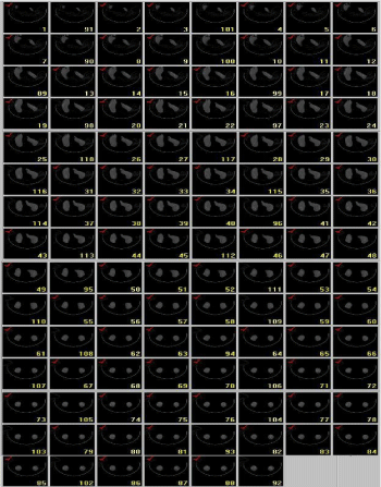
Figure 2: MRI scan data of clubfoot of Patient-I.
For this research two volunteer unilateral clubfoot male patients of age 6 years and 4 months were taken for MRI scan from hospital with the help of orthopaedic surgeon and radiologist, thee patients are here after refer as Patient-I, and Patient II. At MRI scan center these patients were laid down on the scan table one at a time and their both foot were scanned and acquired image data in Dicome formate were taken. One sample MRI scan data set of a clubfoot patient is shown in (Figure 2). These acquired image data were processed in mimics where user can modify the image by definition for computing 3D foot and Talus model by segmentation that is an image enhancement method by which a particular object, organ, or image characteristic is extracted from image data for the purpose of visualization and measurement. The image segmentation of above scanned images were separated into region of interest using following different tools:
i) Data visualization
ii) Thresholding
iii) Image editing
iv) Region growing
v) Boolean operations
vi) 3D image calculation/generation.
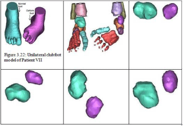
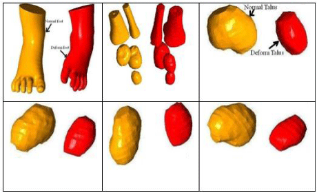
On processing a virtual 3D model of unilateral clubfoot and Talus bones of these patients are created and are shown in (Figure 3,4) respectively. The created 3D represented helps in diagnosis and treatment of Clubfoot and ankle disorders [21-24]. The 3D talus tarsal bone geometry representation will found to be particular useful in correction of CTEV condition [5,1,2].

Figure 3: Developed 3D unilateral clubfoot, skeletal & Talus of Patient-I.

Figure 4: Developed 3D unilateral clubfoot, skeletal & Talus of Patient-II.
Results and Discussion
The 3D model of normal and clubfoot’s internal bony structure of hind foot of two unilateral clubfoot volunteer male patients of age 6 Years and 4 Month developed At tender age of the baby, the bone of the foot is under development stage, therefore the full connectivity of the bone is not observed in modeling. The shape of the bony structure is observed and compared in (Figure 3,4) reconstructed from the sequence of acquired MRI images by the methodology described above. The representation can be rendered for showing outline of the foot and within that outlines the anatomical structure of interest can be selected for visualization of actual shape geometry. This allows the visualization of a source in its context, and should aid in finding the correct structure in which the activity takes place. The representation approximates a clubfoot of live patient that give more information of the whole 3D clubfoot shape and this helps in drawing the physical interpretations about the clubfoot features. From this 3D model the two lower bone of leg namely tibia and fibula meeting with talus, form the ankle joint and in that, joint medial extension of tibia and lateral extension of fibula known as medial and lateral malleolus helps in holding the talus at appropriate place. The back part of the foot having heel bone is the largest bone in the foot known as calcaneus and is connected to the talus. Hence, the reconstructed 3D representations shown in (Figure 3,4) provides a familiar means of viewing ankle joint of clubfoot and is useful for visualization, shape measurement and simulation. The model would be useful in the medical science for non-surgical correction planning of clubfoot.
The research provides an opportunity to view the talus bone of the clubfoot whose upper smooth surface would articulate with the lower surface of tibia, medial malleolus of tibia and lateral malleolus of fibula and form a very important load bearing joint known as ankle joint on growth of babies patients. This ankle joint is responsible for flexion and extension of foot. The lower surface of the talus on growth rests on and articulates with the calcaneus, form the subtalar joints and would be responsible for inversion and eversion motion of the foot. From the figures the anteriorly projected part of talus, known as talus head whose constricted upper part is called talus neck is underdeveloped in clubfoot as compare to normal foot. The geometry of the talus gives an impact to look interestingly into Congenital Talipes Equinovarus (CTEV) and this details may be compare with the normal talus geometry and a better correction idea can be develop for abnormal foot [4,5,21,24]. On studying and comparing of normal foot with clubfoot in figure 3 and 4, following observations were made.
i. The maximum length of deformed foot (hind foot to fore foot) is shorter in clubfoot than the normal foot,
ii. The maximum width of forefoot in deformed foot is more than the normal foot,
iii. In clubfoot, the heal and toe is twisted inwardly as compared to normal foot,
iv. It is observed that, in clubfoot, the forefoot curls towards the heel,
v. In clubfoot, the lateral malleolus is displaced posteriorly as compared to normal foot.
It is observed that the talus is underdeveloped; the talar neck is shortened and is deviated in the medial and plantar direction. The abnormal position of the talus causes the calcaneus to fall into the equinus. Similarly, from Figures, the navicular is smaller than normal and articulates with the medial aspect of the neck of the talus, which forces the forefoot into adduction [21,24].
This research by integration of MRI and image processing tools proposes a novel computer assisted representation of clubfoot. The representation focus on new insights pertaining to the detailed look of talus bone based on patient-specific image data. The approach provides a better understanding of normal and clubfoot. The study helps in understanding the ankle joint anatomy of clubfoot. The model increases their clinical importance, as it depict the true shape geometry of talus of club foot. The outcome gives clear information to an orthopadician about major orthopaedic short coming in clubfoot and he is in a better position to judge the treatment. We believe that the study advances the understanding of talus of clubfoot and helps in its better treatment planning.
As the research is integration of MRI scan with Computer Aided Design (CAD) and Rapid Prototyping (RP) in the domain of clubfoot and in future would be useful for the development of dynamic Ankle Foot Orthosis (AFO) with application of CAD in forth coming research, where we will consider detail study of Pirani’s Group of classification of severity of clubfoot and existing serial plastic casting techniques of clubfoot correction.
As the research is integration of MRI scan with Computer Aided Design (CAD) and Rapid Prototyping (RP) in the domain of clubfoot and in future would be useful for the development of dynamic Ankle Foot Orthosis (AFO) with application of CAD in forth coming research, where we will consider detail study of Pirani’s Group of classification of severity of clubfoot and existing serial plastic casting techniques of clubfoot correction.
Conclusion
In order to study congenital CTEV foot deformity that is also sometime said to be the deformity of talus [1] it is necessary to visualize the geometry of talus from CAD point of view. Hence, the individual 3D view of the talus bone of clubfoot is studied. The presented work contributes to the area of computer – aided surgery planning in particular for orthopaedic application and consequently in carrying out biomechanical study for medical device development [25]. The major outcome of this representation is the details geometrical visualization of talus bone that assists in diagnosis and better treatment of a historical medical condition CTEV [26]. Further down stream application of this representation is finite element analysis that will be helpful for future biomechanics research on human clubfoot.
Acknowledgement
The authors would like to thank professor NN Kishor, Head department of mechanical engineering, Indian Institute of Technology Kanpur, India for providing financial support, from department research grant, in acquiring MRI scanned data from diagnostic centre for this research work.
The authors also thank the radiologist Dr. Gupta and MRI technician Mr. Ledgee of Kanpur city India for extending their technical support in acquiring MRI scan data of clubfoot.
References
- Ponseti IV. Congenital Clubfoot: Fundamentals of Treatment, Oxford University Press, Oxford. 1996.
- Simon GW. The Clubfoot – The Present and a View of the Future, Springer, New York, NY. 1993.
- Arthur J, James VH, Sherman DS, Luciano. Human physiology: the mechanisms of body function. Tata Mc Graw Hill Publishing Co. Ltd, New Delhi. 1991.
- Hering John Anthony, Pediatric Orthopaedics. WB Saunders Company. Third edition 2000; 2.
- Jahss Melvin H. Disorders of the Foot and Ankle - Medical and Surgical management. W. B. Saunders Company, Philadelphia, USA. Second Edition. 1991; 1.
- Asai T, Murkami H. Development and evaluation of a finite element foot model. Proceeding of the 5th Symposium on Footwear Biomechanics, Zuerich/Switzerland. 2001; 10-11.
- Beaugonin M, Haug E. A numerical model of the human ankle/foot under impact loading in inversion and eversion. International Conference on high performance computing in automotive design, Engineering and manufacturing cray research silicon graphics Inc. Enghien France. 1996; 1-13.
- Hirsch BE, Udupa JK, Robert D. Computerized 3-D reconstruction of the foot from computed tomographic scan. J. Ammer. Podiatr. Med. Assos. 1989; 79: 384-394.
- J Kreck, J Duhovnik. Model generation and application in medical domain. International design conference - DSIGN 2002, Dubrovnik. 2002; 1-5.
- P Lasaygues, P Laugier. Bone imaging using compound ultrasonic Tomography. Journe`es Os- Ultrasons. 2002; 24-25.
- LAD Camacho, RW Ledoux, Eic Rohr, JB Sangeorzan, PR Ching. A three-dimensional, anatomically detailed foot model: a foundation for a finite element simulation and means of quantifying foot-bone position. Journal of Rehabilitation Research and Development. 2002; 39: 401-410.
- Jacob S, Patil MK. Three-dimensional foot modeling and analysis of stresses in normal and early stage Hansen’s disease with muscle paralysis. Journal of rehabilitation research and Development. 1999; 36: 252-263.
- Nan Weng, Yee-Hong Yang. Three-dimensional surface reconstruction using optical flow for medical imaging. IEEE Transaction on medical imaging. 1997; 16: 630-641.
- Udupa JK, Hirsch BE, Hillstrom H, Bauer, G Kneelanc B. Analysis of in vivo 3-D internal kinematics of the joints of the foot. IEEE Trans. Biomed. Eng. 1998; 45: 1387-1396.
- Procter P, Paul JP. Ankle joint Biomechanic. Journal of Biomechanics. 1982;15: 627-634.
- Wynarsky GT, Greenwald AS. Mathematical model of the human ankle joint. Journal of Biomechanics. 1983;16: 241-251.
- Ying Ning, Kim Wangdo. Use of dual Euler angles to quantify the three-Dimensional joint motion and its application to the ankle joint complex. Journal of Biomechanics. 2002; 35: 1647-1657.
- Ying N, Kim W, Wang YS. Mathematical model of the ankle joint complex. ICBME Singapore. 2002.
- Manak Lal Jain, Sanjay Govind Dhande, Nalinaksh S. Vyas. A biomechanical control mechanism for correction of clubfoot deformity in babies. Int. J. Biomechatronics and Biomedical robotic. 2009; 1: 51-56.
- ML Jain. Enhancement of Success Rate of Complex Surgical Cases through Biomodeling. Journal of Orthopedics, Rheumatology and Sports Medicine. 2015: 102.
- ML Jain, SG Dhande, Nalinaksh S, Vyas. Computerised rapid prototyping of human foot using computed tomography, International Journal of Design Engineering. 2015; 6.
- ML Jain. Dhande SG. Vyas NS. Computer aided diagnosis of human foot’s bone. International Journal of Biomedical Engineering and Science (IJBES), 2014; 1.
- Manak J, S Dhande, N Vyas. Biomodeling of clubfoot deformity of babies. Rapid Prototyping Journal. 2009; 15: 164-170.
- ML Jain, Dhande SG, Vyas NS. A virtual environment in the healthcare domain for the management of clubfoot deformity in newborn babies: a case study. International Journal of Healthcare Technology and Management. 2008; 9:136-142.
- Leardini A, O’Connor JJ, Catani F, Giannini S. A geometrical model of the human ankle joint. Journal of Biomechanics. 1999; 32: 585-591.
