Abstract
Ankle sprains are one of the most common orthopedic injuries among both athletes and the general population. Persistent lateral ankle instability may require surgical repair. Currently, the gold standard for surgical repair of chronic lateral ankle instability is the modified Broström procedure. Augmentation of damaged ligaments, including the Anterior Yalofibular Ligament (ATFL) has been suggested to help improve patient clinical outcomes after surgery. Several options exist for augmenting the ATFL, including autograft, allograft and synthetic devices. In this case series, we present our experience using two commercially available synthetic devices; suture tape (Arthrex InternalBrace™, Arthrex, Naples, FL, USA) and a polyurethane urea (PUUR) woven matrix (Artelon® FLEXBAND®, Marietta, GA, USA), for augmentation of the ATFL during modified Broström repair.
Level of Clinical Evidence: Level V
Keywords: Lateral Ankle instability; Ankle Reconstruction; Suture Tape; PUUR; ATFL; Augmentation
Abbreviations: ATLF: Anterior Talofibular Ligament; PUUR: Polyurethane Urea; CLAI: Chronic Lateral Ankle Instability; mBP: Modified Broström Procedure; ST: Suture Tape
Introduction
Chronic Lateral Ankle Instability (CLAI) is a common condition that typically develops from an injury affecting the ligaments of the lateral ankle marked by pain, swelling and reduced ankle function persisting at least 12-months post-injury [1]. CLAI is well characterized in the literature as a performance limiting injury accounting for 30%-40% of injuries in athletes [2-4]. CLAI is prevalent in the general population as well, impacting >2 million Americans annually and develops in up to 70% of those who experience an acute ankle sprain [5]. The ATFL is the most common tissue damaged following ankle sprains because, biomechanically it is the weakest of the lateral ligament complex. Failure to resolve damage to the ATFL and ongoing mechanical and functional destabilization of the ankle leads to further chronic complications that may develop into osteoarthritis, [5] Lack of response to conservative treatments results in evaluation for surgical intervention to avoid additional long-term complications. Currently, the gold standard for surgical repair of the ATFL is the modified Broström procedure (mBP) [7-9]. Injured ligaments can be augmented with auto-or allo-genic tendon grafts [10] or synthetic scaffolds, [11] which are anchored approximate to the damaged ligament for biomechanical support during the healing process. While the mBP is generally effective in restoring stability to the ankle joint in most patients, 13-35% of patients have persistent pain [12] and up to 31% [13] report continued instability, resulting in reoperation rates as high as14% [14]. (Baraza N et al.). Issues that can impact the integrity of the mBP include ligament laxity, poor tissue quality, diabetes and high BMI [15]. Data demonstrate that repaired ATFL using auto-/allografts have only approximately 50% strength of native ligaments [16]. Further, tissue grafts can vary in quality and instances of tissue necrosis and resorption have been reported. These clinical challenges have prompted the development of new synthetic, biocompatible structural scaffolds to improve the consistency of mBP results [18]. Historically, synthetic, soft tissue augmentation devices were designed with a strong rigid structure that are unmatched to the elastic modulus of the musculoskeletal tissues targeted for reconstruction. The stiffer profiles of these synthetics can result in the transfer of mechanical load to the device, leading to potential failure due to tissue stress-shielding or device fatigue [8]. Newer developments in synthetic devices employ resorbable materials, designed to optimize tensile properties with mechanical properties that more accurately match the biomechanics of the augmented soft tissue. One such material is comprised of a co-polymer of polycaprolactone based Polyurethane Urea (PUUR) [21]. The enhanced biomechanical properties may more accurately enable load sharing during the tissue healing process, allowing for direct cell infiltration, differentiation, and the production of ECM within the synthetic scaffold. The device then degrades benignly by hydrolysis following tissue repair [22].
In the present case series, we compare two synthetic devices used to augment the ATFL during mBP repair; a polyethylene Suture Tape (ST) (InternalBrace, Arthrex, Naples, FL) and a polyurethane urea woven matrix (PUUR) (Flexband™, Artelon, Sandy Springs, GA). By comparing clinical outcomes, post-operative rehabilitation, and overall recovery trajectories in these patients, this study may provide insights on the efficacy of such devices in impacting patient outcomes after surgery.
Methods
Three patients in this case series were presented to the clinic for evaluation of CLAI. Failing conservative care, surgery was discussed, and all patients opted for surgical reconstruction of the lateral ankle. All surgeries were performed by the same surgeon. Following surgical repair of the ATFL with either ST or PUUR augmentation, patients were followed up in the clinic to assess healing and rehabilitation and return to activities of daily living.
ATFL Augmentation via ST
Repair of the ATFL for all patients was performed using the mBP with augmentation utilizing ST for reinforcement. Initially, two fibertak anchors were inserted into the fibula flanking the origin of the ATFL. The first bone anchor was placed in the talus under the
manufacturer's recommended technique. The second bone anchor drill hole was then placed on the anterior face of the fibula in the orientation of the ATFL ligament origin. The mBP was used with the foot in the dorsiflexed everted position. Finally, the ST was placed into the fibula under tension with continuation of proper reduced position completing the augmentation.
ATFL Augmentation via PUUR
Repair of ATFL for all patients was performed using an mBP with PUUR graft augmentation. A wire was placed into the talar neck in a 45° angle to the body and mid-height. Inspection with a biplanar fluoroscopy view was performed to ensure proper placement. Drilling was performed to proper depth over the wire, followed by insertion of the bone anchor containing the PUUR graft. A second bone anchor drill hole was placed into the anterior face of fibula to proper depth in the orientation of the ATFL origin.
Results
Patient #1
Patient 1, a 47-year-old female former smoker with Type 2 diabetes, hypertension, vitamin D deficiency, Hashimoto's thyroiditis, and high cholesterol, presented initially with bilateral plantar fasciitis with chronic ankle instability (Table 1). Despite initial conservative measures the patient continued to present with pain, particularly on the right side. Examination revealed tenderness along the peroneal tendon, ATFL, including positive anterior drawer and laxity. Subsequent MRI showed prominent tendinosis of the peroneal tendon and edema in the plantar muscles adjacent to the plantar fascia. The patient opted to undergo ankle stabilization consisting of peroneus brevis tendon repair, endoscopic plantar fasciotomy and ATFL reconstruction using ST augmentation.
Summary
Tx
Patient
Age
Gender
PMHx
Social
HistoryAffected
AnkleLeft Ankle
Right Ankle
1
47
Female
Type 2 DM, HTN,
Vitamin D Deficiency, Hashimoto’s thyroiditis,
HLD,Former smoker
Left and Right
Repair with Artelon FLEXBAND.
No revision
surgeryInitial repair with InternalBrace.
Removal of InternalBrace and revision with Artelon
FLEXBAND2
30
Female
HTN
Current every day
smokerLeft
Initial repair with InternalBrace. Removal of InternalBrace and revision with Artelon
FLEXBANDNone
3
51
Female
None
Non-
smokerLeft and
RightRepair with
InternalBraceRepair with Artelon
FLEXBAND
Table 1: Summary of Electronic Medical Record (EMR) notes for all patients.
    Post Rehab


    Summary


    Flexband
    Internal Brace


    Avg Pain Duration (weeks)
    6.75
    30


    Pain Level


    >6 weeks
    0/10
    unknwn


    6 weeks
    1/10
    unknwn


    15 weeks
    0/10
    unknwn


    24 weeks
    0/10
    4/10


    Progression to weight    bearing (weeks)
    4
    8


    PT Initiation
    unknwn
    3


    Recurrent Pain (yrs)
    none
    3 yrs post-op
Table 2: Summary of average post-rehabilitation progress for Artelon Flexband and Internal Brace.
    Post Rehab


    Patients 1 & 2


    Patient 1
    Patient 2


    Left Ankle
    Right Ankle
    Left Ankle


    Flexband
    Flexband
    InternalBrace
    Flexband
    InternalBrace


    Pain Duration (weeks)
    6
    15
    60
    2
    10


    Pain Level


    2 weeks
    unknwn
    4/10
    unknwn
    0/10
    unknwn


    6 weeks
    0/10
    2/10
    unknwn
    0/10
    unknwn


    15 weeks
    0/10
    0/10
    unknwn
    0/10
    unknwn


    24 weeks
    0/10
    0/10
    4/10
    0/10
    unknwn


    PT Initiation (weeks)
    3
    2
    3
    2
    3


    Recurrent Pain (yrs)
    unknwn
    unknwn
    unknwn
    none
    3
Table 3: Summary of post-rehabilitation for patients 1 and 2.
    Post Rehab


    Patient 3


    Right Ankle
    Left Ankle


    Flexband
    Internal Brace


    Pain Duration (weeks)
    4
    20


    PT Initiation (weeks)
    2
    3


    Progression to weightbearing (weeks)
    4
    8


    Pain Improvement


    Percentage Improvement
    100%
    90%


    Time (months)
    3.5
    5
Table 4: Summary of post-rehabilitation for patient 3.
    Post-op Summary


    Patient 1


    Left Ankle
    Right Ankle


    Flexband
    Flexband
    Internal Brace


    Non-weight bearing (weeks)
    2
    1
    2


    CAM boot weight bearing (weeks)
    6
    6
    6


    Time to full weight bearing    (weeks)
    13
    unknwn
    15


    PT Initiation (weeks)
    3
    2
    3


    PT Course (visits)
    15
    11
    17


    Return to activity
    unknown
    Return to pre- injury
    Never went

      back to pre- injury
Table 5: Shows a summary of key points during the patient's post-op recovery and rehabilitation.
    Post-op Summary


    Patient 2


    Left Ankle


    Flexband
    Internal Brace


    Non-weight bearing (weeks)
    1
    2


    CAM boot weight bearing (weeks)
    unknown
    6


    Time to full weight bearing    (weeks)
    unknown
    10


    PT Initiation (weeks)
    2
    3


    PT Course (visits)
    unknown
    17


    Return to activity
    unknown
    Never went

      back to pre- injury level
Table 6: Shows a summary of key points during the patient’s post-op rehabilitation.
Post Rehab
Patient 3
Right Ankle
Left Ankle
Flexband
Internal Brace
Non-weight bearing (weeks)
1
1
CAM boot weight bearing (weeks)
4
5
Time to full weight bearing (weeks)
4
8
PT Initiation (weeks)
2
3
PT Course (visits)
15
16
Return to activity
3.5 months
with 100% improvement5 months with
90%
improvement
Table 7: Shows a summary of key points during the patient's post-op recovery and rehabilitation.
Approximately 5-months after the reconstruction of the right ankle, the patient reported left ankle pain with similar symptoms. MRI showed tendinosis of the peroneal tendon with tenosynovitis and effusions of both the ankle and subtalar joints. With MRI findings and similar clinical presentation, the patient underwent left ankle stabilization with ATFL repair using PUUR augmentation.
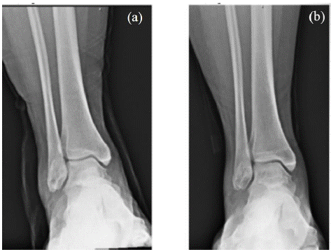
Figure 1: X-ray of patient’s right ankle at different time points: (a) Prior to removal of InternalBrace and revision of lateral ankle reconstruction with secondary repair of ATFL with Artelon FLEXBAND; (b) 1 week post operation displaying good position of the soft tissue anchor.
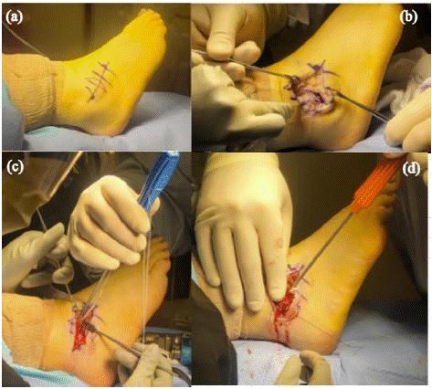
    Figure 2: Shows different points during the patient’s right ankle revision surgery with Artelon Flexband: (a) The initial incision placement; (b) Displays the dissection exposing the residual
Internal Brace and periosteal fibular reflection; (c) Depicts surgeons placing an anchor with the Artelon attachment towards the talar neck; (d) Shows surgeons tightening and anchoring the Artelon graft into the fibula.
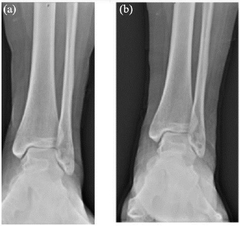
    Figure 3: X-ray of patient’s left ankle at different time points: (a) Prior to removal of InternalBrace and revision of lateral ankle reconstruction with secondary repair of ATFL with Artelon
FLEXBAND; (b) 1-week post-op and depicts good position of the soft tissue anchor.
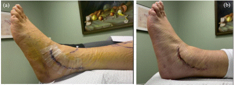
Figure 4: (a) Clinical picture of patient’s left ankle 1-week post-op with no acute signs of clinical infection; (b) Depicts recovery 2 weeks post-op with incision fully scabbed over.
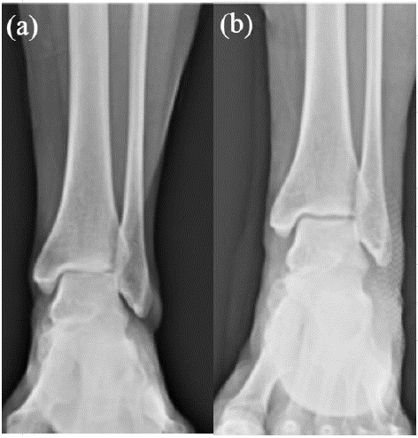
Figure 5: X-ray of the patient’s left ankle: (a) Prior to lateral ankle reconstruction with secondary repair of ATFL with InternalBrace; (b) 1-week post-op and depicts good position of the soft tissue anchor.
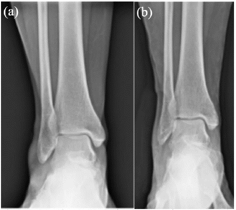
Figure 6: X-ray of the patient’s right ankle: (a) prior to removal of InternalBrace and revision of lateral ankle reconstruction with secondary repair of ATFL with Artelon FLEXBAND; (b) 1 week post operation displaying good position of the soft tissue anchor.
Following the left ankle reconstruction, the patient continued to have pain and discomfort of the right ankle with persistent inflammation which failed to respond to steroid injections. At 15 months post-operative, the patient elected to undergo a revision surgery on the right ankle, requiring the perioperative removal of ST. During the procedure, excessive inflammation and arthrofibrosis surrounding the ST was observed, fibrotic tissue was removed, along with secondary repair of ATFL with PUUR augmentation, Post-operatively, the patient’s recovery on her right ankle was expedited following PUUR augmentation compared to ST augmentation. Weight bearing was started in a CAM immobilizer at one week post operatively instead of 2 weeks as in the primary procedure. When comparing the pain following both primary surgeries, at 24 weeks the patient reported 4/10 pain on the ST augmented ankle. While during the PUUR revisional surgery the patient had a visual analog scale of 0/10 pain at 6 weeks on the left while on the right, she experienced a 2/10 pain on the VAS scale at 6 weeks and 0/10 pain at 15 weeks post-operative. Subjective clinical evaluation by the surgeon noted a reduction in the overall size of the ankle complex, reduced erythema, reduction in edema and overall apprehension of manipulation of the ankle joint complex while under stress exam. Strength was noted as well to be accelerated in comparison to the index procedure with a 5/5 manual muscle testing at 6 weeks postoperatively after the PURR revisional reconstruction.
Patient #2
Patient 2, a 30-year-old female with hypertension and a current every day smoker presented with left ankle pain and swelling. The pain was initially attributed to an acute injury following a slip and fall. Despite immobilization and subsequent physical therapy, the patient had persistent symptoms, positive anterior drawer and inversion stress tests, and reduced eversion strength.
MRI revealed mild peroneal tenosynovitis and edema in the anterolateral recess of the ankle. Conservative treatments failed to provide functional improvement and the patient opted to undergo surgery for lateral ankle stabilization with ST augmentation of the ATFL and peroneal repair.
Post-operatively, the patient progressed to full weight bearing as tolerated with assistance of a CAM boot at 2-weeks. However, six weeks post-operative, the patient continued to report mild discomfort at night and tightness in the ankle and while able to return to work 10 weeks post- operative, reported ongoing mild pain and discomfort.
Approximately 3 years post- ST augmentation, the patient experienced recurrent pain following an acute ankle sprain and was placed in a CAM boot, ultimately transitioning to a brace. The patient progressed to physical therapy with limited improvement, reporting moderate to severe pain after 10 weeks of physical therapy. MRI revealed thickening of the ATFL with possible arthrofibrosis and impingement. Treatment via corticosteroid was successful in temporary pain relief however, pain returned to pre-injection levels within 2 weeks. Taken together with the findings of the MRI, surgical intervention was recommended and selected by the patient. Revisional ankle reconstruction required the removal of ST, secondary repair of the ATFL with PUUR augmentation, and repair of the peronealtendon. Marked inflammation was noted as in the prior patient #1 surrounding the ST during removal. The capsular thickening and hemorrhagic synovitis on the interior of the joint lining was remarkable in comparison to the surgeon's prior experience. A similar postoperative course to patient #1 noted above was utilized with a 1-week period of non-weight bearing immobilization in a CAM immobilizer and the introduction of a course of physical therapy at 2 weeks. After revision, at one week the patient reported mild pain, had negative anterior drawer and talar tilt tests. At the two- and five-week clinical follow-ups, the patient reported 0/10 pain score with no complications at the surgical site.
Patient #3
Patient 3, a 51-year-old female, non-smoker, with no significant medical history, presented initially with plantar fasciitis, left lateral ankle pain, localized swelling, and difficulty performing daily activities. Following attempts at conservative care, an MRI revealed acute chronic plantar fasciitis and peroneal tendinopathy with a partial longitudinal split in the peroneus brevis tendon. Given the history of CLAI she elected to undergo ankle reconstruction, involving ATFL repair with ST-augmentation, peroneus brevis repair and an endoscopic plantar fasciotomy.
The patient reported 50% improvement in pain score and 85% improvement by 2 weeks following ST augmentation of the ATFL in the left ankle. She initiated physical therapy with no complications at the surgical site 3-weeks post-operative. However, by 5-weeks post-operative, the patient remained unable to bear full weight while in a boot and the ankle presented with persistent swelling. The patient transitioned to an airport brace and was permitted to wear shoe gear of choice at 8 weeks. At 5-months post-operative, the patient reported 90% improvement in pain compared to preoperative scores but continued to report continued swelling in the foot/ankle when upright for extended periods of time. Approximately eight-months following the left ankle augmentation with ST, the patient presented with right ankle pain following an acute ankle sprain. Clinical examination revealed edema, tenderness to the lateral ankle ligaments, a positive anterior drawer test, and positive instability on talar tilt. A tear in the ATFL and a low to intermediate sprain of the CFL was demonstrated on MRI. The patient elected to undergo ankle reconstruction with revisional repair of the ATFL via PUUR-augmentation.
In contrast to the ST augmentation of the left ATFL, the patient was weightbearing as tolerated in a CAM boot at 1-week post-PUUR-augmentation and began a full regiment of physical therapy at 2 weeks postoperatively. By 4 weeks post-operative, the patient reported 0/10 pain and was full weight bearing without the assistance of a boot or brace. The patient continued to report improvement in a clinical follow-up 3.5 months post-operative, stating 100% improvement of the right ankle with no stability issues and mobility without limitation on uneven surfaces.
Discussion
The present case series presents our experience with two commercially available synthetic augmentation devices for ATFL repair to treat CLAI. In all cases, the patients received both devices, presenting a unique opportunity to evaluate the clinical differences of each. Immediate post-operative outcomes, allowed for patient mobility at early time points with both devices, two of the three cases in this series receiving ST-augmented ATFLs presented excessive inflammation and pain, which required revision. In the surgeon's experience, overall recovery timeline was reduced with the revisional PUUR augmentation in comparison to the index ST augmentation procedure. Return to ADL’s was noted to be faster in all patients after revision.
Reduction in pain, swelling, erythema and edema were all noted with PURR augmentation. Review of the progression of physical therapy was observed to be at a faster pace among the 3 patients reviewed in this case review. Single leg stance, balance board and functional progressive activities were accelerated in the PUUR revisional patients, even when PUUR was utilized in a revisional nature. One would believe that revisional surgery would, on average, take a longer period of time to recover to full ADL’s.
Suture tapes are typically comprised of non-resorbable materials that are biomechanically stiff [22], which provide the benefit of immediate support tensile strength post-surgically. However, such rigidity in device structure can lead to challenges with appropriate tensioning on implant. Because the device is non-resorbable, overtightening can lead to inappropriate/limited biomechanics of the ankle reducing functionality in the short and long-term, and the potential for ultimate permanent disruption of the native kinematics of the ankle repair [23]. In addition, complications reported with STs in ligament repair include persistent immune responses/inflammation, (arthro)fibrosis, synovitis and osteoarthritis [22-24]. In this case series, we observed some of these reported concerns with suture tape, with long-term persistent pain and inflammation necessitating revision procedures.
The elasticity of PUUR provides the potential for more flexibility in tensioning the device at implant, but also the potential to more accurately mimic ATFL biomechanics during the healing process [25]. The degradation profile of PUUR allows for the device to maintain much of its biomechanical strength for support during the early to mid-healing phases of tissue repair, while allowing the native tissue to gradually take over the biomechanical load once repaired.
Following augmentation of the ATFL, patient pain levels decreased, regardless of whether ST or PUUR was used in ATFL augmentation. However, pain levels were higher, and patient-reported functional quality lower, in ST ATFL augmentations compared to PUUR augmentations. Further, pain reduction was achieved earlier and to a greater extent in PUUR ATFL augmentations compared to ST augmentations. In addition, assessment of post-operative functional rehabilitation showed that PUUR augmentation allowed for quicker return to weight- bearing and normal shoe gear and less limitations long
Conclusion
Augmentation of ATFL with mBP for repair is commonly performed to alleviate pain and improve function in lateral CLAI. In this case report, we presented our experience with two commonly used augmentation devices, ST and PUUR. Both ST and PUUR augmentation devices provided support to the healing ligaments. However, anecdotally, we have noted increased inflammation and pain in ankles reconstructed with suture tape augmentation of the ATFL. To date, use of PUUR has not required revision surgery in any of the cases presented, which are 9 - 15 months post-operative.
References
- Chen ET, Borg-Stein J, McInnis KC. Ankle Sprains: Evaluation, Rehabilitation, and Prevention. Current sports medicine reports. 2019; 18: 217-223.
- Chan KW, Ding BC, Mroczek KJ. Acute and chronic lateral ankle instability in the athlete. Bulletin of the NYU hospital for joint diseases. 2011; 69: 17-26.
- Gibboney MD, Dreyer MA. in Stat Pearls. 2023.
- Fong DT, Hong Y, Chan LK, Yung PS, Chan KM. A systematic review on ankle injury and ankle sprain in sports. Sports medicine (Auckland, N.Z.). 2007; 37: 73-94.
- Herzog MM, Kerr ZY, Marshall SW, Wikstrom EA. Epidemiology of Ankle Sprains and Chronic Ankle Instability. Journal of athletic training. 2019; 54: 603-610.
- Al-Mohrej OA, Al-Kenani NS. Chronic ankle instability: Current perspectives. Avicenna journal of medicine. 2016; 6: 103-108.
- Yoo JS, Yang EA. Clinical results of an arthroscopic modified Brostrom operation with and without an internal brace. Journal of Orthopaedics and Traumatology. 2016; 17: 353-360.
- Pijnenburg AC, Van Dijk CN, Bossuyt PM, Marti RK. Treatment of ruptures of the lateral ankle ligaments: a meta-analysis. J Bone Joint Surg Am. 2000; 82: 761-773.
- Pijnenburg AC, Bogaard K, Krips R, Marti RK, Bossuyt PMM, van Dijk CN. Operative and functional treatment of rupture of the lateral ligament of the ankle. A randomised, prospective trial. The Journal of bone and joint surgery. British. 2003; 85: 525-530.
- Spennacchio P, Seil R, Mouton C, Scheidt S, Cucchi D. Anatomic reconstruction of lateral ankle ligaments: is there an optimal graft option? Knee surgery, sports traumatology, arthroscopy: official journal of the ESSKA. 2022; 30: 4214-4224.
- Wittig U, Hohenberger G, Ornig M, Schuh R, Reinbacher P, Leithner A, et al. Improved Outcome and Earlier Return to Activity After Suture Tape Augmentation Versus Brostrom Repair for Chronic Lateral Ankle Instability? A Systematic Review. Arthroscopy. 2022; 38: 597-608.
- Ahn BH, Cho BK. Persistent Pain After Operative Treatment for Chronic Lateral Ankle Instability. Orthopedic research and reviews. 2021; 13: 47-56.
- Messer TM, Cummins CA, Ahn J, Kelikian AS. Outcome of the Modified Broström Procedure for Chronic Lateral Ankle Instability Using Suture Anchors. Foot & Ankle International. 2000; 21: 996-1003.
- Baraza, N., Hardy, E. & Shahban, S. Re-operation rates following Brostrom repair. JSM Foot Ankle. 2017; 2: 1019.
- Xu DL, Gan KF, Li HJ, Zhou SY, Lou ZQ, Wang Y, et al. Modified Broström Repair With and Without Augmentation Using Suture Tape for Chronic Lateral Ankle Instability. Orthopaedic surgery. 2019; 11: 671-678.
- Waldrop NE, Wijdicks CA, Jansson KS, LaPrade RF, Clanton TO. Anatomic suture anchor versus the Broström technique for anterior talofibular ligament repair: a biomechanical comparison. Am J Sports Med. 2012; 40: 2590-2596.
- Lan R, Piatt ET, Bolia IK, Haratian A, Hasan L, Peterson AB, et al. Suture Tape Augmentation in Lateral Ankle Ligament Surgery: Current Concepts Review. Foot & ankle orthopaedics. 2021; 6: 24730114211045978.
- Li J, Qi W, Yun X, Wei Y, Liu Y, Wei M. Comparison of Modified Broström Procedure with or without Suture Tape Augmentation Technique for the Chronic Lateral Ankle Instability. BioMed research international. 2022; 2022: 6172280.
- Velasco BT, Patel SS, Broughton KK, Frumberg DB, Kwon JY, Miller CP. Arthrofibrosis of the Ankle. Foot Ankle Orthop. 2020; 5: 2473011420970463.
- Yoo JS, Yang EA. Clinical results of an arthroscopic modified Brostrom operation with and without an internal brace. J Orthop Traumatol. 2016; 17: 353-360.
- McKenna BJ, McCoy AM, McKenna BJ, Rushing CJ, Hyer CF, Berlet GC, et al. The Anatomic Reconstruction for Chronic Lateral Ankle Instability Utilizing Absorbable Synthetic Graft (ATLAS Procedure): Description and Short Term Follow Up. Foot & Ankle Orthopaedics. 2021; 7: 2473011421S2473000360.
- Shiroud Heidari B, Ruan R, Vahabli E, Chen P, De-Juan-Pardo E, Zheng M, et al. Natural, synthetic and commercially-available biopolymers used to regenerate tendons and ligaments. Bioactive Materials. 2023; 19: 179-197.
- Pettit DP, Munjal V, Alvarez PM, Barker T, Martin KD. Arthroscopic Assisted, Lateral Ligament Reconstruction with Suture Tape Augmentation and Knotless All Suture Anchors: A Technique Guide. Arthroscopy Techniques. 2023; 12: e2099-e2103.
- Zheng T, Cao Y, Song G, Li Y, Zhang Z, Feng Z, et al. Suture tape augmentation, a novel application of synthetic materials in anterior cruciate ligament reconstruction: A systematic review. Frontiers in bioengineering and biotechnology. 2023; 10: 1065314.
- Peterson L, Eklund U, Engstrom B, Frossblad M, Saartok T, Valentin A. Long-term results of a randomized study on anterior cruciate ligament reconstruction with or without a synthetic degradable augmentation device to support the autograft. Knee Surgery, Sports Traumatology, Arthroscopy. 2014; 22: 2109-2120.
- Kulwin R, Watson TS, Rigby R, Coetzee JC, Vora A. Traditional Modified Broström vs Suture Tape Ligament Augmentation. Foot Ankle Int. 2021; 42: 554-561.
