Research Article
Austin J Gastroenterol. 2014;1(2): 1007.
Correlation of Clinical, Ultrasound and CT Findings in Patients with Acute Pancreatitis
Tomislav Tasic1*, Saša Grgov1 and Aleksandar Nagorni1,2
1Department of gastroenterology and hepatology, General hospital Leskovac, Serbia
2Clinic for gastroenterology and hepatology, Clinical center Niš, Serbia
*Corresponding author: :Tomislav Tasic, Department of gastroenterology and hepatology, General hospital Leskovac, Rade Koncara 9, 16000 Leskovac, Serbia
Received: May 31, 2014; Accepted: June 23, 2014; Published: June 25, 2014
Abstract
Acute pancreatitis represents a set of dynamic and systematic and pathophysiological changes which are a result of autodigestive activation of pancreatic proenzyme, within gland parenchyma itself. The goal of the project included to determine the frequency of acute pancreatitis according to sex, age groups and severity of clinical picture. The goal was also to determine an correlation of obtained clinical, biohumoral, ultrasound, and CT (computed tomography) changes in acute pancreatitis, and the course and prognosis of the examined patient`s illness. The project also deals with the correlation of etiologic factors with the course and prognosis of the disease.
Methods: This study included 273 patients with acute pancreatitis, classified according Ranson’s criteria, with their clinical, ultrasound, endoscopy, radiology, and CT findings also classified and compared according the severity rate.
Results: No differences by the frequency, severity of clinical picture, course, and outcome of the disease between the sexes, (p>0,01), the differences in distribution of frequency are significant when it comes to the etiological factor (p<0,01). A significant correlation is established between severity of a disease by Ranson’s score and ultrasound findings by Balthazar’s score (r= 0,448 P-vrednost=0,0001). The high degree correlation (r=0,778 p=0,0001) is also proved between ultrasound and CT findings by Balthazar’s score in acute pancreatitis. CT finding correlates (r=0,415 p=0,001) with clinical picture of acute pancreatitis.
Conclusion: There is a significant correlation of severity a disease by Ranson’s score and ultrasound and CT findings by Balthazar’s score, and Clinic and ultrasound in acute pancreatitis.
Keywords: Acute pancreatitis, ultrasound, correlation
Introduction
Acute pancreatitis represents a set of dynamic and systematic and pathophysiological changes that are a result of autodigestive activation of pancreatic proenzyme, within gland parenchyma itself. In Europe, the incidence of acute pancreatitis is between 17.5 and 73.4 to 100.000 people, which indicates to epidemiological social significance of this disease [1,2]. The incidence of acute pancreatitis is significantly rising within the last few years, and the reason could be the routine testing of pancreas enzymes by the urgent condition with acute abdominal pain and in the raise of incidence of biliary lithiasis and obesity in the population [1]. There are mild and severe forms of acute pancreatitis. Mild forms, which occur in 80 to 90% of cases, correspond to so-called acute edematous pancreatitis, with the moderate edema of parenchyma, which ends with no major complications after the conservative therapy. There are major complications in severe hemorrhagic necrotic form of this disease, which occurs in 10 to 20% cases, which are threatening to vital functions and cause possible death due to the shock, hydroelectrolytic disorder, sepsis, metabolic disorders and multiple organ failure. Despite to the progress in diagnostic and therapy 10-25% of patients with severe form of acute pancreatitis end with lethal outcome. Two most common causes of acute pancreatitis, in 60-90% of cases, are biliary lithiasis and chronical consuming of alcohol. In an urban environment, more common cause is the consuming of alcohol, while the dominant cause in other environments is biliary calculosis. There are several theories which explain pathophysiological mechanism of the formation of the acute pancreatitis, but the most significant theories are the theory of primary lesion of acinic cells and theory of ductal obstruction with the bile reflux. The activated enzymes of pancreas (trypsin, chemotropsin, kallikrein, elastase, phospholipase A) enter the systematic circulation and cause the shock using different mechanisms. That causes a higher production and the release of inflammatory cytokines from neutrophils, macrophages and lymphocytes, like II-, II-6, II-8 and the tumor necrosis factor alpha (TNF--α). That causes the syndrome of systematic inflammatory response (SIRS) which requires at least 2 of the next criteria: a puls above 90/min, number of respirations above 20/min, or PCO2 under 32mmHg, number Le under 4.000 or above 12.000 by mm cube, rectal temperature under 36 or above 38 degrees by Celsius and syndrome of multiorganic failure (MOF) [2,3,4], like systolic pressure under 90mmHg, PaO2 under 60mmHg, serum creatinine above 177μmol/l. Bleeding in Gl tract above 500ml/24h. Different grading systems are used for estimation of clinical picture and the disease prognosis, like Ranson’s score, Glazgov’s score, APACHE II score and others. Besides clinical, different morphological scoring systems are used (ultrasound, CT- Computed Tomography and NMR-Nuclear Magnetic Resonance) [5]. The aim is the estimation of the severity degree and the prognosis of acute pancreatitis by the correlation of obtained clinical, ultrasound, and CT analysis, as well as the review of the etiological factors correlation with the course and prognosis of the disease.
Material and Methods
The retrospective-prospective study has included 273 patients (137 females and 136 males), with the average age of 58, 08 ± 0, 79 (between the age of 18 and 85), treated on the Clinic for gastroenterology and hepatology, from 2009 to 2012, with the diagnosis of acute pancreatitis. During the first 48 hours of hospitalization, patients had upper abdominal organ ultrasound examination (pancreas ultrasound examination). Ultrasound examinations were performed with real time devices SIEMENS ACUSION X300 with color Doppler, and also with TOSHIBA ECOSEE 75 with color Doppler, with sector and convex probes with the frequency of 3 and 3,5MHz. Clinical parameters were the level of blood pressure and the pulse frequency. The certain number of patients (61) was subjected to CT examination of the upper abdomen with contrast, and the ultrasound examination of upper abdomen, and they are compared to each other in the same patients. Clinical parameters, by the use of Ranson’s score and ultrasound examination, were the criteria for the classification of patients in the group of mild of the group of severe acute pancreatitis form. By the level of Ranson’s score, patients with the level of 0-2 are classified in the group of mild acute pancreatitis form, and patients with the score 3 or higher, are classified in the severe form group. The Ranson’s criteria within 48h of hospitalisation include: the age of the patient (the age above 70 is significant), the value of glycemia (the value above 10mnol/l is significant, except for diabetics), the value of ALT (aspartate of aminotransferase, significant values are above 200 IU/ml), the LDH value (lactate dehydrogenase, significant values are above 600 IU/ml), number of leukocytes in peripheral blood (the significant enlargement is above 15000/mm3).
The Balthazar’s grading system of ultrasound changes and changes on CT of the examined patients is used for examined patients, whose reports on pancreas and abdomen are classified from degree A to E. The numeric version of 0-4 is associated to each of these degrees: normal pancreas corresponds to the score of 0, focal or diffuse enlargement without peripancreatic lesions with smaller or larger intra pancreatic liquid collection, corresponds to the score of 1. Lesions from the previous stage plus enlargement plus peripancreatic inflammatory changes correspond to the score of 2, lesions from the previous stages plus enlargement with per pancreatic liquid collection, corresponds to the score of 3, lesions from the previous stages extensive liquid per pancreatic formations, correspond to the score of 4. Besides Balthazar’s system in CT acute pancreatitis classification, the necrosis score is also used. No necrosis, corresponds to the score of 0, necrosis is found on 1/3 of gland, corresponds to the score of 2, necrosis is found on ½ of gland, corresponds to the score of 4, necrosis is found on ½ of gland, corresponds to the score of 6. We have only compared the obtained values of Balthazar score using the same criteria for both morphological methods. Necrosis score could not be compared between US and CT because of limited capability of ultrasound to distinguish the necrosis and other changes like liquid area. Clinical hemodynamic indicators such as pulse frequency and value of high blood pressure are graded from 0 to 2. The clinical outcome in these groups is compared, in terms of average length of hospitalization and the outcome of treatment. The degree of the correlation between clinical, ultrasound and CT examination, and benefit of these diagnostic methods for predicting the course and disease outcome. To evaluate the severity and presence of complications, some patients underwent X-rays of the chest (pleural effusion presence, infiltration, etc.), with the grading of the findings to the appropriate score. Native X-rays of the abdomen was carried out and the in the standing position in order to exclude the presence of intestinal obstruction or pneumoperitoneum as the cause of pain, but also to define better the diagnostic pain in the abdomen, where the findings also graded according to the appropriate score. Proximal endoscopy was performed in some patients in whom there was a suspicion of gastrointestinal bleeding, peptic ulcer disease, or damage to the digestive tract within multiorgan failure. We used the video gastroscope PENTAX A -120 663, as well as light source Pentax EPK 1000 with LCD Monitor Sony LMD-1950 MD.
The processed results of examination are showed graphically and in table. The results analysis is done with standard statistic tests such as the arithmetic mean, standard deviation, Student’s t test, Fisher’s test, the test of linear correlation by Spearman and χ2 test.
Results
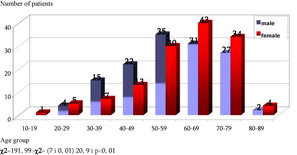
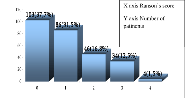
A statistically significant difference in the distribution of acute pancreatitis frequency is determined, depending on age, with the highest expression in the seventh decade (p<0, 01), and it’s also determined that the highest expression frequency by males is in slightly younger age (sixth decade). In younger age groups males are prevalent and in older age groups females are prevalent. The value obtained by test of frequency distribution is χ2=191, 99> χ2 (7 and 0, 01) =20, 9 and p<0, 01 (Chart 1). It is determined that there is no difference in the structure of patients by the severity degree of acute pancreatitis clinical appearance between sex: χ2=1,05<χ2 =6,63 (1 and 0,01) p>0,01, 37,7% of total patients had Ranson’s score 0,31,5% had Ranson’s score 3 and 1,45% patients had Ranson’s score 4 (Chart 2). The average value of Ranson’s score was statistically higher in the group of patients with severe compared to the group with mild form (3, 11±0,05 and 0,76±0,05, respectively) acute pancreatitis (t=4,24>2,58 p<0,01). The ratio of the average values of Ranson’s score between the groups of alcoholic (Table 1) and biliary pancreatitis (Table 2), didn’t show statistically significant difference (t=0,0025<1,96 p>0,01), 235 (86,1%) of total number of patients were in the group of mild, and 38(13,9%) were in the group of severe acute pancreatitis form t=4, 38>2,58, if p<0,01. In the biliary pancreatitis group, the average age is significantly higher than the age in the alcoholic pancreatitis group, t=4, 97>2,58, p<0,01. According to the structure of patients by etiology, in our group the most common were patients with biliary pancreatitis, 157 (57,51%), then with alcohol 78 (28,57%), idiopathic or unknown etiology pancreatitis 26 (9,52%), caused by hyperlipidemia 10(3,67%), caused by ERCP of other causes 2(0,73%) (Table 3). We have determined that there is no statistically significant difference in the number of treated patients yearly in the period of monitoring from 2009 to 2012, χ2=10,39< χ2(3 and 0,01)=11,34, p>0,01 the frequency distribution of hospitalized patients doesn’t depend on age of monitoring.
Chart 1: Frequency of acute pancreatits according the age and sex differences.
Chart 2: Distribution of frequency of acute pancreatitis according Ranson’s score value (0-4).
    Variable
    N
    ean
    StDev
    SE Mean
    95% CI


    age
    78
    52.12
    13.85
    1.57
    (48.99;55.2)


    Lenght of    hospitalisation-days
    78
    6.91
    4.78
    0.54
    (5.83;7.99)


    Glycemia    mmol/l
    78
    6.71
    2.67
    0.30
    (6.11;7.31)


    LDH value    IU/l
    78
    520.3
    305.1
    34.5
    (451.6;589.)


    AST value    IU/l
    78
    91.1
    123.6
    14.0
    (63.3;119.0)


    Le value/109
    78
    12.54
    9.68
    1.10
    (10.36;14.7)


    CRP value    mg/l
    27
    162.6
    152.8
    29.4
    (102.1;223.)


    Ca++value    mmol/l
    78
    2.30
    0.16
    0.018
    (2.27;2.34)


    Triglic.value    mmol/l
    78
    1.87
    1.11
    0,12
    (1,62;2,12)


    Cholesterol    mmol/l
    78
    5.15
    1.66
    0.18
    (4.77;5.52)


    SAmylase    IU/l
    78
    877.9
    809.6
    91.7
    (695.3;1060.4)


    U/U Amylase    IU/l
    46
    10903
    13255
    1954
    (6967;1483)


    ALT value    IU/l
    78
    109.4
    159.9
    18.1
    (73.3;145.4)


    GGT Value    IU/l
    78
    255.9
    263.5
    29.8
    (196.5;315.)


    AST/ALT ratio
    78
    1.22
    0.91
    0.10
    (1.02;1.43)
Table 1: Represents the most important variables in the group of alcoholic pancreatitis.
    Variable
    N
    Mean
    StDev
    SE Mean
    95% CI


    age
    157
    61.7
    13.7
    1.1
    (59.5;63.8)


    lenght of hospitalisation-days
    157
    6.36
    3.22
    0,25
    (5.8;6.8)


    glycemia value mmol/l
    157
    7.17
    3.34
    0.26
    (6.6;7. 7)


    LDH value IU/l
    157
    593.4
    442.2
    35.3
    (523.7;663.1)


    AST value IU/l
    157
    186.3
    195.8
    15.6
    (155.4;217.2)


    Le value/109
    157
    10.66
    4.50
    0.35
    (9.95;11.37)


    CRP value mg/l
    59
    91.8
    112.6
    14.7
    (62.5;121.2)


    Ca++ value mmol/l
    157
    2.29
    0.17
    0.01
    (2.26;2.31)


    Triglic.value mmol/l
    157
    1.64
    1.82
    0.14
    (1.3;1.9)


    Cholesterol mmol/l
    157
    9.28
    50.52
    4.03
    (1.31;17.24)


    S Amylase IU/l
    157
    1225
    1420
    113
    (1001;1449)


    U/UAmylase IUI/l
    69
    11299
    14519
    1748
    (7811;14787)


    ALT value IU/l
    157
    287,8
    253,0
    20,2
    (247,9;327,7)


    GGT value IU/l
    157
    308.0
    313.8
    25.0
    (258.5;357.5)


    AST/ALT    ratio
    157
    0.86
    1.43
    0.11
    (0.63;1.08)
Table 2: Represents the most importnant variables in the group of biliary pancreatitis.
Etiology
Sex
Year of hospitalization
total
2009
2010
2011
2012
Num.
%
Num.
%
Num.
%
Num.
%
Number
1
AlcoholMale
5
6, 8%
26
35, 6%
21
28, 8%
21
28, 8%
73
Female
0, 0%
2
40, 0%
0, 0%
3
60, 0%
5
Total
5
6, 4%
28
35, 9%
21
26, 9%
24
30, 8%
78
2
BliaryMale
9
18, 0%
15
30, 0%
13
26, 0%
13
26, 0%
50
Female
23
21, 5%
26
24, 3%
27
25, 2%
31
29, 0%
107
Total
32
20, 4%
41
26, 1%
40
25, 5%
44
28, 0%
157
3 Hyperlipidemia
Male
3
37, 5%
2
25, 0%
0, 0%
3
37, 5%
8
Female
1
50, 0%
0, 0%
1
50, 0%
0, 0%
2
Total
4
40, 0%
2
20, 0%
1
10, 0%
3
30, 0%
10
4 Unknwn
Male
0, 0%
2
40, 0%
1
20, 0%
2
40, 0%
5
Female
6
28, 6%
2
9, 5%
5
23, 8%
8
38, 1%
21
Total
6
23, 1%
4
15, 4%
6
23, 1%
10
38, 5%
26
5
Post ERCP and autherMale
0
0
0
0
0
Female
0, 0%
2
100, 0%
0, 0%
0, 0%
2
Total
0
0, 0%
2
100, 0%
0
0, 0%
0
0, 0%
2
Total
Male
17
12, 5%
45
33, 1%
35
25, 7%
39
28, 7%
136
Female
30
21, 9%
32
23, 4%
33
24, 1%
42
30, 7%
137
Total
47
17, 2%
77
28, 2%
68
24, 9%
81
29, 7%
273
Table 3: Structure of patients with acute pancreatitis according to etiology, sex and age.
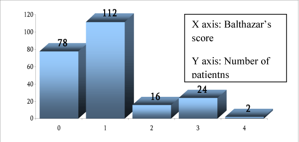
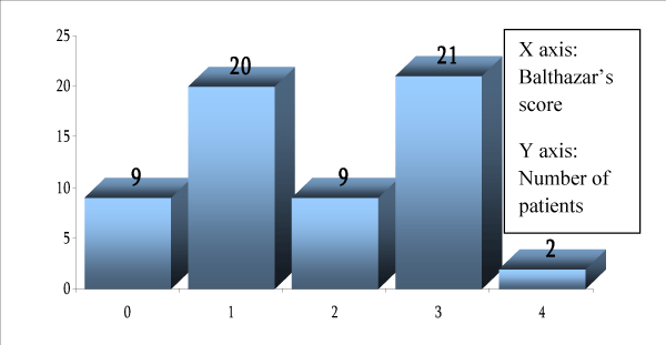
In our study by initial ultrasound examination, the pancreas is successfully perceived in 84, 9% of patients. The presence of statistically significant medium degree correlation between the value of Ranson’s score as a parameter of clinical appearance severity and the value of ultrasound score by Balthazar (r= 0,448, p=0,0001) (Chart 3) is determined. Also, the statistically significant high degree correlation between the values of ultrasound and CT score by Balthazar is determined. (r=0,778, p=0,001) (Chart 4).
Chart 3: Distribution of frequency of patients with acute pancreatitis according to echosonography criteria by Balthazar score. (0-4).
Chart 4: Distribution of frequency of acute pancreatitis according CT score by Balthazar (0-4).
Comparing the value of Ranson`s score and the score of native X-ray of abdomen, low level of positive correlation was found, r= 0,134, p value= 0,09. Also, comparing the value of Ranson`s score and the score of upper gastrointestinal endoscopy no statistically significant correlation was found, r=0,089, p value=0,04. Between the values of Ranson`s score and the score of chest radiography low level of correlation was found, r= 0,203, p value= 0,01.
When it comes to basic hemodynamic parameters, as well as their predictive prognostic relationship with clinical course and outcome, our study found a negative correlation between the value of Ranson`s score and rank values of arterial blood pressure, r = -0,182, p = 0,003 . Ranson`s score also correlate with pulse-frequency in the sense of a low degree of positive correlation, r = 0,261, p <0,0001, Pearson’s test.
There is no statistically significant connection between major etiological factors and the treatment outcome of patients with acute pancreatitis χ2 =0,52< χ2 (1 and 0,01)=6,63, p >0,01, and there is no significant connection between etiological factor and mortality, (p=1,0 > 0,05) (Table 4). There is no statistically significant connection between the sex affiliation (χ2=0,1<(1 and 0,01) χ2 =6,63 p>0,01) and the treatment outcome and mortality (p=1,0>0,05) (Table 5).
    Etiology
    End point of treatment - mortality
    Summary


    Released
    Died


    Number
    %
    Number
    %
    Number


    Alcoholic
    63
    100.0%
    0
    0.0%
    63


    Billiary
    131
    98.5%
    2
    1.5%
    133


    Summary
    194
    99.0%
    2
    1.0%
    196
Table 4: Relation between the most frequent etiology groups and mortality
    Sex
    End point of    treatment-mortality
    Summary


    Released
    Died


    Number
    %
    Number
    %
    Number


    Men
    135
    99.3%
    1
    0.7%
    136


    Women
    136
    99.3%
    1
    0.7%
    137


    Summary
    271
    99.3%
    2
    0.7%
    273
Table 5: Relation between sex and mortality in acute pancreatitis.
Discussion
Epidemiological data show the enlargement of acute pancreatitis frequency. Biliary pancreatitis is more common by females; alcohol is more common by males, idiopathic equally in both males and females [6]. Our results are in accordance with the structure of patients by sex in literature data. Also, we have determined that there is no difference in terms of structure of patients by the severity of acute pancreatitis clinical appearance depending on sex and etiology. This cognition is in accordance with literature data. De Campos and his associates [7] said that there is no significant difference between the groups of patients with or without systematic complications, as well as between the groups of survivors and deaths when it comes to age and sex, as well as etiological factor. Kaya and associates [8], as well as Papachristou and associates [9] obtained similar results.
We established that there is no statistically significant difference in the number of treated patients yearly in period from 2009 to 2012. According to some authors, such as Tonsy and associates [10], in the last few years there is the enlargement of acute pancreatitis incident ion as the consequence of modern diagnostic methods application and enlargement of biliary acute pancreatitis frequency.
According to etiology, the most frequent pancreatitis in our group of patients was biliary, then alcoholic, idiopathic, caused by hyperlipidaemia, ERCP, or other causes. In their series of 199 patients, Kaya and associates [8], found 53% of those with biliary etiological factor, and 26% of those with idiopathic pancreatitis. Similar results showed some other studies [11]. In the series of 153 patients, Chazicostas and associates [12] found biliary etiology in 67,3%, alcoholic in 9,2%, idiopathic in 17%, and in 6,5% of cases different mixed etiological factors.
The frequency of acute pancreatitis by our patients is not equally distributed by age groups, with the top expression in seventh decade of lifetime. It is also observed that the top acute pancreatitis frequency by men is in slightly younger age group (sixth decade of lifetime), as well as that, in younger age groups, males are prevalent, and in older groups females are prevalent, which is consistent with literature data [12,13].
Banks and associates [13] cite that about 85% cases concerned mild form of interstitial pancreatitis, and in the rest 15% of cases concerned severe form of necrotic pancreatitis. According to the same authors, rates of total mortality in acute pancreatitis are from 3-5% by interstitial and 17% by necrotizing pancreatitis (from 12% by sterile necrosis to 30% by infective necrosis). In our series of patients with acute pancreatitis, there were 235 (86,1%) of patients with mild and 38 (13, 9%) of patients with severe form of the disease according to Ranson’s criteria, which is consistent to literature data [12,13]. We found the significant difference in the medium values of Ranson’s score between the population with mild and severe form of acute pancreatitis. Ranson’s score, according to Soumitra and associates [11] can be considered as reliable factor in prediction of acute pancreatitis patient’s clinical course and outcome, and is completely comparable with other systems of evaluation (APACHE score). The mortality, according to these authors, is in absence of organs failure 0%, by the one organ system failure to 3% (from 0-8%), and by more organ systems failure 47% (from 28-69%). In our population of patients, the mortality rate was 0,73% (2 patients). Other authors [9, 12-15] mention the mortality rate of 2-4%.
Literature data show that it is not always possible to adequately perceive pancreas because of the abundance of gases, but also because of poor preparation and cooperation of patients. According to Jeremic and associates [16], the percentage of pancreas non perceivement during the initial ultrasound examination is from 20-50%. In our study, the pancreas is successfully perceived by initial ultrasound examination in 84,9%. The distribution of acute pancreatitis patients frequency according to Ranson’s score shows progressive decrease of number going from lower to higher scores of Ranson’s score. However, the distribution of patient’s frequency is similar by ultrasound examination degree according to Bathazar’s score. This fact is in accordance with literature data of Rickes and associates [17], who question the correlation of clinical status, CT examination and ultrasound in their study with 31 patients with acute pancreatitis. Pandey and associates [18] also, in their study with 110 acute pancreatitis patients reach similar conclusions.
When it comes to the relation between ultrasound and CT results by our acute pancreatitis patients, the high degree positive correlation is found. This fact is in accordance with the other author`s results [17,18]. The value of Ranson’s score and CT score according to Balthazar also correlate in our study, with the existence of medium degree positive correlation, as well as according to the literature data [17,19].
Plain abdominal X-ray film, often applied as a diagnostic tool in acute pancreatitis, the Pearson`s test showed a positive low-level correlation between Ranson`s score value and plain abdominal X-ray score. This fact is explained by the ileus, which can be seen with plain film of the abdomen, and can be a sign of easing vital organs, according to the Hirota and associates [19].
Between gastroscope findings and Ranson`s score, Pearson`s test shows no statistically significant correlation. This data is also consistent with some literature data. Hirota and associates [19] cite a study with 17 patients with a severe form of acute pancreatitis, which is measured by the level of gastric pH within the first 48 h of admission, where he established higher mortality rate in patients with a low pH value, as well as many failure of organ systems in these patients, but emphasizes, however, that gastric pH cannot be taken as a parameter for the diagnosis and differentiation between severe and mild clinical form of pancreatitis.
Findings of X-rays of the chest, graded and compared with Ranson`s score, indicates a low degree of positive correlation. According to Hirota and co-workers [19], early pleural effusion in patients with acute pancreatitis can be a sign of inflammation and enlarged unilateral or bilateral effusions may be associated with poor outcome.
Our study found a low-grade negative correlation between the value of Ranson`s score and rank values arterial blood pressure. Value of Ranson`s score also correlates with pulse frequencies in terms of a positive correlation of low degree, as already described in other studies [5,13,20,21].
When it comes to the clinical course of the disease, we have compared the length of hospitalisation between the groups with severe and mild acute pancreatitis form and we haven’t found a statistically significant difference. This result is not in accordance with the most literature data, where there is statistically significantly longer hospitalization of patients with severe form of acute pancreatitis [18- 21]. However, De Campos and associates [7], and the others [22], also cite that there is no statistically significant difference between the groups with and without systematic complications and groups of survivors and deaths when it comes to the length of hospitalization in days. According to our tests, there is no statistically significant difference between the length of hospitalization and acute pancreatitis etiology (biliary or alcoholic). We also established that the average age in the group of patients with severe form of acute pancreatitis is higher compared to the group the group with mild form, which is in accordance with the study of Nojgaard and associates [23], which shows higher mortality in older population.
Conclusion
The acute pancreatitis is equally represented in both males and females. There is a significant difference of the indention between males and females when it comes to the etiology of the disease, biliary pancreatitis is more frequent by females, and alcoholic pancreatitis is more frequent in males. There is no significant difference if the clinical appearance and outcome between the sexes. The frequency distribution is significantly different depending on the age of patients, with the top expression in older ages, provided that in the younger age groups there are more male cases, and in the older more female cases. Average age is significantly higher in the severe form of acute pancreatitis. According to etiology, the most often are biliary and alcoholic pancreatitis. The mild form of acute pancreatitis prevails compared to the severe form. The values of Ranson’s score are significantly higher in the group of patient with severe form. There is a significant correlation between acute pancreatitis severity degree and ultrasound and CT results according to Balthazar’s score. CT and ultrasound result significantly correlates with the acute pancreatitis clinical appearance severity by Ranson’s score. There is a high degree of correlation between ultrasound and CT findings in patients with acute pancreatitis, which indicates the diagnostic value of ultrasound performed by trained ultrasonographer. The clinical picture of pancreatitis by Ranson`s score correlates with plain film of the abdomen score. Between gastroscope findings and Ranson`s score there`s no statistically significant correlation.
X-ray of the chest score and value of pulse frequencies correlate by positive low level. Also, arterial blood pressure correlates with Ranson`s score by negative low level, and the other hemodynamic parameter, pulse, correlates with the value of Ranson`s score by positive low level.
References
- Young SP, Thompson JP. Severe Acute Pancreatitis. Cont Edu Anaesth Crit Care& pain. 2008; 8: 125-128.
- Cappell MS. Acute pancreatitis: etiology, clinical presentation, diagnosis, and therapy. Med Clin North Am. 2008; 92: 889-923, ix-x.
- Kasimu H, Jakai T, Qilong C, Jielile J. A brief evaluation for pre-estimating the severity of gallstone pancreatitis. JOP. 2009; 10: 147-151.
- Werner J, Feuerbach S, Uhl W, Büchler MW. Management of acute pancreatitis: from surgery to interventional intensive care. Gut. 2005; 54: 426-436.
- Balthazar EJ. Acute pancreatitis: assessment of severity with clinical and CT evaluation. Radiology. 2002; 223: 603-613.
- Pezzilli R, Zerbi A, Di Carlo V, Bassi C, Delle Fave GF; Working Group of the Italian Association for the Study of the Pancreas on Acute Pancreatitis. Practical guidelines for acute pancreatitis. Pancreatology. 2010; 10: 523-535.
- De Campos T, Cerqueira C, Kuryura L, Parreira JG, Soldá S, Perlingeiro JA, et al. Morbimortality indicators in severe acute pancreatitis. JOP. 2008; 9: 690-697.
- Kaya E, Dervisoglu A, Polat C. Evaluation of diagnostic findings and scoring systems in outcome prediction in acute pancreatitis. World J Gastroenterol. 2007; 13: 3090-3094.
- Papachristou GI, Muddana V, Yadav D, O'Connell M, Sanders MK, Slivka A, et al. Comparison of BISAP, Ranson's, APACHE-II, and CTSI scores in predicting organ failure, complications, and mortality in acute pancreatitis. Am J Gastroenterol. 2010; 105: 435-441.
- Tonsi AF, Bacchion M, Crippa S, Malleo G, Bassi C. Acute pancreatitis at the beginning of the 21st century: the state of the art. World J Gastroenterol. 2009; 15: 2945-2959.
- Eachempati SR, Hydo LJ, Barie PS. Severity scoring for prognostication in patients with severe acute pancreatitis: comparative analysis of the Ranson score and the APACHE III score. Arch Surg. 2002; 137: 730-736.
- Chatzikostas C, Roussomoustakaki M, Vardas E, Romanos J, Kouroumalis EA. Balthazar computed tomography severity index is superior to Ranson criteria and APACHE II and III scoring systems in predicting acute pancreatitis outcome. J Clin Gastroenterol. 2003; 36: 253-260.
- Banks PA, Freeman ML; Practice Parameters Committee of the American College of Gastroenterology. Practice guidelines in acute pancreatitis. Am J Gastroenterol. 2006; 101: 2379-2400.
- Eachempati SR, Hydo LJ, Barie PS. Severity scoring for prognostication in patients with severe acute pancreatitis: comparative analysis of the Ranson score and the APACHE III score. Arch Surg. 2002; 137: 730-736.
- Simchuk EJ, Traverso LW, Nukui Y, Kozarek RA. Computed tomography severity index is a predictor of outcomes for severe pancreatitis. Am J Surg. 2000; 179: 352-355.
- Jeremic M, Stojanovic M. Surgery of pancreas. Special surgery I. In: Jeremic M, editor. Pelikan print, Niš. 2001.
- Rickes S, Uhle C, Kahl S, Kolfenbach S, Monkemuller K, Effenberger O, et al. Echo enhanced ultrasound: a new valid initial imaging approach for severe acute pancreatitis. Gut. 2006; 55: 74-78.
- Pandey L, Milicevic M, Grbic R, Bulajic M, Krstic R, Golubovic G, et al. The value of ultrasound in staging the severity of acute pancreatitis. Acta Chir Iugosl. 1997; 44-45: 63-7.
- Hirota M, Takada T, Kawarada Y, Hirata K, Mayumi T, Yoshida M, et al. JPN Guidelines for the management of acute pancreatitis: severity assessment of acute pancreatitis. J Hepatobiliary Pancreat Surg. 2006; 13: 33-41.
- Gürleyik G, Emir S, Kiliçoglu G, Arman A, Saglam A. Computed tomography severity index, APACHE II score, and serum CRP concentration for predicting the severity of acute pancreatitis. JOP. 2005; 6: 562-567.
- Feldman M, Friedman LS, Brandt LJ. Sleisenger & Fordtran’s Gastrointestinal and Liver Disease, 8th edition. Elsevier Science. 2006.
- Kim YS, Lee BS, Kim SH, Seong JK, Jeong HY, Lee HY, et al. Is there correlation between pancreatic enzyme and radiological severity in acute pancreatitis? World J Gastroenterol. 2008; 14: 2401-2405.
- Nøjgaard C, Matzen P, Bendtsen F, Andersen JR, Christensen E, Becker U, et al. Factors associated with long-term mortality in acute pancreatitis. Scand J Gastroenterol. 2011; 46: 495-502.


