
Case Report
Austin J Gastroenterol. 2014;1(3): 1015.
Splenosis Presenting as Pancreatic Neoplasm: Report of Two Cases
Beltrame V, Merigliano S and Sperti C*
Department of Surgery, Oncology and Gastroenterology, University of Padua, Italy
*Corresponding author: :Cosimo Sperti, Department of Surgery, Oncology and Gastroenterology, 3rd Surgical Clinic, University of Padua, via Giustiniani 2, 35128 Padova, Italy
Received: August 04, 2014; Accepted: August 18, 2014; Published: August 20, 2014
Abstract
plenosis is the auto transplantation of splenic tissue generally seen after traumatic injury or elective splenectomy. Although this condition affects one to two thirds of patients submitted to splenectomy for trauma, Splenosis is rarely diagnosed preoperatively, and sometimes, it is mistaken for a neoplasm.
We report two consecutive patients, incidentally discovered to have a mass in the tail of the pancreas, during a work-up for other pathology. Both patients had undergone splenectomy, 12 and 14 years before, respectively. Abdominal ultra sonography, computed tomography, and magnetic resonance imaging demonstrated a round, solid mass with low contrast enhancement in the pancreatic tail: both clinical and radiologic findings suggested for the first patient, a solid-cystic or neuroendocrine tumour of the pancreas. After distal pancreatectomy, microscopic examination revealed heterotrophic splenic tissue. The second patient presented with two solid, round masses in the tail, and in the peripancreatic tissue of the head of the pancreas, respectively. Tc-99 sulphur colloid scintigraphic showed intense tracer uptake in both lesions, suggesting the presence of splenic tissue. So, surgical operation was avoided. Splenosis should be considered in the differential diagnosis of incidental pancreatic masses in previously splenectomised patients.
Keywords: Accessory spleen; Pancreas; Pancreatic neoplasm; Splenectomy; Splenosis
Case Presentation
Case 1
A 57-year-old woman was referred to our Department for a 1-year history of a solid mass in the tail of the pancreas. Her medical history included hysterectomy 14 years before, and anterior resection of sigmoid colon cancer with splenectomy for spleen injury, 12 years before. The pancreatic mass was incidentally discovered by abdominal ultra sonography during follow-up evaluation after endoscopic excision of bladder polyps. She had no symptoms or any remarkable physical signs. Routine laboratory tests were within normal range, including carcinoembrionic antigen (CEA) and carbohydrate antigen, CA 19-9. Magnetic resonance imaging (MRI), confirmed the presence of 4 x 3 cm, slightly contrast enhanced mass in the tail of the pancreas. The enhancement pattern was homogeneous, with regular margins. An integrated positron emission tomography with computed tomography (CT) acquisition (PET/CT) using 18-Fluorodeoxyglucose (FDG) revealed a 4-cm, low-density, lesion with low FDG uptake in the tail of the pancreas (SUV=1.5): the mass had some internal septa, but did not show contrast enhancement (Figure 1).
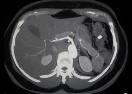
Figure 1 Case 1: Computed tomography (CT) of the abdomen showing a well-defined, 4-cm mass in the tail of the pancreas (arrow).
A suspect of solid- cystic or non-functioning endocrine tumour of the pancreas was made, and the patient underwent exploratory laparotomy. A rubbery mass was palpated posteriorly to the pancreatic tail; then, distal pancreatectomy was performed. The gross pathologic findings showed a 4, 5x3x2, 5 cm red, well-circumscribed nodule abutting the posterior surface of the pancreas, with normal surrounding pancreatic parenchyma. . Histologic examination of the mass revealed splenic tissue without hilum, attached to unremarkable pancreatic parenchyma (Figure 2). The postoperative course was uneventful, and the patient is alive, and well 24 months after surgery.
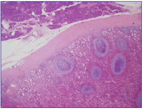
Figure 2 Case 1: Histopathological detection of splenic tissue and normal pancreatic parenchyma. (H&E x 10).
Case 2
A 70-year old man presented a 1-month history of abdominal masses discovered during a work-up for new onset diabetes mellitus. His medical history included post-traumatic splenectomy and left nephrectomy for vehicle accident 43 years before, hypertension and chronic alcoholic hepatitis from ten years. Routine laboratory tests were in the normal range, including CEA, CA 19-9 and alpha-fetoprotein. Abdominal ultrasound showed a 1.6 cm hypoechoic node anteriously to the head of the pancreas, and a 5-cm mass in the left hypochondrium. Abdominal CT confirmed the small (up to 2 cm in diameter) peripancreatic lesion and a 5-cm solid lesion in continuity with the tail of the pancreas (Figure 3). A suspect of multiple Splenosis in the peripancreatic region was made: the patient underwent 99-Tc sulphur colloid scintigraphic and SPECT that demonstrated a 2-cm area at the hepatic haulm and a 5-cm mass in the splenic area, compatible with splenic tissue (Figure 4). So, routine follow-up was scheduled, and the patient is alive and well 20 months after discharge.
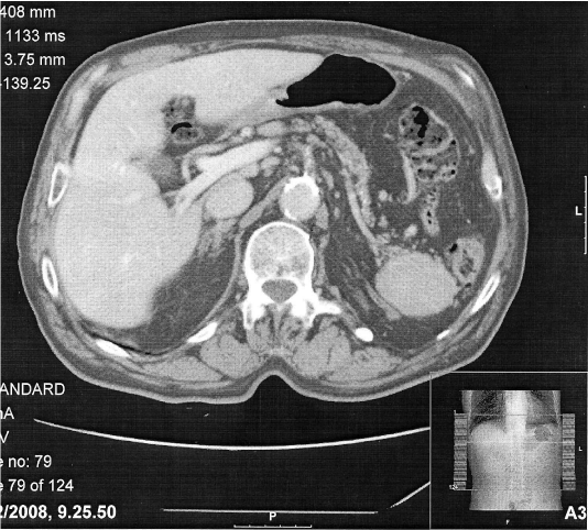
Figure 3 Case 2: Computed tomography of the abdomen showing a welldefined, 5-cm mass in continuity with the tail of the pancreas (arrow).
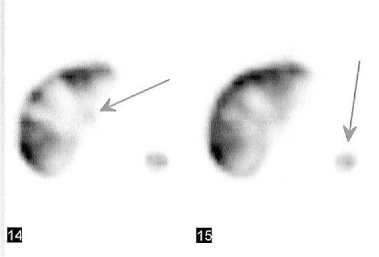
Figure 4 Case 2: Tc-99 sulphur colloid scintigraphy showing uptake of tracer in the mass located in the splenic bed and a small area in the hepatic hilum/arrows).
Discussion
Splenosis is the hetero topic auto transplantation of splenic tissue generally after traumatic rupture of the spleen. The incidence of Splenosis in patients splenectomised for trauma range between 44% -76% [1]: it has been reported also in rare cases of elective open splenectomy for haematological disorders [2-4]. Generally, Splenosis is clinically silent, being an incidental discovery at radiographic examination. However, it may be clinically relevant because almost invariably, it is misdiagnosed as neoplasm. On the other hand, differentiating an accessory spleen from other neo plastic lesions is crucial, and can avoid unnecessary laparotomy.
Our cases had similar presentation and conventional imaging features, but substantially differed for their clinical management. The first one underwent surgical resection for presumed pancreatic neoplasm; the second one had a correct diagnosis of splenic tissue and was followed-up. The lesson from the first case helped us to avoid unnecessary operation in the second case. These lesions are usually discovered incidentally on radiologic imaging performed for an unrelated reason, as in our cases. Most patients are asymptomatic, and, commonly, ectopic spleens are multiple and variable in size. Although Splenosis is usually found in peritoneal cavity, it has been reported in the retro peritoneum, the thorax, and distant sites as the brain [5]. Isolated Splenosis has been rarely reported, and involved the liver [6], kidney [7], mesoappendix [8], and stomach [9]. Since splenic tissues mimic abdominal tumours, the major problem when encountering such lesions, is to distinguish them from other neoplasms, avoiding unnecessary operation in asymptomatic patients. Isolated intra pancreatic accessory spleen forms a well-defined nodule within the tail of the pancreas and is commonly mistaken by imaging studies (CT, MR and US) as hyper vascular pancreatic tumour, such as a neuroendocrine neoplasm or an acinar cell carcinoma [3,10].
Although usual imaging modalities (US, CT, MRI) are helpful to localize and determine the size, structure and relations with adjacent organs, they are not specific. More specific and diagnostic studies using agents that are sequestered by reticuloendothelial tissue, like 99mTechnetium sulphur colloid [4] and recently ferumoxide-enhanced MRI [11] have been used. Tc-99 sulphur colloid scintigraphic represents a suitable preliminary screening test for the detection of residual splenic tissue in splenectomised patients with equivocal abdominal masses [12]. In our case 2, Tc-99 scintigraphic confirmed the clinical diagnosis of Splenosis, sparing unnecessary operation. This benign lesion is often asymptomatic; therefore surgical excision is not warranted if a certain diagnosis can be made. To avoid unnecessary surgery some authors [7] suggest performing a diagnostic laparoscopy to obtain an accurate pathologic diagnosis.
In conclusion, isolated Splenosis should be considered in asymptomatic patients who underwent splenectomy. Frequently, it is discovered close to the splenic hilum and the pancreas is the second most frequent location [13]. Heterotypic spleen tissue has always to be considered in the differential diagnosis of distal pancreatic masses in splenectomised patients, in order to avoid useless surgery because of their benign nature.
References
- Barbaros U, Dinççağ A, Kabul E Minimally invasive surgery in the treatment of splenosis. Surg Laparosc Endosc Percutan Tech. 2006; 16: 187-189.
- Celiloglu M, Dogan E, Kocoglu S, Sarihan E Splenosis presenting with adnexal mass: a case report. Arch Gynecol Obstet. 2004; 270: 129-130.
- Matonis LM, Luciano AA. A case of splenosis masquerading as endometriosis. Am J Obstet Gynecol. 1995; 173: 971-973.
- Khosravi MR, Margulies DR, Alsabeh R, Nissen N, Phillips EH. Morgenstern L Consider the diagnosis of splenosis for soft tissue masses long after any splenic injury. Am Surg. 2004; 70: 967-970.
- Young JT, Jaibaji MM, Vandermolen R. Splenosis: a remote consequence of traumatic splenectomy. J Am Coll Surg. 2004; 199: 500-501.
- D'Angelica M, Fong Y, Blumgart LH. Isolated hepatic splenosis: first reported case. HPB Surg. 1998; 11: 39-42.
- Kiser JW, Fagien M, Clore FF. Splenosis mimicking a left renal mass. AJR Am J Roentgenol. 1996; 167: 1508-1509.
- al-Ahmadi M, Brundage S, Brody F, Jacobs L, Sackier JM. Splenosis of the mesoappendix: case report and review of the literature. J R Coll Surg Edinb. 1998; 43: 200-202.
- Deutsch JC, Sandhu IS, Lawrence SP. Splenosis presenting as an ulcerated gastric mass: endoscopic and endoscopic ultrasonographic imaging. J Clin Gastroenterol. 1999; 28: 266-267.
- Lehtinen SJ, Schammel CM, Devane M, Trocha SD. Intrapancreatic accessory spleen presenting as a pancreatic mass. J Gastrointest Oncol. 2013; 4: E23-26.
- Berman AJ, Zahalsky MP, Okon SA, Wagner JR. Distinguishing splenosis from renal masses using ferumoxide-enhanced magnetic resonance imaging. Urology. 2003; 62: 748.
- Castellani M, Cappellini MD, Cappelletti M, Fedriga E, Reschini E, Cerino M, et al Tc-99m sulphur colloid scintigraphy in the assessment of residual splenic tissue after splenectomy. Clin Radiol. 2001; 56: 596-598.
- Katuchova J, Baumohlova H, Harbulak P, Stofcikova M, Svajdler M, Repovsky A, et al. Intrapancreatic accessory spleen. A case report and review of literature. JOP. 2013; 14: 261-263.