Case Report
Austin J Gastroenterol. 2023; 10(2): 1127.
Small Intestine Lymphangioma – A Rare Cause of Gastrointestinal Bleeding: Case Report
Foroogh Alborzi Avanaki¹; Asieh Shakib²*; Samaneh Sardari³
1Department of Gastroentrology, Imam Khomeini Hospital Complex, Colorectal Reserch Center, Tehran University of Medical Science, Tehran Iran
2Division of Gastroenterology, Imam Khomeini Hospital, School of Medicine, Tehran University of Medical Sciences, Tehran, Iran
3Division of Cardiology, Imam Khomeini Hospital, School of Medicine, Tehran University of Medical Sciences, Tehran, Iran
*Corresponding author: Asieh Shakib Division of Gastroenterology, Imam Khomeini Hospital, School of Medicine, Tehran University of Medical Sciences, Tehran, Iran. Email: drash_tums@yahoo.com
Received: November 08, 2023 Accepted: December 22, 2023 Published: December 29, 2023
Clinical Report
The patient was a 20-year-old man, barber, with a past medical history of suspected SMA disease (difficulty in walking), presented with chief complaint of malaise, weakness, dizziness, loss of appetite, and weight loss in the 3rd month of year 2022. He was admitted to hospital because of severe symptomatic anemia (Hb: 5.5).
The patient's past medical history included no significant gastrointestinal conditions or surgeries. Upper GI endoscopy showed mucosal atrophy in D2 without fissure and nodularity. Pathology of duodenal mucosa revealed mild chronic gastritis, negative for celiac disease.

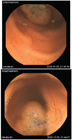
Figure 1:
In colonoscopy, there was one small livid lesion in 60 cm in the transverse colon, three irregular polypoid lesions, about 22mm, without a pedicle and livid in color in the ascending colon and cecum. Spiral abdominopelvic CT scan was unremarkable except, horse shoe kidneys were seen. In a second look colonoscopy in a tertiary center, three vascular polypoid lesions were seen in the transverse colon (about 10mm). Also, two vascular polypoid lesions were seen in the ascending colon (20 mm and 30 mm) which all was removed and sent for pathology. For detailed small intestine inspection, in order to find any other lesions in the small intestine video capsule endoscopy was done, which revealed two 10 and 15mm bluish color polypoid lesions in the distal part of the ileum , One of them was ulcerated. In pathology, benign angiomatous lesion suggestive of lymphangioma was reported. The patient was managed conservatively with regular follow-up to monitor for any recurrence or complications. Patient experienced no flare in 1 year follow up.
Discussion
The diagnostic and treatment challenges encountered in this case highlight the importance of a multidisciplinary approach to diagnose and manage rare and complex conditions such as colonic lymphangioma and suspected neurologic diseases. The initial diagnosis of severe anemia required further investigation to identify the underlying cause, which ultimately resulted in the identification of multiple hemangiomas in the gastrointestinal tract, including colonic lymphangioma [1].
The identification and characterization of the colonic lymphangioma posed a diagnostic challenge, as lymphangiomas are rare and often asymptomatic. The incidence of Lower GI lymphangioma in literature reported as much as 0.18 %. The diagnosis was confirmed through histopathological examination of the lesion, but the identification of the lesion required a second look colonoscopy [1,3].
The treatment of the colonic lymphangioma also posed a challenge. Radical excision is the preferred treatment for lymphangiomas, but it can be difficult to achieve complete resection without causing complications such as bleeding or perforation. In this case, the lymphangioma was removed using a hot snare, and hemoclips were deployed to prevent bleeding. The rationale for the chosen treatment approach was to minimize the risk of complications while still removing as much of the lesion as possible. The importance of regular follow-up was emphasized to monitor for any potential complications or recurrence [2-4].
Additionally, the patient's suspected neurologic disease, specifically Spinal Muscular Atrophy (SMA), posed both diagnostic and treatment challenges. The diagnosis was not confirmed, and further evaluation may have been needed to definitively diagnose the condition [5-7]. The association between vascular defects and SMA raised the possibility of a relationship between the patient's neurologic condition and the gastrointestinal lymphangiomas, but further research is needed to understand the potential link between these conditions and the implications for treatment [5,7,8].
Overall, this case report provides valuable insights into the diagnosis and management of colonic lymphangioma and highlights the need for further research to better understand this rare condition and its potential associations with other diseases.
In summary, this case report highlights the importance of a thorough diagnostic workup and individualized management plan for patients with rare and complex conditions such as colonic lymphangioma and suspected neurologic diseases. A multidisciplinary approach involving gastroenterologists, pathologists, neurologists, and other specialists may be necessary to effectively diagnose and manage these conditions [1-8].
References
- Gupta R, Khosla R, Sharma R, et al. Colonic lymphangioma mimicking intussusception: a case report and review of literature. J Pediatr Surg. 2009; 44: e1-3.
- Kuru S, Yilmaz B, Yildiz S, et al. Colonic lymphangioma: a rare cause of lower gastrointestinal bleeding. Turk J Gastroenterol. 2010; 21: 202-5.
- Lacoste C, Ginestet N, Cassagnau E, et al. Colonic lymphangioma: a rare cause of gastrointestinal bleeding. Gastroenterol Clin Biol. 2009; 33: 955-8.
- Teng A, Lee S, Kim S. Colonic lymphangioma presenting as acute abdomen in an adult. Int J Surg Case Rep. 2016; 28: 233-6.
- Wang Y, Li J, Liu C, et al. Spinal muscular atrophy and vascular defects: a systematic review. Orphanet J Rare Dis. 2021; 16: 82.
- Pearn J. Incidence, prevalence, and gene frequency studies of chronic childhood spinal muscular atrophy. J Med Genet. 1978; 15: 409-13.
- Wirth B, Garbes L, Riessland M. How genetic modifiers influence the phenotype of spinal muscular atrophy and suggest future therapeutic approaches. Curr Opin Genet Dev. 2013; 23: 330-8.
- Wang CH, Finkel RS, Bertini ES, Schroth M, Simonds A, Wong B, et al. Consensus statement for standard of care in spinal muscular atrophy. J Child Neurol. 2007; 22: 1027-49.