Clinical Image
Austin J Gastroenterol. 2015;2(1): 1033.
Rare Cause of Uncomplicated Gas-forming Pyogenic Liver Abscess
Nijhawan S*, Singh B, Kumar A, Gupta G
Department of Gastroenterology, SMS Medical College, Jaipur, India
*Corresponding author: Sandeep Nijhawan, Professor, Department of Gastroenterology, SMS Medical college, Jaipur, India
Received: January 29, 2015; Accepted: February 03, 2015; Published: February 05, 2015
Clinical Image
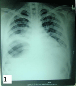
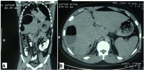
A 24 years old male presented with complaint of continuous pain in right upper abdomen and fever associated with chills for 12 days. He was also complaining of breathlessness for three days. He was non-alcoholic, non-diabetic and without any history of biliary surgery in the past. On examination patient had tachycardia and tender hepatomegaly. Laboratory data showed increased leukocyte count (26,800/μL) with normal liver and kidney function tests. Chest plain film showed a huge round cavity with air fluid level on the right-upper abdomen (figure 1). Ultrasound abdomen showed a 90 x 87mm hypoechoic area below right diaphragm. A contrast enhancenced computed tomography (CECT) of abdomen was done, showing a 120 x 104 mm cavity with air fluid level in right lobe of liver (Figure 2a and b). Yellow-white pus was aspirated from the cavity under ultrasound guidance which on culture showed Escherichia coli (E. coli) ESBL strain. He was treated with imipenem and amikacin based on in vitro sensitivity, symptoms subsided and was discharged after 2 weeks of intravenous antibiotics.
Gas-forming Pyogenic Liver Abscess (GPLA) is a rare entity with higher mortality and usually occurs in immunocompromised conditions. GPLA has more chances of rupturing into pleural or peritoneal cavities. However a close watch and conservative management, as in our case, can suffice. Klebsiella is the most common organism but rarely Clostridium or Salmonella have been reported [1, 2]. Our case was caused by E. coli which has hardly been reported [3].

Figure 1: Chest - X-ray showing large cavity with air fluid level below right diaphragm.

Figure 2A and B: CECT abdomen [coronal and axial view] showing gas containing liver abscess.
References
- Lee CC, Poon SK, Chen GH. Spontaneous gas-forming liver abscess caused by Salmonella within hepatocellular carcinoma: a case report and review of the literature. Dig Dis Sci. 2002; 47: 586-589.
- Law ST, Lee MK. A middle-aged lady with a pyogenic liver abscess caused by Clostridium perfringens. World J Hepatol. 2012; 4: 252-255.
- Chong VH, Yong AM, Wahab AY. Gas-forming pyogenic liver abscess. Singapore Med J. 2008; 49 : 123-125.