
Research Article
Austin J Gastroenterol. 2015;2(4): 1045.
The Percutaneous ‘Pick and Fix’ Suture: A Surrogate Port for Paediatric Laparoscopic Procedures
Bajpai M* and Mitra A
Department of Paediatric Surgery, All India Institute of Medical Sciences, India
*Corresponding author: Bajpai M, Department of Paediatric Surgery, All India Institute of Medical Sciences, India
Received: February 11, 2015; Accepted: March 24,2015; Published: March 27, 2015
Abstract
Objective: To apply and expand the utility of the traditional percutaneously placed stay suture during laparoscopy.
Materials and Methods: Children undergoing laparoscopic surgery from the period, December 2012 to November 2013 were included in this study. The use of the percutaneously stay suture (‘pick and fix’) in facilitating surgery was studied. The ease of placement, enhanced visualization as noted by the operating surgeon, total number of ports placed during surgery, total number of sutures placed and per-operative complications were all noted. Procedure related morbidity and eventual patient/care-giver satisfaction with cosmetic outcome were also noted.
Results: 58 children underwent laparoscopic procedures during the aforementioned duration and included nephroureterectomies visualization and decrease the total number of ports across the variety of procedures to which it was applied. Cosmetic results were satisfactory.
Conclusion: The versatility of this technique allows its application to various laparoscopic procedures. It enhances the minimally invasive nature of laparoscopy itself and may, thus, act as a substitute for conventional ports, the ultimate result being good cosmetic outcomes and patient satisfaction.
Keywords: Less; Paediatric laparoscopy; Percutaneous hitch stitch; Cholecystectomy; Appendicectomy; Lumboscopy
Introduction
The advent of Minimal Access Surgery (MAS) has given rise to a plethora of applications in paediatric surgery. The emerging techniques in MAS include multiport/single-incision procedures, Natural Orifice Transluminal Endoscopic Surgery (NOTES) and more recently single-port/single-incision procedures. The evolution of modes of access reflects the desire to cause minimal trauma to the patient [1]. There is a need [2] to introduce techniques such as percutaneous retraction sutures and modifications of the standard laparoscopes to minimize instrumentation.
Patients and Methods
Children undergoing laparoscopic surgery from the period, December 2012 to November 2013 and operated by the senior author (MB) were included in this study. Standard operating techniques were used while giving primacy to the safety of the procedure. The use of the percutaneous hitch suture in facilitating surgery was studied. The ease of placement, enhanced visualization as noted by the operating surgeon, total number of ports placed during surgery, total number of sutures placed and per-operative complications were noted. Procedure related morbidity and eventual patient/care-giver satisfaction with cosmetic outcomes were also observed.
The procedures carried out in this time frame can be broadly divided into:
- Laparoscopic
- Cholecystectomy
- Appendectomy
- Exploration for DSD
- Retroperitoneoscopy (including lumboscopic approach)
- Nephroureterectomy
- Lumboscopy assisted Anderson-Hyne’s pyeloplasty
- Lumboscopic uretero/pyelolithotomy
- Thoracoscopic
- Repair of eventration of diaphragm
The Approach
Laparoscopy: The operative laparoscope (Figure 1) (the 0°, all in one laparoscope with parallel eyepiece) necessitates the insertion of a 12 mm port. Our standard approach is intra-umbilical insertion i.e. through the umbilical cicatrix, with meticulous reconstruction at the time of port closure for maximum aesthetic benefit. The 6 mm working channel of this “co-axial” scope allows one to dispense with one of the additional 5 mm ports.
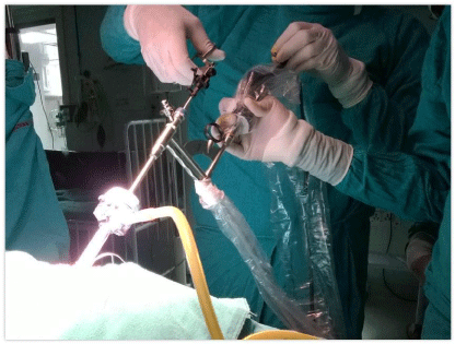
Figure 1: The operative laparoscope.
The first few cholecystectomies were done by the standard four-port approach. As one ascended the learning curve and the comfort level with the co-axial scope increased, the total number of ports was gradually decreased. Currently, the Single External Port Cholecystectomy (SEPC) technique (unpublished data) developed by the senior author allows us to achieve the same results with the aid of puppet sutures and a single multi-purpose instrument, the ultrasonic shears (HARMONIC®, Ethicon Endosurgery).
The technique of “suture-less” appendicectomy developed at our institution utilized a single external intraumbilical port with the second port inserted via the same skin incision but a different site in the rectus sheath. The procedure consists of anchoring the mesoappendix to the anterior abdominal wall by an extracorporeally inserted ‘pick and fix’ stitch followed by dissection and division of mesoappendix and appendix only with the harmonic scalpel [3]. The results of this preliminary communication suggested that the use of the ‘pick and fix’ suture to stabilize the appendix (in place of the conventional grasper) is technically feasible. As we gradually ascend the learning curve, even the aforementioned second port seems superfluous. With the advent of the operative laparoscope, we are now performing appendicectomies with great ease using only the 12mm intraumbilical port and the percutaneous hitch stitch.
Retroperitoneoscopy: The lumbosopic approach with the patient in prone position is an elegant solution to renal access. A port inserted at the cusp of the sacrospinalis and the abdominal wall muscles beneath the tip of the 12th rib allows direct access to the kidney with minimal dissection. With the help of the hitch-stitch on the redundant portion of the renal pelvis, exposure can be facilitated in preparation for pyeloplasty or the hilar structures as a prelude to nephrectomy. This versatile suture also allowed stabilization of a grossly dilated ureter prior to ureterolithotomy in a child with multiple, impacted ureteral calculi prior to ureterotomy.
Thoracoscopic (Figure 2): The repair of eventration of the diaphragm was envisioned to be suture less and the 45 mm Endostapler was the perfect instrument to achieve this. The tenting of the diaphragm with well-placed trans-thoracic hitch stitches and the application of the jaw of the stapler to the redundant diaphragm while gradually milking the contents (bowel loops) downwards allowed safe excision under vision and a technically sound correction.
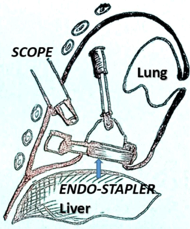
Figure 2: Thoracoscopic repair of eventration of diaphragm.
The various ways in which the hitch stitch was applied included:
The “Pick and Fix” Suture: Entails a simple pass with the suture (on a needle with a large chord length) through the body wall (transabdominal or transthoracic), the organ of interest (gall bladder, mesoappendix, large bowel, gubernaculum of testis, renal pelvis or redundant diaphragm as the case maybe) and out through the containing wall again (Figures 3, 4). Tagging the two ends with a pair of artery forceps allowed differential traction to be applied in order to facilitate dissection.
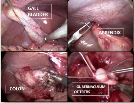
Figure 3: Composite picture of various applications of the hitch stitch.
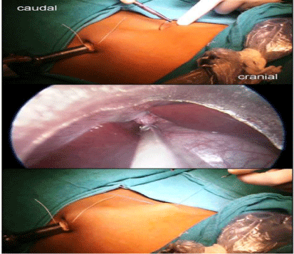
Figure 4: Inserting the ‘pick and fix’ suture.
The Hammock Suture: This involves the application of the wellestablished principles of puppeteering and was especially useful in the context of laparoscopic Cholecystectomy. The insertion of a loop of 1-0 Prolene suture (carried within an 18 G spinal needle) was done under vision through the right upper quadrant and into the fundus of the gall-bladder (Figure 5). This resulted in a loop of suture emerging out of the fundus. Another 1-0 Nylon suture was introduced in the same manner (colour differential for ease of recognition during surgery) through the body wall and then through the Hartmann’s pouch with the needle tip directed into the loop of Prolene as a sling. The Nylon thread was then pushed in while simultaneously withdrawing the needle. The Prolene loop was subsequently pulled extracorporeally which brought out the Nylon suture akin to a noose. On placing traction to these two sutures (fundal and Hartmann’s pouch), the Calot’s triangle opened up and allowed further dissection of both artery and duct under vision.
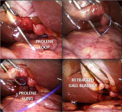
Figure 5: The hammock suture being inserted in the gall bladder.
Results
A total of 58 laparoscopic procedures were done between November 2012 and December 2013 (Table 1). The age range was from 1.5 to 16 years (mean 14.4) with 40 boys and 18 girls in the group of children operated for various indications.
    Procedure
    Single Port
    Multi-Port
    Total (%)


    Cholecystectomy
    11
    7
    18 (31.0)


    Appendectomy
    7
    10
    17 (29.3)


    Laparoscopic exploration for DSD
    0
    3
    3 (5.1)


    Lumboscopic nephroureterectomy
    1
    6
    7 (12.0)


    Retrperitoneoscopic uretero/pyelolithotomy
    0
    3
    3 (5.1)


    Lumboscopy assisted Anderson-Hyne’s pyeloplasty
    6
    0
    6 (10.3)


    Thoracoscopic repair of eventration of diaphragm
    1
    3
    4 (6.8)


    Total
    26
    32
    58
Table 1: Procedures done.
Laparoscopic cholecystectomy and appendectomy accounted for the majority of procedures (60.3%) Of the total number of procedures, almost 42% were single port procedures, demonstrating an obvious comfort level with the use of percutaneous traction sutures as surrogates for graspers and retractors.
The average duration of surgery for single port Cholecystectomy was 85 minutes, of which suture placement accounted for only 3% of the total duration of surgery. Time required for suture placement ranged from 1.0 to 2.6 minutes and was influenced by the nature of the procedure and type of stitch applied i.e. simple “pick/fix” vs. hammock-type. The hammock suture being an intrinsically more complex technique had a relatively steep learning curve and required an additional simple traction suture in the first two cases (Table 2). Thereafter, the insertion of two components of the hammock could be mastered to adequately sling the fundus and the Hartmann’s pouch.
    Procedure
    Number
    Total Number of Ports
    Traction Sutures Placed
    Total Avg. Duration
      (Min)
    Avg. Time for Suture Placement (Min)
    Port “Rescue”


    Cholecystectomy (single-port)
    11
    1
    1 (9)
      2 (2)
    85
    2.6
    2


    Cholecystectomy (multi-port)
    7
    4 (3)
      2 (4)
    0 (3)
      1 (4)
    80
    2.5
    None


    Appendicectomy (single-port)
    7
    1
    1 (5)
      2 (2)
    45
    1.9
    None


    Appendicectomy (multi-port)
    10
    2
    1
    40
    1.5
    None


    Laparoscopy for DSD
    3
    3
    1
    78
    1.2
    1


    Nephroureterectomy (single-port)
    1
    1
    2
    70
    1.5
    None


    Nephroureterectomy (multi-port)
    6
    2
    1 (5)
      2 (1)
    89
    1.5
    2


    Uretero/pyelolithotomy
    3
    3 (2)
      2 (1)
    2
    120
    2.1
    1


    Pyeloplasty
    6
    1
    1
    75
    2.0
    1


    Eventration repair (single-port)
    1
    1
    3
    120
    1.7
    None


    Eventration repair (multi-port)
    3
    2
    2
    110
    1.5
    None
Table 2: Parameters studied.
All single-port procedures utilized the benefits of the operative laparoscope. The traction sutures were not counted as “ports”. The total number of traction sutures placed by the operating surgeon was found to vary according to the difficulty in exposure and length of the tissue to be retracted.
Port “rescue” was defined as the need for additional ports beyond those initially placed, as per unanticipated requirement during surgery. Approximately 12% (7 of 58 procedures) required the insertion of an additional port for various reasons, including two single-port Cholecystectomy procedures (in view of dense pericholecystic adhesions). One of single port appendectomy procedures required the insertion of another port due to a retrocaecal buried appendix with dense adhesions. Conversion to open surgery was required in only one case of lumboscopy-assisted Anderson- Hyne’s pyeloplasty due to inadvertent injury to an arcuate artery at the hilum with resultant need for haemostasis (1.6%). Subsequently, we have restricted our dissection to regions away from the hilum and no conversion has been needed.
All but one of the Retroperitoneoscopy procedures were multiport because of the restricted anatomy of the region, high density of vital vascular structures and limited space available for the placement of percutaneous traction sutures. The incidence of port “rescue” was also higher in Retroperitoneoscopy procedures (25%) as opposed to the laparoscopic group (7.3%).
The patient and care-giver satisfaction with the cosmetic outcome was good. The intra-umbilical approach coupled with the imperceptible points of entry of the needle of the hitch stitch, especially in the single port Cholecystectomy and appendectomy, resulted in the appearance of an unscarred abdomen on follow-up.
There were no complications noted during placement of the hitch/ puppet sutures. Surgeon-perceived enhancement of visualization was good with additional sutures being placed as per requirement and a low incidence of port rescue.
Discussion
The practitioners of MAS are constantly striving for minimal percutaneous access and the “one-instrument, one access port” notion is now being challenged. To achieve this goal, a variety of novel methods have been devise over the years to do away with conventional ports. While LESS (Laparoendoscopic Single-Site Surgery) is becoming more of a reality everyday with innovative modes of access and instrumentation, the issues of good triangulation and lack of space are crucial, especially in the paediatric age group. Further, the development of consensus statements such as the 2010 Laparoendoscopic Single-Site Surgery Consortium for Assessment and Research (LESSCAR), further the cause of a more widespread adoption of this approach and uniformity in nomenclature and the reporting of results [4].
Cholecystectomy and appendectomy accounted for the bulk of the laparoscopic procedures done by our team and is arguably the most appropriate springboard from which one can increase one’s skill set. The minimization of port number and the appropriate placement of hitch stitches were best done in these operations as the comfort level with their conventional counterparts was high. Traction/retraction sutures and the string-puppet technique have all been developed as surrogates for port-based instrument access and may well prove to be inexpensive substitutes for the multitude of SILS ports and curved instruments [5-8].
Ganpule et al. provided an exhaustive list of manoeuvres which they employed to avoid instruments from clashing in their quest for LESS urological procedures in children which included the use of the hitch stitch and the Harmonic scalpel [9]. The percutaneous sling which had been described at the turn of the millennium in two and three port laparoscopic Cholecystectomy [10] and subsequently for one-trocar salpingectomy for tubal pregnancy [5], now finds a multitude of applications. It can be used anywhere in the abdomen (including the retroperitonuem) and even the thorax, as seen in our series. A well-placed stitch can retract to increase the field of vision and expose vital structures, provide traction to aid dissection and division, stabilize tubular organs in preparation for procedures and act as a rein for removal of the specimen from the body.
Another tool which aids in the effort to minimize the number of ports is the operative laparoscope, which has been in use for many years in gynaecology, neurosurgery and urology. In the two decades since the introduction of Begin’s technique of single-port, transumbilical, extracorporeal appendectomies using the operative laparoscope [11], this tool has almost been forgotten by the general surgeons. Also, descriptions of its use in children are few possibly due to the lack of commercially available scopes. The senior author reinvented its use in children which, thus, became the workhorse of all single-port and most of the multi-port laparoscopic procedures done by us.
The total number of ports placed was not invariable or standardized due to the gradual increase in experience and the introduction of the operative laparoscope during the time-frame of this particular series. The number of traction sutures placed was customized according to the requirement perceived during the particular procedure and the average time of placement was noted. The application of a suture like the hammock type required considerably more time due to its innate complexity and passage through regions requiring extreme caution such as the Calot’s triangle. The surgeon was also prepared to place additional unanticipated sutures for a long appendix or a hammocktype suture for a particularly dilated ureter during ureterolithotomy.
Anticipation of possible complications due to difficult dissection in two of the single port Cholecystectomy procedures allowed us to insert another port in time. This underscores the importance of knowing when to add conventional ports or even convert to the open technique (as was required in a pyeloplasty in our series). A review by Antoniou et al. showed that 9.3% of the SILC were unsuccessful primarily due to the improper identification of the Calot’s triangle [12].
Except for the pyeloplasty procedures which were performed by a novel technique developed at our institution (unpublished data), most of our urological procedures were multi-port because of the need for extensive and delicate dissection in the confines of the retroperitoneum. As described, the incidence of port “rescue” was also greater. The initial attempts by Hirano et al. [13] with single port adrenalectomy, paved the way for LESS in urology. The number of urological procedures for which single site access is being used has grown exponentially [14]. However, the technique still remains in the process of development and especially in the paediatric context, awaits extensive trials to prove its benefit.
The average duration of surgery was slightly longer for both single port Cholecystectomy (85 vs. 80 minutes) and single port appendectomy (45 vs. 40 minutes) and hitch stitch application
accounted for 3% and 8% of total time, respectively. Although greater numbers are required to comment on the significance of this observation, it compares well with previous studies [15,16] in single incision Cholecystectomy. The operative time lessened with experience and familiarization with technique within our series.
It has been proven that the younger age and smaller size of the paediatric patient do not affect the efficacy of commonly performed laparoscopic procedures such as appendicectomy and orchidopexy [17]. The latter study is encouraging and further justifies the development of LESS in the paediatric age group. The proven benefits of better outcomes, aesthetic benefits and decreased psychological impact due to the barely visible scars [18,19], cannot be overemphasized.
In the adult context, Navarra et al. [20] were the first to report the transumbilical Single-Incision Laparoscopic Cholecystectomy (SILC) in 1997. They surmised that SILC might be associated with less pain and reduced hospitalization. Since those early days, the jury has still been out regarding the relative superiority of conventional (four-port) laparoscopic Cholecystectomy vs. SILC. One of the most recent meta-analysis on this subject concluded that a better cosmetic score, length of incision, and less postoperative pain within 12 h were found with SILC [21] whereas CLC (Conventional Laparoscopic Cholecystectomy) was associated with a shorter operative time and required fewer additional instruments. The authors do acknowledge that patient heterogeneity and the lack of double-blind randomized controlled trials underscores the need for cautious interpretation of results.
Conclusion
While no comparative analysis is possible because of the observational nature of the study the “pick and fix” stitch was found to be a very versatile tool with numerous applications across most paediatric laparoscopic procedures. Its role in the reduction of the total number of ports during surgery is obvious and further studies on a larger scale will possibly establish this statistically as well. An amalgamation of the use of the operative laparoscope, the Harmonic scalpel and the appropriate use of the ‘pick and fix’ suture will hopefully allow rapid expansion of paediatric LESS in the near future.
References
- Cuschieri A. Single-incision laparoscopic surgery. J min access surg. 2011; 7.1: 3.
- Udwadia, Tehemton E. Single-incision laparoscopic surgery: An overview. J minimal access surg. 2011; 7: 1-2.
- Bajpai M. Technique of ‘suture less’ appendicectomy by laparoscopy in children: Preliminary communication. J Indian Assoc Pediatr Surg. 2014; 19: 28-30.
- Gill IS, Advincula AP, Aron M, Caddedu J, Canes D, Curcillo PG, et al. Consensus statement of the consortium for laparoendoscopic single-site surgery. Surgical endoscopy. 2010; 24: 762-768.
- Ghezzi F, Cromi A, Fasola M, Bolis P. One trocar salpingectomy for the treatment of tubal pregnancy: A marionette like technique. BJOG. 2005; 112: 1417–1419.
- Fausto D, Tsin DA, Dominguez G Ulises Davila, Ramiro Jesús, Adriana Gomez de Arteche. Transvaginal cholecystectomy without abdominal ports. JSLS. 2009; 13: 213–216.
- Tsin DA, Colombero LT. Laparoscopic Leash: A simple technique to prevent specimen loss during laparoscopy. Obstet Gynecol. 1999; 94: 628–629.
- Puntambekar SP, Patil AM, Rayate NV, Puntambekar SS, Sathe RM, Kulkarni MA. A novel technique of uterine manipulation in laparoscopic pelvic oncosurgical procedures: ‘‘The uterine hitch technique’’. Minim Invasive Surg. 2010; 1–5.
- Ganpule A, Sheladiya C, Mishra S Sabnis R, Desai M. Laparoendoscopic Single-Site Urologic Surgery in Children Less Than 5 Years of Age. Korean J Urol. 2013; 54: 541-546
- Endo S, Souda S, Nezu R, Yoshikawa Y, Hashimoto J, Mori T, et al. A new method of laparoscopic cholecystectomy using three trocars combined with suture retraction of gallbladder. J Laparoendosc Adv Surg Tech A. 2001; 11: 85-88.
- Begin GF. Appendectomy in children by simple port laparoscopy. Chir Endosc. 1993; 2: 6-9.
- Antoniou SA, Pointner R, Granderath FA. Single-incision laparoscopic cholecystectomy: a systematic review. Surgical endoscopy. 2011; 25: 367-737.
- Hirano D, Minei S, Yamaguchi K, Yoshikawa T, Hachiya T, Yoshida T, et al. Retroperitoneoscopic adrenalectomy for adrenal tumors via a single large port. J Endourol. 2005; 19: 788-792.
- Symes A, Rane A. Urological applications of single-site laparoscopic surgery. J Min Access Surg. 2011; 7: 90-95.
- Marks J, Tacchino R, Roberts K. Prospective randomized controlled trial of traditional laparoscopic cholecystectomy versus single-incision laparoscopic cholecystectomy: report of preliminary data. Am J Surg. 2011; 201: 369-373.
- Phillips MS, Marks JM, Roberts K, Tacchino R, Onders R, DeNoto G, et al. Intermediate results of a prospective randomized controlled trial of traditional four-port laparoscopic cholecystectomy versus single-incision laparoscopic cholecystectomy. Surgical endoscopy. 2012; 26: 1296-1303.
- Teckchandani N, Bajpai M, Baidya DK. Efficacy of Laparoscopic Approach for Appendicectomy and Orchiopexy across the Pediatric Age and Size Spectrum. Open J Ped. 2014; 4: 247-251.
- Piaggio LA, Franc-Guimond J, Noh PH, Wehry M, Figueroa TE, Barthold J, et al. Transperitoneal laparoscopic pyeloplasty for primary repair of ureteropelvic junction obstruction in infants and children: comparison with open surgery. J Urol. 2007; 178: 1579-1583.
- Broder HL, Smith FB, Strauss RP. Effects of visible and invisible orofacial defects on self-perception and adjustment across developmental eras and gender. Cleft Palate Craniofac J. 1994; 31: 429-436.
- Navarra G, Pozza E, Occhionorelli S, Carcoforo P, Donini I. One-wound laparoscopic cholecystectomy. Br J Surg. 1997; 84: 695-696.
- Geng L, Sun C, Bai J. Single Incision versus Conventional Laparoscopic Cholecystectomy Outcomes: A Meta-Analysis of Randomized Controlled Trials. Plos One. 2013; 8: e76530.