Clinical Image
Austin J Gastroenterol. 2016; 3(1): 1060.
An Unusual Intestinal Intussusception Detected by Single-Balloon Enteroscopy
Chen BC¹, Chan DC² and Huang TY¹*
¹Division of Gastroenterology, Department of Internal Medicine, Tri-Service General Hospital, National Defense Medical Center, Taiwan
²Division of General Surgery, Department of Surgery, Tri-Service General Hospital, National Defense Medical Center, Taiwan
*Corresponding author: Tien-Yu Huang, Division of Gastroenterology, Department of Internal Medicine, Tri-Service General Hospital, National Defense Medical Center 325, Sec 2, Cheng-Kung Road, Neihu 114, Taipei, Taiwan
Received: March 15, 2016; Accepted: May 25, 2016; Published: May 27, 2016
Clinical Image
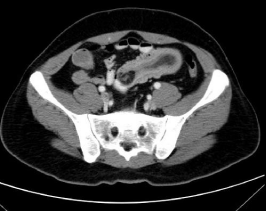
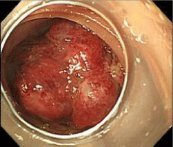
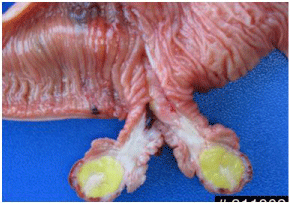
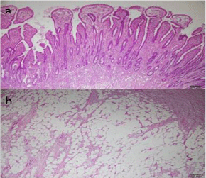
A 41-year-old man presented with passage of dark red stool, nausea, vomiting and abdominal crampy pain. Physical examination revealed pale conjuntivae and local tenderness over the periumbilical region of abdomen, without peritoneal signs. Laboratory studies showed normocytic anemia (hemoglobin, 9.6 g/dL). Serum carcinoembryonic antigen (CEA) level was within normal limits. Abdominal computed tomography revealed a “target sign” in the left middle abdomen, and ileoileal intussusception with a leading lipoma about 2.4 cm was suspected (Figure 1A). Single-balloon enteroscopy demonstrated one irregular hyperemic firm tumor about 3 cm in diameter located at the distal ileum with impaction of the lumen (Figure 1B). Mucosaderived tumor or malignant submucosal tumor such as liposacroma was suspected initially. Laparoscopic assisted segmental resection of small intestine was performed (Figure 2A), fortunately, pathological examination of the surgical specimen revealed tubular adenoma with moderate dysplasia and underlying submucosal lipoma (Figure 2B). The patient recovered well after surgical intervention. Both benign and malignant tumors have also been reported to be associated with enteric intussusception [1,2,3]. One retrospective study revealed 45.5% of the intussusceptions were enteric type, of which 30% were malignant tumor and 20% were benign tumor [4]. Among enteric intussusception, lipoma and Peutz–Jegher adenoma were the two most common etiologies, while malignant lymphoma, metastatic melanoma and gastrointestinal stroma tumor were the three most common malignancy [3]. Abdominal pain was the most common clinical presentation, followed by intestinal obstructive syndrome (nausea, vomiting), gastrointestinal bleeding and abdominal palpable mass. With the signs of target or sausage, mesenteric fat and vessels, abdominal computerized tomography was the most useful preoperative diagnostic modality, which was superior to revealing the site, level, and cause of intestinal obstructions [4,5]. Our patient’s abdominal CT demonstrated enteric intussusception resulted from benign lipoma, however, the image from enteroscopy did not show typical lipoma pattern. In this rare case, the intestinal tumor showed unusual and atypical endoscopic manifestations via balloon-assisted enteroscopy. It involved the coexistence of mucosal adenoma and submucosal lipoma, which resulted in ileal intussusception.

    Figure 1A:  Axial contrast-enhanced abdominal CT showed a welldemarcated
cylindrical mass, the leading point of which was a homogenous
fat density lesion in the lumen.

    Figure 1B:  Enteroscopy revealed a single, irregular, hyperemic, firm,
ulcerative tumor that occupied the lumen of the distal ileum with ileal
intussusception.

Figure 2A: Gross picture of this resected small intestinal tumor.

    Figure 2B:  Histological examination of the surgical specimen showed a
55-mm polypoid lesion protruding into the lumen with focal ulceration on
the surface. Cross-section of the polypoid lesion showed a circumscribed
yellowish nodule in the submucosa and a fibrous stalk. Histological
examination revealed a tubular adenoma with moderate dysplasia (a) and
underlying submucosal lipoma (b).
References
- Turan M, Karadayi K, Duman M, Ozer H, Arici S, Yildirir C, et al. Small bowel tumors in emergency surgery. Ulus Travma Acil Cerrahi Derg. 2010; 16: 327-333.
- Liu H, Cheng YJ, Chen HP, Hwang JC, Chang PC. Multiple bowel intussusceptions from metastatic localized malignant pleural mesothelioma: a case report. World J Gastroenterol. 2010; 16: 3984-3986.
- Chiang JM, Lin YS. Tumor spectrum of adult intussusception. J Surg Oncol. 2008; 98: 444-447.
- Wang N, Cui XY, Liu Y, Long J, Xu YH, Guo RX, et al. Adult intussusception: A retrospective review of 41 cases. World J Gastroenterol. 2009; 15: 3303–3308.
- Beattie GC, Peters RT, Guy S, Mendelson RM. Computed tomography in the assessment of suspected large bowel obstruction. ANZ J Surg. 2007; 77: 160-5.