
Case Report
Austin J Gastroenterol. 2017; 4(1): 1077.
Clinical Cases of Crohn‘s Disease in Pediatric Hirschprung‘s Patients
Cerniauskaite R¹, Statkuviene J², Labanauskas L², Urbonas V³, Bagdzevicius S4, Adamonis K5, Rokaite R², Janciauskas D6 and Kucinskiene R²*
¹Department of Radiology, Vilnius University Hospital Santariskiu Clinics, Lithuania
²Department of Pediatric gastroenterology, Hospital of Lithuanian University of Health Sciences, Lithuania
³Department of Pediatric gastroenterology, Vilnius University Hospital Santariskiu Clinics, Lithuania
4Department of Pediatric surgery, Hospital of Lithuanian University of Health Sciences, Lithuania
5Department of Gastroenterology, Hospital of Lithuanian University of Health Sciences, Lithuania
6Department of Pathology, Hospital of Lithuanian University of Health Sciences, Lithuania
*Corresponding author: Kucinskiene R, Department of Pediatric Gastroenterology, Hospital of Lithuanian University of Health Sciences, Eiveniust. 2, LT-50009, Kaunas, Lithuania
Received: January 27, 2017; Accepted: February 28, 2017; Published: March 02, 2017
Abstract
Crohn’s and Hirschsprung’s diseases are two different conditions of intestinal tract though both of them are genetically predisposed. Each disease have genetic mutations in some genes, however there are no evidence that there are mutations common to both conditions.
We present 3 clinical cases of patients who underwent surgery in infancy for Hirschsprung’s disease. Later in early childhood all the patients developed clinical symptoms of inflammatory bowel disease and Crohn’s disease was diagnosed. Both conditions were confirmed histologically. After introducing the treatment for Crohn’s disease positive effect was shown. These cases raise the hypothesis that the two conditions may have similarities in etiology and pathogenesis and may represent a spectrum of intestinal inflammatory diseases in children.
Keywords: Hirschsprung’s disease; Crohn’s disease; Pediatrics
Case Presentation
Case 1
Normally born healthy boy was constipated from birth and meconium ileus was present. He started to regurgitate frequently and suffered from chronic abdominal distention when he was 1 month old. At the age of 7 months biopsy from rectum was done and histopathology showed findings typical for Hirschsprung’s disease (Figure 1); thus hemicolectomy was performed. Being 4 years old patient suffered from abdominal pain and more frequent stools, as well unexplained fever episodes. The diagnosis of cystic fibrosis and tuberculosis were not confirmed. He constantly had low levels of hemoglobin (Hb), elevated rates of erythrocyte sedimentation (ESR) as well as C-reactive protein (CRP) and platelets (PLT). From 7 till 11 years old patient wasn’t gaining weight, his bone age was delayed. Several times positive effect of antibacterial treatment (metronidazole and cefuroxime) was observed when the boy had sub-febrile temperature, abdominal pain and loose stools. Any possible infection wasn’t confirmed thus colonoscopy with biopsy was performed. Colonoscopy showed inflammatory changes in transversal, ascending and cecum parts of colon mucosa and edematous stenosis of ileocecal part; histopathology confirmed active chronic colitis with reactive lymphoid hyperplasia (Figure 2) - Crohn’s disease was diagnosed. Treatment with steroids and azathioprine was initiated, though despite the treatment, relapses with diarrhea, abdominal pain and inflammatory blood changes (CRP - >140 mg/l, PLT - > 700 x 109/l) occurred. 7 months later after the diagnosis of Crohn‘s disease Infliximab was started. 3 weeks after the last induction dose symptoms of intestinal obstruction occurred - patient was vomiting after greater meal, suffered from abdominal pain and lost weight.
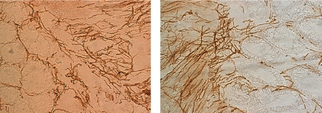
    Figure 1: Patient No 1 - boy, 7 months old.
Enzyme histochemistry of the biopsy from intestine show in gaberrantacetyl
choline esterase (ACHE)-positive fibres (brown) in the lamina propriamucosae.
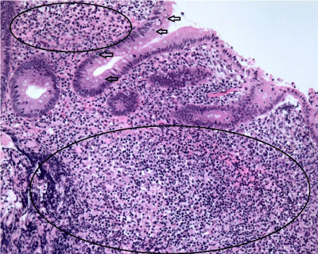
    Figure 2: Patient No 1 - boy, 11 years old. Histopathology showing chronic
active colitis (arrows) with reactive lymphoid hyperplasia (delineated).
Boy was admitted to the hospital and bowel X-ray with contrast imaging showed intestinal loop dilatation (Figure 3) although contrast passed to rectum. Additionally computed tomography of bowel showed dilatation of terminal ileum. During the surgery it was discovered that 80 cm of distal ileum was dilated, but terminal part which enters cecum was stenotic with thickened walls. There was also inflammatory infiltration in cecum with enlarged lymph nodes (5 cm of diameter), thus terminal ileum (around 60 cm) and cecum were resected and anastomosis ileum to colon was made. Histopathology displayed patchy active chronic inflammation with fissuring ulcers in the intestine (Figure 4). The main diagnosis was stenotic Crohn’s disease, Azathioprine was continued. Patient was genotyped for the 3 common NOD2/CARD15 polymorphism - all negative. After surgery patient had good appetite, normal stools and no abdominal pain episodes, started to gain weight. Now he is in clinical remission for 3 years.
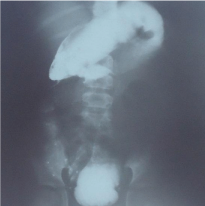
    Figure 3: Patient No 1 - boy, 12 years old. Bowel X-ray with barium showing
intestinal loop dilatation.
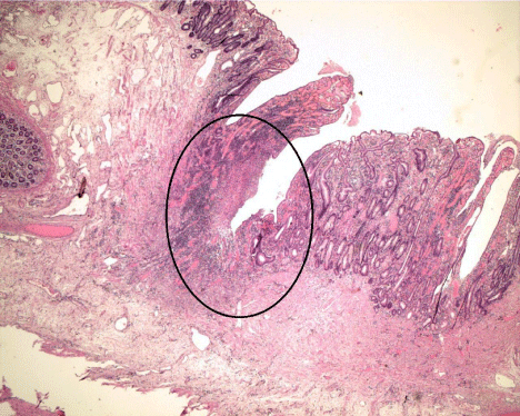
    Figure 4: Patient No 1 - boy, 12 years old. Histopathological Figure displays
patchy active chronic inflammation with fissuring ulcer (delineated) in the
intestine.
Case 2
Second patient was born from normal gestation and delivery, yet on the first day of life she developed bilious vomiting. Necrotic enterocolitis was suspected, thus parenteral nutrition and adequate antibiotic therapy was initiated. At the age of 7 days the condition of the patient deteriorated, she developed clinical signs of partial ileus and on emergent laparotomy the changes specific to Hirschsprung’s disease were observed: significantly dilated ascending part of the colon, narrowing at the hepatic and splenic flexures, descending and sigmoid colon were narrow with thickened walls. Transversostoma at the splenic flexure was formed and pathological specimens were taken. The biopsy results showed the absence of ganglion cells and left hemicolectomy was performed because of the long segment involvement. After surgery the general condition of the patient improved but she wasn’t gaining weight properly. At the age of 5 she started suffering from irregular, but quite common loose stools episodes and at the age of 7 she was admitted for examination having loose stools up to 10 times a day, abdominal distension and weight loss. Blood tests showed increasing rates of CRP, colonoscopy of 20 cm of colon (after hemicolectomy) showed no visible changes, but terminal ileum (20 cm in length) was with ulcers specific to Crohn’s disease. Biopsy results showed patchy active chronic ileocolitis with reactive lymphoid hyperplasia and granulation tissue proliferation due to ulceration (Figure 5). Crohn’s disease was diagnosed and treatment with Prednisone was initiated at once with the Azathioprine introduction in the future plans. The patient had normal stools no abdominal distention and clinical remission was achieved soon.
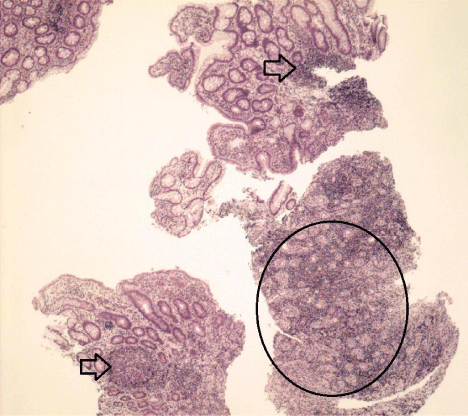
    Figure 5: Patient No 2 - girl, 7 years old. Patchy active chronic ileocolitis with
reactive lymphoid hyperplasia (arrows) and granulation tissue (delineated)
proliferation due to ulceration.
Case 3
Third patient - a female with delayed meconium pass, who started vomiting bile on the 6th day of life and was admitted to hospital suspecting intestinal obstruction. Laparotomy was required during which meconium ileus was found and appendicostomy was formed. Nevertheless the patient continued to vomit, wasn’t gaining weight, and was defecating only after enema. The patient was genotyped for cystic fibrosis, but CFTR gene wasn’t found. Irigo scopy showed typical radiological findings for Hirschsprung’s disease. Due to long segment involvement and many complications patient had undergone 7 surgeries and hemicolectomy was performed at last. The histopathology confirmed the absence of ganglia in the segments of colon. After surgical treatment patient still wasn’t gaining weight properly, had difficulties in defecation. At the age of 4 she was admitted to the hospital due to long lasting (2-3 weeks) fever without clear location of infection. During this period she was treated with antibacterial therapy however without positive effect. Blood tests showed increased levels of CRP (82 mg/l) and ESR (87 mm/h). Normally patient had 1-2 bowel movements per day with slightly loose stools, however it was decided to perform colonoscopy during which the ulcers covered with fibrin layer was observed, biopsy was taken and the results showed chronic active inflammation with ulceration. The diagnosis of Crohn’s disease was made and treatment with Prednisone orally was initiated. Patient relapsed several times thus the colonoscopy was repeated however the colon was inspected only in a short segment up to stenosis. Azathioprine was added to the treatment. Patient started gaining weight and developing normally.
Discussion
Hirschsprung’s disease (HD) is the commonest congenital gut motility disorder and is characterized by a lack of ganglion cells in a variable length of distal gut [1]. Clinical symptoms of Hirschsprung’s disease manifest in neonatal or early childhood period when, because of lack of innervation in some part of the colon, intestinal obstruction is caused. The predominant gene affected is the RET proto-oncogene [2].
Crohn’s Disease (CD) is an immune-mediated disorder characterized by recurrent chronic uncontrolled inflammation on the intestinal mucosa affecting any part of the gastrointestinal tract. Dysregulation of the immune interaction between enteric antigens and the enteral mucosa results in chronic, immune-mediated inflammation [3].
We have checked the online data base and have found only 3 articles with case reports (10 cases in total) referring diagnosis of Hirschsprung’s and Crohn’s diseases to the same patient [4-6]. We didn’t find any information or proof that those two conditions might be connected or predispose each other.
It is not clear why those two diseases could occur fore-and-aft in one patient. However there is consideration about the link in pathogenesis of those two conditions: impaired mucosal barrier, mucin production and changes in immune response [7]. The most likely hypothesis why Crohn’s disease occurs in patients with previous Hirschsprung’s disease is changes in microbiota. Gut microbiome can be affected after surgery and alterations in colonization predisposes to chronic inflammation. More research is needed to investigate this connection.
We present 3 clinical cases from Lithuania of patients who were diagnosed with Hirschsprung’s disease in newborn period or infancy and later developed Crohn’s disease. All 3 patients had normal perinatal and gestational anamnesis and there was negative family history for inherited chronic intestinal tract diseases in all cases. All patients were diagnosed with long segment Hirschsprung’s disease and were operated several times because of complications. Crohn’s disease was diagnosed in early childhood (4 to 7 years old). Diagnosis was delayed due to poor gain of weight since birth and loose stools as postoperative Hirschsprung’s disease outcome. According to literature up to 40% of patients develop Hirschsprung’s-associated enterocolitis [8]. This pathology may mimic Crohn’s disease symptoms and delay the diagnosis. According to our data suspicion of Crohn’s disease in Hirschsprung’s disease post-operative patients should be raised when diarrhea, fever, weight loss and signs of systemic inflammation appear some years later after the surgery. The main diagnostic investigation is ileocolonoscopy with obligatory visualization and biopsies of terminal ileum. In all our presented cases Crohn’s disease was confirmed based on intestinal biopsy and histopathology data. The positive clinical effect after initiation of steroids impacted the final diagnosis as well. The investigation of NOD2 mutations may be important to differentiate Crohn’s disease with postoperative enterocolitis yet it is not always identified [9]. Lacking the possibility to test the patients genetically 2 of the presented patients were not genotyped.
There are too little cases reported to make a reliable guidelines or conclusions however patients who underwent surgery for Hirschsprung’s disease should be followed up keeping in mind the inflammatory bowel disease possibility in the future.
References
- Kenny SE, Tam PK, Garcia-Barcelo M. Hirschsprung’s disease. Semin Pediatr Surg. 2010; 19: 194-200.
- Parisi MA, Kapur RP. Genetics of Hirschsprung disease. Curr Opin Pediatr. 2000; 12: 610-617.
- Hsu YC, Wu TC, Lo YC, Wang LS. Gastrointestinal complications and extraintestinal manifestations of inflammatory bowel disease in Taiwan: A population-based study. J Chin Med Assoc. 2016; 80: 56-62.
- Levin DN, Marcon MA, Rintala RJ, Jacobson D, Langer JC. Inflammatory bowel disease manifesting after surgical treatment for Hirschsprung disease. J Pediatr Gastroenterol Nutr. 2012; 55: 272-277.
- Ikeuchi H, Kusunoki M, Yamamura T, Utsunomiya J. A case of Crohn’s disease following Hirschsprung’s disease. The journal of Japanese Practical Surgeon Society. 1997; 58: 622-625.
- Kessler, Bradley H, Henry B, Becker, Jerrold M. Crohn’s disease mimicking enterocolitis in a patient with an endorectal pull-through for Hirschsprung’s disease. J Pediatr Gastroenterol Nutr. 1999; 29: 601-603.
- Mattar AF, Coran AG, Teitelbaum DH. MUC-2 mucin production in Hirschsprung's disease: possible association with enterocolitis development. J Pediatr Surg. 2003; 38: 417-421.
- Menezes M, Puri P. Long-term outcome of patients with enterocolitis complicating Hirschsprung's disease. Pediatr Surg Int. 2006; 22: 316-318.
- Lacher M, Fitze G, Helmbrecht J, Schroepf S, Berger M, Lohse P, et al. Hirschsprung-associated enterocolitis develops independently of NOD2 variants. J Pediatr Surg. 2010; 45: 1826-1831.