
Special Article - Crohn’s Disease and Colitis
Austin J Gastroenterol. 2017; 4(1): 1078.
Surgical Management of Primary Crohn’s Disease
Makni A*, Magherbi H, El Héni A, Daghfous A, Rebai W, Chebbi F, Fterich F, Ksantini R, Jouini M, Kacem M and Ben Safta Z
Department of General Surgery ‘A’, La Rabta Hospital, Tunisia
*Corresponding author: Makni Amin, Department of General Surgery ‘A’, La Rabta Hospital, Jabbari 1007, Tunis, Tunis El Manar University, Faculty of Medicine of Tunis, 15 Rue Djebel Akhdhar, Tunisia
Received: February 14, 2017; Accepted: March 13, 2017; Published: March 24, 2017
Abstract
Inflammatory bowel disease is a chronic gastrointestinal condition that is characterized by chronic gastrointestinal inflammation. The management of Crohn’s disease is complex and requires skill, knowledge and experience with current advances in the field. Over the past several years, there have been a number of achievements and progress made in the care and management of this disorder. The diagnostic tools have greatly improved. The therapeutic armamentarium has expanded. The genetics of IBD has become more detailed and the role of the gut microbiome has been better defined. The evolution of biological agents has revolutionized the way we approach this disease. However, surgery is still required in more than 80% of patients with Crohn’s disease (CD). This article aims to study the epidemiological, anatomical and therapeutic principles of surgical forms of CD.
Keywords: Crohn’s disease; Surgery; Recurrence; Stricture; Fistula
Introduction
Surgery is required in more than 80% of patients with Crohn’s disease (CD) [1]. The aim of surgery is not to cure the disease, which evolves in most cases to the recurrence of the remaining intestine [2]. Surgical treatment of intestinal lesions caused by CD is guided by two main criteria: operate only complicated forms, and refractory to medical treatment, and perform an intestinal resection as limited as possible, removing only lesions responsible for the symptoms observed. Indeed, perineal CD is problematic as regards to the diagnostic, prognostic and specific management. Over the past several years, there have been a number of achievements and progress made in the care and management of this disorder. However, surgery is still required in more than 80% of patients with Crohn’s disease (CD). This article aims to study the epidemiological, anatomical and therapeutic principles of surgical forms of CD.
The indications of surgical management
Multidisciplinary approach is now mandatory to discuss the therapeutic strategy and the time for surgery. The indication for surgery in CD depends on a number of factors-complications, clinical course, relapse and location. We could say that surgery is timely in any of the following situations: failure of medical treatment, onset of specific complications related to the disease or to pharmacological treatment, dysplasia or cancer and stagnated or retarded growth in children [3,4].
Chronic complications of Crohn’s disease
Surgery in the era of medical management: Today’s, medical management (immunosuppressants, biological therapies) has been used increasingly and was initiated much earlier during the course of CD. However, this evolving therapeutic strategy was not associated with a decrease in the need for surgery or in a decrease of the occurrence of intestinal complications. The real benefit is that large intestinal resections became more unusual [5].
Time for surgery: Identifying the best time for surgery is not always an easy task. In order to determine the best time for surgery we should assess the severity and type of symptoms, failure of medical treatment, the onset of adverse effects and surgical risk/ benefit. All this together will enable gastroenterologists, surgeons and patients to agree on optimal time for surgery. Those advocating early surgery argue that if medical treatment does not achieve substantial improvement there is no reason to await the onset of a serious, potentially life-threatening complication, or to increase surgeryrelated risk. On the other hand, authors critical of early surgery argue that since relapse and re-operation rates are high, the chances of short bowel syndrome are very low. This argument does not hold because since small resections and strictureplasties are being conducted this syndrome is highly unlikely to occur [3].
Type and topography of lesions: Crohn’s disease (CD) is a very heterogeneous disease with a relatively unpredictable clinical course. Nevertheless, important prognostic information is provided by classification based on the anatomic location, disease behavior, surgical history, and response to corticosteroid treatment. The Vienna Classification, a simple phenotypic classification structured on a combination of age at diagnosis, location (upper gastro-intestinal, terminal ileon, ileo-colon) and behavior of disease (stricturing, penetrating, non-stricturing non-penetrating), provides distinct definitions to categorize Crohn’s patients into various subgroups [6- 8].
The most common location of lesions which required surgery is the terminal ileum [9] and the ileocecal junction [10-14]. In this case, the most common indications are symptomatic stricture (Figure 1) or mixed forms (which causes intra-abdominal abscess or complex fistulas) [9]. Followed by the colorectal location, which the most common indication is the resistance to medical treatment of non-stricturing non penetrating form followed by colonic stricture (Figure 2). Rarely, we can observe proximal lesions (duodenal, jejunal and ileal), which the most common indication is small bowel obstruction caused by strictures. Chronic fibrosis and scarring that do not respond to conservative management necessitates [3,15].
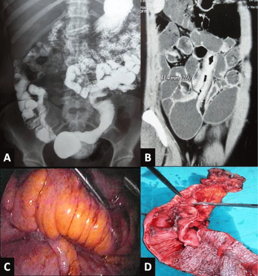
    Figure 1: Stenosis of the distal ileum. A- Small bowel opacification: Stenosis
extended last two ileum loops. B- Abdominal CT-scan: inflammatory stricture
of the distal ileal that takes intensely the contrast. C- Intraoperative view: the
handle is inflammatory with a sclero-lipomatosis D- Surgical specimen: The
ileal stenosis.
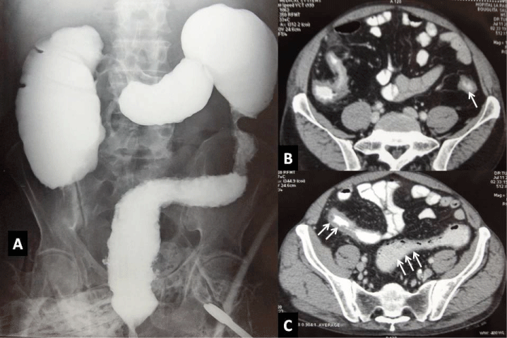
    Figure 2: Colonic Crohn’s disease. A- Barium enema: presence of multiple
colonic stenosis with inter-stenotic dilatation. B- Abdominal CT-scan: stenosis
of descending colon C- Abdominal CT-scan: Inflammation of the ileum (two
arrows) and sigmoid colon (three arrows).
Table 1 describes the clinical presentation and the indication of surgery depending on location and type of lesion.
    Behaviour

      Location
    Stricturing
    Penetrating
    Non-stricturing non penetrating


    Upper gastrointestinal
      Frequency*
      Indications of surgery

      ++
      Intestinal obstruction

      +/-
      Digestive by-pass

      -


    Ileo-colon/terminal ileon
      Frequency*
      Indications of surgery
    Mixed form
      +++
      Intestinal obstruction, intra-abdominal abscesss**


      -


    Colon and rectum
      Frequency*
      Indications of surgery

      +
      Intestinal obstruction

      -

      ++
      Resistance to medical treatment


    Frequency* (-) extremely rare (+/-) rare  (+) possible (++)    frequent (+++) very common
      **Complex fistula: ileo-ileal,    ileo-cecal, ileo-vesical and/or entero-cutaneous fistula.
Table 1: Description of the clinical presentation and the indication of surgery depending on location and type of lesion.
The acute complications of Crohn’s disease
Acute peritonitis: Acute peritonitis (Figure 3) caused by free perforation of a lesion in the peritoneal cavity is very rare [13]. It requires resection of the perforated segment and performing a stoma [16] that we prefer to transformation of the perforation in stoma which is difficult to perform because of the abondance of sclerolipomatosis.
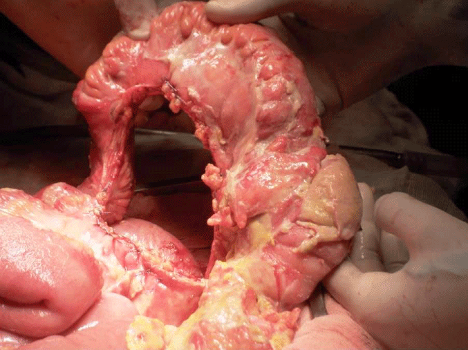
    Figure 3: Acute peritonitis. Intra-operative view: acute peritonitis related to
the peritoneal rupture of an intra-abdominal abscess complicating Crohn’s
disease.
The intra-abdominal abscesses: They are more common and can be managed sequentially starting first by percutaneous drainage (Figure 4) under radio logical guidance rather than surgical drainage [17]. Jawhari, et al. has demonstrated that drainage was technically possible in half of patients [17]. This radiological technique allowed to surgery to be performed in better conditions: patient better prepared, the possibility of immediate digestive anastomosis, a lower operative morbidity and the opportunity to offer the laparoscopic approach [17-19].
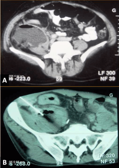
    Figure 4: Intra-abdominal abscess. Abdominal CT-scan: A- Presence of
a collection at the right iliac fossa. B- Radiological-guided percutaneous
drainage of the abscess.
The acute mechanical bowel obstruction: It is the complication of stenosing lesions. However, the indication of surgery in the emergency is a rare situation. Indeed, patients restored usually quickly, their bowel under resuscitation because in the majority of cases, stenosis is of the inflammatory rather than the sclerotic type.
Massive gastrointestinal bleeding: This complication is rare, occurring in 1-2% of patients. Treatment is usually based on the medical intensive care including administration of vasopressin, in case of failure; arteriography with embolization is an effective treatment [20]. Surgery is rarely indicated, only in case of cataclysmic bleeding or failure of previous treatment [21].
Acute severe colitis: When it is resistant to medical treatment (first-and second-line) or complicated (perforation or toxic megacolon). Surgery is less common that ulcerative colitis. The lifesaving gesture is a subtotal colectomy without anastomosis.
The isolated appendiceal Crohn’s disease: It is rare with a frequency of 0.2%. The most common clinical features are those of complicated acute appendicitis (abscess, peritonitis). Only the pathological examination of specimen could confirm the appendicular Crohn’s disease [22]. On the other hand, the achievement of the appendix during the ileocecal Crohn’s disease is much more common (21%) and offers no clinical features [23].
Degeneration of Crohn’s disease
Two meta-analyzes [24,25] have demonstrated excess risk of digestive cancers occurred in patients with Crohn’s disease. The overall relative risk is about 1.9 (confidence interval 95%: 1.4 to 2.5). The degeneration in the small bowel requires an oncological digestive resection. However in the case of degeneration in the colon, the choice between segmental colectomy and total colectomy is a real dilemma and current data do not suggest that oncologic segmental colectomy is sufficient or colorectal proctectomy is necessary.
The Operative Procedures
The principles of surgery in Crohn’s disease are: intra operative exploration allowing an inventory of lesions and ultimately measure the length of the small intestine, economic resection in macroscopically healthy tissue in order to avoid short bowel syndrome, not the need of lymph node dissection, and not need of intraoperative frozen section slices [26]. Can be distinguished:
The intestinal resection
The intestinal resections are the most frequently performed procedures. The kind of resection will depend upon the location of lesions. For small bowel lesions: ileal, rarely jejunal or often ileocecal resection in case of simultaneous achievement in the cecum and terminal ileum. After ileocolic resection, ileocolic anastomosis could be: side to end, end to end or side to side, stapled or handsewn. Side to side stapled anastomosis is as safe as conventional sutured end-to-end anastomosis and results in a lower incidence of symptomatic recurrent Crohn’s disease and need for reoperation. [27,28]. For colonic lesions: segmental colonic, sub-total or total colonic resection. For segmental colonic lesions, recent data suggests that segmental colectomy is preferable than total colectomy, indeed for a similar recurrence rate, segmental colectomy offers a better functional outcome [29].
Proctectomy, is a mutilating procedure in young patients. It is needed in less than 10% of patients [30]. This gesture is indicated in cases of microrectie, an ano-perineal major disrepair resistant to medical treatment. The rectal resection is difficult because of the local inflammation and the sclerolipomatosis in the mesorectum. The resection has to be performed flush with the rectal wall to preserve the pelvic innervations.
The total proctocolectomy is indicated in case of diffuse colorectal disease. It is admitted by most teams that this procedure ends with a permanent ileostomy. Continent ileostomy carries a significant risk of non-severe complications. In selected patients, it represents a valuable alternative to an end ileostomy. However, in recent years, this dogma has been abandoned by some teams, who proposed ileo-anal anastomosis for patients with Crohn’s disease provided to respect strict selection criteria, namely: the absence of perineal lesion and free ileum reached. The good functional results above 50% make this approach a promising one [31,32] in the future in young patients thus avoiding permanent ileostomy.
Stricturoplasty
This procedure is a bowel-sparing alternative to resection in the treatment of stricturing Crohn’s disease. This technique is most often indicated in cases of multiple stenosis and especially recurrent Crohn’s disease. Depending on the length of the stenosis, strictureplasty may be Heineke-Mikulicz type in case of short strictures (<5 cm), Finney type in case of stenosis of 5 to 20 cm long or Michelassi type (sideto- side strictureplasty technique) for extended stenosis (>20 cm). Strictureplasty is a safe and effective procedure for jejunoileal Crohn’s disease, including ileocolonic recurrence, and it has the advantage of protecting against further small bowel loss. However, the place for strictureplasty is less well defined in duodenal and colonic diseases [33,34]. The recurrence rate is equivalent to the intestinal resection with morbidity of around 12%. This technique leaves in place an inflammatory diseased tissue and does not spare the patient from escalating therapy postoperatively to stop the inflammation [21,22].
Digestive derivations
We can distinguish schematically, external and internal diversions. External derivations (ileostomy, colostomy) are indicated to protect an anastomosis or more frequently to derive the fecal stream in case of major rectal or perineal lesions [16]. The internal derivations (i.e., gastro-entero-anastomosis) are indicated in case of lesion localized in the duodenum.
Management of complex internal fistula and enterocutaneous fistula
For ileo-ileal (Figure 5) fistula which has the appearance of a knot of vipers, we prefer to extend the resection and taking the diseased segment and the victim segment that is usually the adjacent segment. For entero-cutaneous fistula (Figure 6), we perform: disconnection of entero-cutaneous fistula, freshening of the edges of the fistula, curettage of its path and resection of the diseased segment.
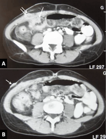
    Figure 5: Management of a complex internal fistula: A- Fistula-in node
viper; extended resection of the diseased segment to segment victim. BDisconnection
of the fistula, resection of the diseased segment and suturing
of the victim segment. C- Resection of the diseased segment and the
segment victim.
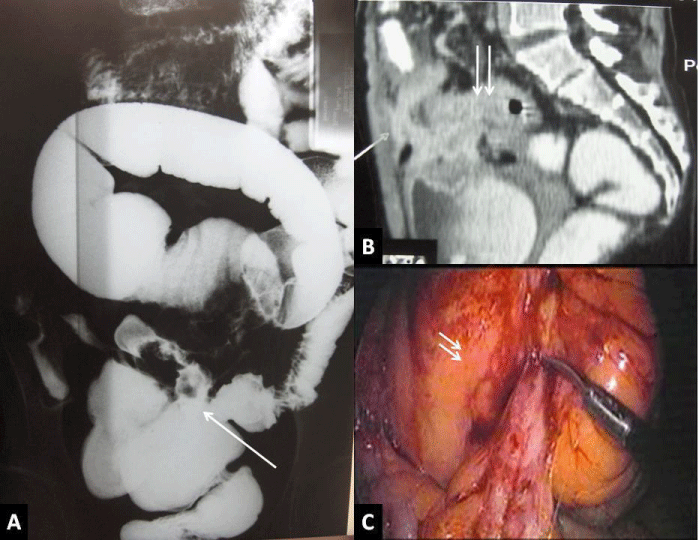
    Figure 6: The ileo-sigmoid fistula. A- Small bowel opacification: precocious
opacification of recto-sigmoid. B- Abdominal CT-scan: the distal intestinal
loop diseased (one arrow), ileo-sigmoid fistula (two arrows). C- Intraoperative
view: fistula between the sigmoid colon and the last ileal loop.
For ileo-sigmoid fistula (Figure 7), or ileo-ileal (with an intermediate segment that is long healthy) attitude is controversial, between a radical attitude (resection of two segments) and a conservative approach (resection of the diseased segment, disconnect- freshening of the edges and closure of sigmoid or ileum). The published data do not demonstrate the superiority of one over the other [35,36]. Laparoscopic treatment is acceptable in select cases without added morbidity [36].
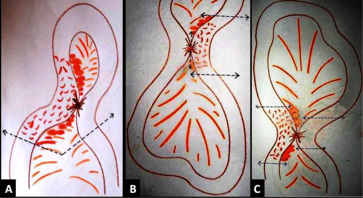
    Figure 7: Entero-cutaneous fistula. Abdominal CT-scan: A- The last loop
frankly pathological (one arrow), with the presence of a fistula into wall (two
arrows). B- The cutaneous orifice of entero-cutaneous fistula.
For ileo-vesical fistula (Figure 8), the disconnection of the fistula and resection of the diseased segment is required. At this gesture, we associate a closure of the bladder outlet that is not required if the fistula orifice was not found, and a trans-urethral catheter for a period of 10 days [37,38].
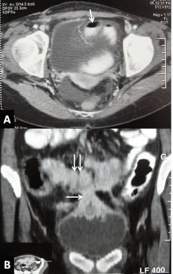
    Figure 8: The ileo-vesical fistula. Abdominal CT-scan A- Presence of air in
the bladder and passage of the contrast from the ileum to the bladder. BPresence
of a direct fistula between a last ileal loop diseased and the bladder.
The place of laparoscopy
Crohn’s disease is an ideal indication for the laparoscopic surgical approach as they are basically benign diseases not requiring lymphadenectomy and extended mesenteric excision. Inflammatory alterations and fragility of the bowel and mesentery, however, may demand a high level of laparoscopic experience. A broad spectrum of operations from the rather easy enterostomy formation for anal CD to total proctocolectomies may be managed laparoscopically [39]. It may be assisted laparoscopic surgery where the mobilization of the bowel segment to be resected is done laparoscopically, subsequently, via a mini-laparotomy, extraction takes part, resection and anastomosis extracorporeally. It can be also a full-laparoscopic surgery, three stages: mobilization, resection and anastomosis are done laparoscopically. All kind of procedures are feasible laparoscopically. The most common procedure performed by laparoscopy is ileo-colic resection.
Even in patients with recurrent Crohn’s disease, the laparoscopic management is a safe and technically feasible option, even in those patients with prior history of Crohn’s related abdominal surgery, with a low complication rate and low conversion rate [40]. The advantages of laparoscopy are: economy parietal, early rehabilitation, fewer wound complications, aesthetic gain in young patients who must undergo repeated surgery [41-43]. Even better, as related to the risk of recurrence, Makni, et al. demonstrated that laparoscopy may reduce the risk of relapse of the disease; this is probably related with a lesser stimulation of the inflammatory process compared to laparotomy [44]. The conversion rate varies among studies from 10 to 30%, the risk factors of laparoscopic conversion are essentially: inflammatory masses, the severity of the disease, the presence of abscess or fistula at surgery [45-49,19].
The Postoperative Results
The early postoperative results
The postoperative results were particular by a low mortality rate from 0 to 0.3%, however, a relatively high morbidity of between 9 and 16%. This high rate of morbidity is in relationship with long-term medical treatment: corticosteroid-therapy and immunosuppressive drugs. Risk factors for septic complications occurred post-operatively are: presence of abscess discovered during surgery, the severity of disease, perioperative malnutrition, long-term corticosteroidtherapy, Penetrating type, operation time >180 minutes, and handsewn anastomoses [27,50,51].
The long-term outcomes
The postoperative recurrence is a major problem especially common in the management of this disease. The risk of surgical recurrence is about 20% at 5 years and 35% at 10 years [50,52].
Risk factors of surgical recurrence
Risk factors for recurrence, different from one series to another, but overall we can say that the severity of the disease, fistulizing form (for some enterocutaneous fistulas), the multifocal nature of the infringement, the early age of onset, reaching proximal (duodenojejunal) and smoking are risk factors most implicated.
New Developments
Single-incision laparoscopic surgery decreases abdominal wall trauma by reducing the number of abdominal incisions, possibly improving postoperative results in terms of pain and cosmetics. The resected specimen can be extracted through the single-incision site, the future stoma site or the natural orifices (i.e. transcolonic/ transanal). In patients with extensive perianal disease or rectal involvement, transperineal completion proctectomy is often feasible, thereby avoiding relaparotomy. By using a close rectal intersphincteric resection, damage to the pelvic autonomic nerves is avoided [19,53- 59].
Conclusion
The type of lesion depends of digestive localization. Complications which reveal the disease are rare. Surgery is intended to limit the escalation therapy by resecting the diseased segment. Surgery does not cure the disease which major risk in long-term is recurrence that rate is low in our series. Therapies currently available are primarily intended to delay recurrence with favorable results.
References
- Higgens CS, Allan RN. Crohn's disease of the distal ileum. Gut. 1980; 21: 933-940.
- Rutgeerts P, Geboes K, Vantrappen G, Kerremans R, Coenegrachts JL, Coremans G. Natural history of recurrent Crohn's disease at the ileocolonic anastomosis after curative surgery. Gut. 1984; 25: 665-672.
- Alós R, Hinojosa J. Timing of surgery in Crohn's disease: a key issue in the management. World J Gastroenterol. 2008; 14: 5532-5539.
- Cosnes J, Nion-Larmurier I, Beaugerie L, Afchain P, Tiret E, Gendre JP. Impact of the increasing use of immunosuppressants in Crohn's disease on the need for intestinal surgery. Gut. 2005; 54: 237-241.
- de Buck van Overstraeten A1, Wolthuis A, D'Hoore A. Surgery for Crohn's disease in the era of biologicals: a reduced need or delayed verdict? World J Gastroenterol. 2012; 18: 3828-3832.
- Gashe C, Scholmerich J, Brynskov J, D'Haens G, Hanauer SB, Irvine EJ, et al. A simple classification of Crohn’s disease: report of the Working Party for the World Congresses of Gastroenterology, Vienna 1998. Inflamm Bowel Dis. 2000; 6: 8-15.
- Louis E, Collard A, Oger AF, Degroote E, Aboul Nasr E Yafi FA. Behaviour of Crohn's disease according to the Vienna classification: changing pattern over the course of the disease. Gut. 2001; 49: 777-782.
- Silverberg MS, Satsangi J, Ahmad T, Arnott ID, Bernstein CN, Brant SR, et al. Toward an integrated clinical, molecular and serological classification of inflammatory bowel disease. Report of a Working Party of the 2005 Montreal World Congress of Gastroenterology. Can J Gastroenterol. 2005; 19: 5A-36A.
- Oberhuber G, Stangl PC, Vogelsang H, Schober E, Herbst F, Gasche C. Significant association of strictures and internal fistula formation in Crohn's disease. Virchows Arch. 2000; 437: 293-297.
- Whelan G, Farmer RG, Fazio VW, Goormastic M. Recurrence after surgery in Crohn's disease. Relationship to location of disease (clinical pattern) and surgical indication. Gastroenterology. 1985; 88: 1826-1833.
- Lapidus A. Crohn's disease in Stockholm County during 1990-2001: an epidemiological update. World J Gastroenterol. 2006; 12: 75-81.
- Valiulis A, Currie DJ. A surgical experience with Crohn's disease. Surg Gynecol Obstet. 1987; 164: 27-32.
- Greenstein AJ. The surgery of Crohn's disease. Surg Clin North Am. 1987; 67: 573-96.
- Hurst RD, Molinari M, Chung TP, Rubin M, Michelassi F. Prospective study of the features, indications, and surgical treatment in 513 consecutive patients affected by Crohn’s disease. Surgery 1997; 122: 661-667.
- Kolar B, Speranza J, Bhatt S, Dogra V. Crohn's disease: Multimodality Imaging of Surgical Indications, Operative Procedures, and Complications. J Clin Imaging Sci. 2011; 1:37.
- Post S, Betzler M, von Ditfurth B, Schürmann G, Küppers P, Herfarth C. Risks of intestinal anastomoses in Crohn's disease. Ann Surg. 1991; 213: 37-42.
- Jawhari A, Kamm MA, Ong C, Forbes A, Bartram CI, Hawley PR. Intra-abdominal and pelvic abscess in Crohn's disease: results of noninvasive and surgical management. Br J Surg. 1998; 85: 367-371.
- Nanakawa S, Takahashi M, Takagi K, Takano M. The role of computed tomography in management of patients with Crohn disease. Clin Imaging. 1993; 17: 193-198.
- Wu JS, Birnbaum EH, Kodner IJ, Fry RD, Read TE, Fleshman JW. Laparoscopic-assisted ileocolic resections in patients with Crohn's disease: are abscesses, phlegmons, or recurrent disease contraindications? Surgery. 1997; 122: 682-688.
- Quandalle P, Gambiez L. Surgical treatment of Crohn disease of the small intestine. Ann Chir. 1997; 51: 303-313.
- Robert JR, Sachar DB, Greenstein AJ. Severe gastrointestinal hemorrhage in Crohn's disease. Ann Surg. 1991; 213: 207-211.
- Prieto-Nieto I, Perez-Robledo JP, Hardisson D, Rodriguez-Montes JA, Larrauri-Martinez J, Garcia-Sancho-Martin L. Crohn's disease limited to the appendix. Am J Surg. 2001; 182: 531-533.
- Ripollés T, Martínez MJ, Morote V, Errando J. Appendiceal involvement in Crohn's disease: gray-scale sonography and color Doppler flow features. AJR Am J Roentgenol. 2006; 186: 1071-1078.
- Canavan C, Abrams KR, Mayberry J. Meta-analysis: colorectal and small bowel cancer risk in patients with Crohn's disease. Aliment Pharmacol Ther. 2006; 23: 1097-1104.
- Jess T, Gamborg M, Matzen P, Munkholm P, Sørensen T. Increased risk of intestinal cancer in Crohn's disease: a meta-analysis of population-based cohort studies. Am J Gastroenterol. 2005; 100: 2724-2729.
- Wolff BG, Beart RW Jr, Frydenberg HB, Weiland LH, Agrez MV, Ilstrup DM. The importance of disease-free margins in resections for Crohn's disease. Dis Colon Rectum. 1983; 26: 239-243.
- Kanazawa A, Yamana T, Okamoto K, Sahara R. Risk factors for postoperative intra-abdominal septic complications after bowel resection in patients with Crohn's disease. Dis Colon Rectum. 2012; 55: 957-962.
- Muoz-Juárez M, Yamamoto T, Wolff BG, Keighley MR. Wide-lumen stapled anastomosis vs. conventional end-to-end anastomosis in the treatment of Crohn's disease. Dis Colon Rectum. 2001; 44: 20-25.
- Andersson P, Olaison G, Hallböök O, Sjödahl R. Segmental resection or subtotal colectomy in Crohn's colitis? Dis Colon Rectum. 2002; 45: 47-53.
- Post S, Herfarth C, Schumacher H, Golling M, Schürmann G, Timmermanns G. Experience with ileostomy and colostomy in Crohn's disease. Br J Surg. 1995; 82: 1629-1633.
- Panis Y, Poupard B, Nemeth J, Lavergne A, Hautefeuille P, Valleur P. Ileal pouch/anal anastomosis for Crohn's disease. Lancet. 1996; 347: 854-857.
- Sagar PM, Dozois RR, Wolff BG. Long-term results of ileal pouch-anal anastomosis in patients with Crohn's disease. Dis Colon Rectum. 1996; 39: 893-898.
- Yamamoto T, Fazio VW, Tekkis PP. Safety and efficacy of strictureplasty for Crohn's disease: a systematic review and meta-analysis. Dis Colon Rectum. 2007; 50: 1968-1986.
- Michelassi F, Taschieri A, Tonelli F, Sasaki I, Poggioli G, Fazio V, et al. An international, multicenter, prospective, observational study of the side-to-side isoperistaltic strictureplasty in Crohn's disease. Dis Colon Rectum. 2007; 50: 277-284.
- Saint-Marc O, Vaillant JC, Frileux P, Balladur P, Tiret E, Parc R. Surgical management of ileosigmoid fistulas in Crohn's disease: role of preoperative colonoscopy. Dis Colon Rectum. 1995; 38: 1084-1087.
- Melton GB, Stocchi L, Wick EC, Appau KA, Fazio VW. Contemporary surgical management for ileosigmoid fistulas in Crohn's disease. J Gastrointest Surg. 2009; 13: 839-845.
- Yamamoto T, Keighley MR. Enterovesical fistulas complicating Crohn's disease: clinicopathological features and management. Int J Colorectal Dis. 2000; 15: 211-215.
- Schraut WH, Chapman C, Abraham S. Operative treatment of Crohn’s ileocolitis complicated by ileosigmoid and ileovesical fistula. Ann Surg 1988; 207: 48-51.
- Kessler H, Mudter J, Hohenberger W. Recent results of laparoscopic surgery in inflammatory bowel disease. World J Gastroenterol. 2011; 17: 1116-1125.
- Huang R, Valerian BT, Lee EC. Laparoscopic approach in patients with recurrent Crohn's disease. Am Surg. 2012; 78: 595-599.
- Sardinha TC, Wexner SD. Laparoscopy for inflammatory bowel disease: pros and cons. World J Surg. 1998; 22: 370-374.
- Bemelman WA, Dunker MS, Slors JF, Gouma DJ. Laparoscopic surgery for inflammatory bowel disease: current concepts. Scand J Gastroenterol Suppl. 2002; 54-59.
- Eshuis EJ, Polle SW, Slors JF, Hommes DW, Sprangers MA, Gouma DJ, et al. Long-term surgical recurrence, morbidity, quality of life, and body image of laparoscopic-assisted vs. open ileocolic resection for Crohn's disease: a comparative study.Dis Colon Rectum. 2008; 51: 858-67.
- Makni A, Chebbi F, Ksantini R. Laparoscopic-assisted versus conventional ileocolectomy for primary Crohn's disease: Results of a comparative study. J Visc Surg. 2012; 150: 137-143.
- Alves A, Panis Y, Bouhnik Y, Marceau C, Rouach Y, Lavergne-Slove A et al. Factors That Predict Conversion in 69 Consecutive Patients Undergoing Laparoscopic Ileocecal Resection for Crohn’s Disease: A Prospective Study. Dis Colon Rectum. 2005; 48: 2302-2308.
- Moorthya K, Shaul T, Hons BS, Foleya RJ. Factors that predict conversion in patients undergoing laparoscopic surgery for Crohn’s disease. Am J Surg. 2004; 187: 47-51.
- Bauer JJ, Harris MT, Grumbach NM, Gorfine SR. Laparoscopic-assisted intestinal resection for Crohn's disease. Which patients are good candidates? J Clin Gastroenterol. 1996; 23: 44-46.
- Pokala N, Delaney CP, Brady KM, Senagore AJ. Elective laparoscopic surgery for benign internal enteric fistulas: a review of 43 cases. Surg Endosc. 2005; 19: 222-225.
- Moorthy K, Shaul T, Foley RJ. Factors that predict conversion in patients undergoing laparoscopic surgery for Crohn's disease. Am J Surg. 2004; 187: 47-51.
- Bauer JJ, Harris MT, Grumbach NM, Gorfine SR. Laparoscopic-assisted intestinal resection for Crohn's disease. Which patients are good candidates? J Clin Gastroenterol. 1996; 23: 44-46.
- Pokala N, Delaney CP, Brady KM, Senagore AJ. Elective laparoscopic surgery for benign internal enteric fistulas: a review of 43 cases. Surg Endosc. 2005; 19: 222-225.
- Watanabe M, Ohgami M, Teramoto T, Hibi T, Kitajima M. Laparoscopic ileocecal resection for Crohn's disease associated with intestinal stenosis and ileorectal fistula. Surg Today. 1999; 29: 446-448.
- Michelassi F, Balestracci T, Chappell R, Block GE. Primary and recurrent Crohn's disease. Experience with 1379 patients. Ann Surg. 1991; 214: 230-238.
- Alves A, Panis Y, Bouhnik Y, Pocard M, Vicaut E, Valleur P. Risk factors for intra-abdominal septic complications after a first ileocecal resection for Crohn's disease: a multivariate analysis in 161 consecutive patients. Dis Colon Rectum. 2007; 50: 331-336.
- Mirow L, Hauenschild L, Hildebrand P, Kleemann M, Keller R, Franke C, et al. Recurrence of Crohn's disease after surgery--causes and risks. Zentralbl Chir. 2008; 133: 182-187.
- Gardenbroek TJ, Tanis PJ, Buskens CJ, Bemelman WA. Surgery for Crohn's disease: new developments. Dig Surg. 2012; 29: 275-280.
- Isik A, Gursul C, Peker K, Aydın M4, Fırat D3, Yılmaz İ3. Metalloproteinases and Their Inhibitors in Patients with Inguinal Hernia. World J Surg. 2017; .
- Isik A, Idiz O, Firat D. Novel Approaches in Pilonidal Sinus Treatment. Prague Med Rep. 2016; 117: 145-152.
- Watanabe M, Ohgami M, Teramoto T, Hibi T, Kitajima M. Laparoscopic ileocecal resection for Crohn's disease associated with intestinal stenosis and ileorectal fistula. Surg Today. 1999; 29: 446-448.