Case Report
Austin J Gastroenterol. 2017; 4(3): 1088.
Advanced Hepatocellular Carcinoma
Soldera J1,2*, Balbinot SS1,3, Balbinot RA1,3, Furlan RG4 and Terres AZ4
1Clinical Gastroenterology, Faculty of Medicine of Universidade de Caxias do Sul, Brazil
2Universidade Federal de Ciências da Saúde de Porto Alegre, Brazil
3Universidade de São Paulo, Brazil
4Department of Gastroenterology, Hepatology and Digestive Endoscopy of Hospital Geral de Caxias do Sul, Brazil
*Corresponding author: Jonathan Soldera, Clinical Gastroenterology, Faculty of Medicine of Universidade de Caxias do Sul, Brazil
Received: July 19, 2017; Accepted: August 08, 2017; Published: September 25, 2017
Abstract
Each year, hepatocellular carcinoma is diagnosed in over half a million people worldwide. In this paper, it is reported the case of a patient with a late diagnosis of advanced hepatocellular carcinoma, with poor prognosis. It was used Barcelona-Clínic Liver Cancer protocol for diagnosis, staging and management. Understanding diagnosis and therapeutic approach to this disease is essential, especially if we keep in mind the quintessential basis of prevention and early detection.
Keywords: Hepatocellular carcinoma; Cirrhosis; Palliative care
Introduction
Hepatocellular carcinoma (HCC) is diagnosed in over half a million people worldwide every year [1]. It has become 5th most common cancer in men and the 7th in women. The burden of this disease is highest in developing countries, where hepatitis B is still endemic: Southeast Asia and Sub-Saharan Africa [2]. Although its incidence is rising worldwide and new therapies have been developed, the 5-year survival rate is still low. In the United States, it has remained below 12% [3].
Since it is highly prevalent in patients with cirrhosis or advanced fibrosis, screening with an ultrasonography (US) each 6 months is recommended as standard-of-care: early diagnosis means more effective treatment and increased survival. The measurement of serum alpha-fetoprotein levels remains controversial due to its low sensitivity and is not widely recommended currently [4]. A Chinese study showed that the combination of US and measurement of alphafetoprotein translates into a 37% reduction in mortality due to HCC [5].
The objective of this paper is to report the management of a case of locally advanced HCC.
Case Presentation
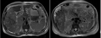
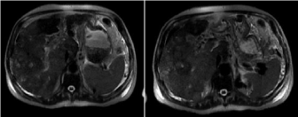
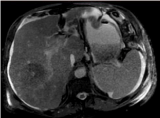
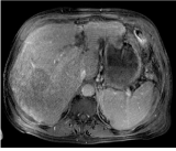
Male patient, 68-years-old, was referred from a small city hospital to ours to investigate jaundice, weight loss and a liver nodule with portal venous thrombosis (PVT) identified in a prior US. He had a previous diagnosis of cirrhosis due to alcohol after an episode of alcoholic hepatitis fifteen years earlier. He was lost to follow-up by his won will afterwards, because of clinical improvement and abstinence. He was admitted with a poor general condition, with overt hepatic encephalopathy (West-Haven 3) [6], ascites and jaundice. Laboratory creatinine 2.2 g/dL, pallets 193,000/mm³, INR 1.25, Hemoglobin 14, 7 g/dL, Leukocytes 9,970/mm³, total bilirubin 17.2 g/dL, albumin 3.09 g/dL. Child-Pugh classification C [7]. A diagnostic and therapeutic paracentesis was performed, with neutrophil count compatible with spontaneous bacterial peritonitis. It was ordered a dynamic contrasted enhanced magnetic resonance imaging (DCE-MRI) and it was began treatment with piperacillin-tazobactam, lactulose and albumin infusion, with no improvement of his general condition. Five days later, he presented hematemesis and an upper digestive endoscopy was performed, with small esophageal varices and duodenal ulcers Forrest IIc [8]. MRI showed moderate ascites and a heterogeneous liver with multiple nodules, some with pseudo-capsules and a PVT, suggesting locally advanced HCC with malignant PVT (Figures 1-4). He was therefore diagnosed with HCC staging Barcelona-Clinic Liver Cancer (BCLC) D [4]. Palliative care was discussed with the family, and although with some resistance, it was began. He died after thirteen days of the hospital admission, in the infirmary, with his family, receiving opioids to attenuate suffering.

Figure 1:

Figure 2:

Figure 3:

Figure 4:
Discussion
The vast majority of HCC cases occur in patients with cirrhosis. At the diagnosis, the clinical features are variable; patients can be asymptomatic or only present the manifestations of decompensated liver cirrhosis [9].
The American Association for the Study of Liver Diseases (AASLD) practice guideline to the management of HCC reviews the approach to liver nodules in cirrhotic patients [4,10]. Due to its unique radiologic characteristics to the dynamic study, most diagnosis is made without the need of a liver biopsy.
Nodules with a size of 10 mm or higher are investigated primarily with a 4-phase multidetector computerized tomography (4-p MDCT) or DCE-MRI. In the cirrhotic or advanced liver fibrosis-patient, if arterial hypervascularity AND venous or delayed phase washout is present in one of this dynamic studies, it is diagnosed HCC. If the first study is negative, it is recommended to move on to the other one. If the two exams are negative, it might be necessary to perform a liver biopsy. Although, sometimes, analyzing each case individually, depending of the patient and the nodule, it is sometimes better to repeat each 3 months a dynamic study instead of deciding in favor of a liver biopsy.
Forner, et al. in a prospective study of 89 cases, showed that noninvasive diagnostic criteria has a specificity of 100% with a 30% loss in sensitivity for nodules up to 20 mm detected in screening programs (about 2/3 of the nodules required histological confirmation) [11]. Sangionvanni, et al. suggested that the sequential algorithm published in the guideline has absolute specificity and increased sensitivity, reducing the amount of biopsies in nodules ranging from 10 to 20 mm [12].
The next step to define management and prognosis after diagnosis is staging the patient. It is globally used the BCLC protocol, which describes five stages for the disease, ranging from 0 to D [4,10]. After staging, it also suggests the subsequent therapy.
Patients with a single nodule and preserved liver function are generally candidates for surgical resection or radiofrequency ablation (RFA). The decision between therapies must weight two studies published in 2015 addressing the subject of resection X RFA, which have found resection to be superior. Therefore, it seems adequate to always strive for the first, and leave the later for the cases with prohibitive surgical risk [13,14]. It has also been studied the adjuvant use of sorafenib to prevent recurrence after resection, with some promising results [15].
Patients with
1. A single nodule up to 50 mm with portal hypertension or reduced liver function; or 2. Up to 3 nodules being the largest up to 30 mm (if your referral liver transplant center uses Milan Criteria) or the sum of the size of the largest nodule plus the total number of nodules resulting in a number up to 7 (if your referral liver transplant center uses Up-To-7 criteria) should be submitted for curative therapies, preferably liver transplantation; otherwise, RFA. Each 3 months 4-p MDCT or DCE-MRI are to be performed for follow-up while the patient waits liver transplantation. In countries where the wait list exceeds 6 months, bridging therapy is recommended to prevent dropout because of disease progression, which can be performed with RFA, alcohol injection or transarterial chemoembolization (TACE). The choice of a specific therapy should consider the size and localization of the nodule [15,16].
Patients with HCC that exceeds the Milan Criteria or the Up- To-7 criteria, with preserved liver function and good performancestatus, but in the absence of metastasis or vascular invasion, should be referred for palliative therapies, such as TACE. In this scenario, although is not yet consensus, patients could undergo TACE, alcohol injection or RFA with the objective of downstaging to the Milan Criteria. Nevertheless, some studies suggest that, after a period of observation of how the disease progresses, these patients could be referred to liver transplantation with no loss in survival [17-19].
Patients with nodules of any size and number, with vascular tumor invasion, lymph node or long-distance metastasis, with good to moderate performance status, should be referred for palliative therapy with sorafenib, which is an oral chemotherapy that increases survival in an average of 2.8 months [20].
Patients with nodules of any size and number with severely affected liver function (Child-Pugh class C) and poor performancestatus, like the patient in the reported case, should receive palliative support care. The patient and their family should be promptly oriented regarding the poor prognosis and lack of effectiveness of the available therapies in this context.
Conclusion
The focus must always be early detection and prevention. Early diagnosis of hepatic diseases and early treatment are essential prevention strategies, such as screening of asymptomatic risk population for HBV and HCV. Worldwide HBV vaccination is yet another main prevention strategy. And, of course, for those with cirrhosis or advanced liver-fibrosis, the availability of ultrasonography screening (with an experienced ultrasonographist) is necessary for early detection strategies, and it reduces HCC-related mortality.
Therefore, understanding diagnosis and therapeutic approach to this disease is essential, especially if we keep in mind the quintessential basis of prevention and early detection.
References
- Ferlay J, Bray F, Pisani P, Parkin DM. GLOBOCAN 2000: cancer incidence, mortality and prevalence worldwide, version 1.0. International Agency for Research on Cancer CancerBase no. 5. Lyon, France: IARC Press. 2001.
- World Health Organization, International. Agency for Research on Cancer. GLOBOCAN 2008.
- Surveillance, Epidemiology, and End Results (SEER) Program. SEER*Stat database: incidence - SEER 9 Regs researchdata, Nov 2009 Sub (1973-2007). Bethesda, MD: National Cancer Institute. April 2010.
- Bruix J, Sherman M. American Association for the Study of Liver Diseases. Management of hepatocellular carcinoma: an update. Hepatology. 2011; 53: 1020-1022.
- Zhang BH1, Yang BH, Tang ZY. Randomized controlled trial of screening for hepatocellular carcinoma. J Cancer Res Clin Oncol. 2004; 130: 417-422.
- Vilstrup H, Amodio P, Bajaj J, Cordoba J, Ferenci P, Mullen KD, et al. Hepatic encephalopathy in chronic liver disease: 2014 Practice Guideline by the American Association for the Study of Liver Diseases and the European Association for the Study of the Liver. Hepatology. 2014; 60: 715-735.
- Child CG, Turcotte JG. Surgery and portal hypertension. In: The liver and portal hypertension. Edited by CG Child. Philadelphia: Saunders. 1964; 50-64.
- Gralnek IM, Dumonceau JM, Kuipers EJ, Lanas A, Sanders DS, Kurien M, et al. Diagnosis and management of nonvariceal upper gastrointestinal hemorrhage: European Society of Gastrointestinal Endoscopy (ESGE) Guideline. Endoscopy. 2015; 47: a1-a46.
- Carrilho FJ, Kikuchi L, Branco F, Goncalves CS, Mattos AA; Brazilian HCC Study Group. Clinical and epidemiological aspects of hepatocellular carcinoma in Brazil. Clinics (Sao Paulo). 2010; 65: 1285-1290.
- Heimbach J, Kulik LM, Finn MDR, Sirlin CB, Abecassis M, Roberts LR, et al. AASLD guidelines for the treatment of hepatocelluar carcinoma.
- Forner A, Vilana R, Ayuso C, Bianchi L, Solé M, Ayuso JR, et al. Diagnosis of hepatic nodules 20 mm or smaller in cirrhosis: Prospective validation of the noninvasive diagnostic criteria for hepatocellular carcinoma. Hepatology. 2008; 47: 97-104.
- Sangiovanni A, Manini MA, Iavarone M, Romeo R, Forzenigo LV, Fraquelli M, et al. The diagnostic and economic impact of contrast imaging techniques in the diagnosis of small hepatocellular carcinoma in cirrhosis. Gut. 2010; 59: 638-644.
- Gory I, Fink M, Bell S, Gow P, Nicoll A, Knight V, et al. Radiofrequency ablation versus resection for the treatment of early stage hepatocellular carcinoma: a multicenter Australian study. Scand J Gastroenterol. 2015; 50: 567-76.
- Liu PH, Hsu CY, Hsia CY, Lee YH, Huang YH, Chiou YY, et al. Surgical Resection Versus Radiofrequency Ablation for Single Hepatocellular Carcinoma ≤2 cm in a Propensity Score Model. Ann Surg. 2016.
- Wang SN, Chuang SC, Lee KT. Efficacy of sorafenib as adjuvant therapy to prevent early recurrence of hepatocellular carcinoma after curative surgery: A pilot study. Hepatol Res. 2014; 44: 523-531.
- Cescon M, Cucchetti A, Ravaioli M, Pinna AD. Hepatocellular carcinoma locoregional therapies for patients in the waiting list. Impact on transplantability and recurrence rate. J Hepatol. 2013; 58: 609-618.
- Clavien PA, Lesurtel M, Bossuyt PM, Gores GJ, Langer B, Perrier A; OLT for HCC Consensus Group. Recommendations for liver transplantation for hepatocellular carcinoma: an international consensus conference report. Lancet Oncol. 2012; 13: e11-e22.
- Lei J, Wang W, Yan L. Downstaging advanced hepatocellular carcinoma to the milan criteria may provide a comparable outcome to conventional Milan criteria. J Gastrointest Surg. 2013; 17: 1440-1446.
- Yu CY, Ou HY, Huang TL, Chen TY, Tsang LL, Chen CL, et al. Hepatocellular carcinoma downstaging in liver transplantation. Transplant Proc. 2012; 44: 412-414.
- Llovet JM, Ricci S, Mazzaferro V, Hilgard P, Gane E, Blanc JF, et al. SHARP Investigators Study Group. Sorafenib in advanced hepatocellular carcinoma. N Engl J Med. 2008; 359: 378-390.