
Research Article
Austin J Gastroenterol. 2017; 4(4): 1089.
Effect of Bile Acid and Pancreatic Juice in Reflux Esophagitis after Total Gastrectomy in Rat Model
Hashimoto N*, Yamanaka S and Imamoto H
Department of Surgery, Kindai University, Life Science Institute, Japan
*Corresponding author: Naoki Hashimoto, Department of Surgery, Kindai University, Life Science Institute, Japan
Received: July 20, 2017; Accepted: August 18, 2017; Published: October 03, 2017
Abstract
Aim: Reflux of duodenal contents contributes to the development of esophageal mucosal lesion. Esophagitis after total gastrectomy has been associated with the reflux of duodenal content (biliary and pancreatic juice) into the esophagus. This study is to determine which fraction of the duodenum content reflux, pancreatic juice or bile acids contributes to the development of reflux esophagitis.
Methods: 8 week Wistar Rat was used. 1. Reflux of Pancreatic juice and Bile (TG): End-to-end esophago-duodenostomy with total gastrectomy (n=8) was performed to produce pancreatic juice and bile reflux. 2. Reflux of Pancreatic Juice (TG+B): End-to-end esophago-duodenostomy with total gastrectomy. Then, a bypass operation of the upper bile duct was made 25 cm below the esophagoduodenostomy anastomosis to produce only pancreatic reflux. Choledocho-jejunostomy was performed (n=6). 3. Sham group (n=5) Three weeks after operation, all rats were euthanized and the esophagus was evaluated histologically. Esophageal injury was evaluated by macroscopic and microscopic findings.
Results: 1. Macroscopic finding: In TG rats, the esophageal wall was thickened and covered with whitish nodular patches that gave it a cobblestone appearance. Together with the changes, longitudinal ulcerations located primarily in the middle and lower thirds of the esophagus were observed. However, the gross appearance of the esophagus from TG+B group all showed only scattered erosions. The ulcer score in TG+B was significantly lowered compared to TG. 2. Microscopic finding: TG group developed large and long longitudinal ulcers, sever infiltration of inflammatory cells and hyperplasia of the epidermis and basal cell in the lower and middle portions of the esophagus. The inflammatory cell infiltration scores and hyperplasia scores were significantly decreased in the TG+B group compared to TG group.
Conclusion: The reflux of pancreatic juice alone is probably not significant development of reflux esophagitis after total gastrectomy compared to the reflux of bile and pancreatic juice.
Keywords: Reflux esophagitis; Pancreatic juice; Bile acid; Total gastrectomy
Introduction
Reflux of duodenal contents contributes to the development of esophageal mucosal lesion. Esophagitis after total gastrectomy has been associated with the reflux of duodenal content (biliary and pancreatic juice) into the esophagus. Cam stat mesylate is commonly used in medical therapy for reflux esophagitis after total gastrectomy [1]. However, cam stat mesylate therapy alone may not result in complete recovery of reflux esophagitis after total gastrectomy. This study is to determine which fraction of the duodenum content reflux, pancreatic juice or bile acids contributes to the development of reflux esophagitis.
Methods
Eight week old male Wistar Rats weighing 200-250g were used. The animal care and use committee of Kindai University prospectively approved all procedures.
Surgical procedures
The rats were permitted to acclimate for 2 weeks before surgery. Prior to surgery, the animals were fasted for 24 hour. An esophagoduodenal anastomosis and choledochojejunostomy were performed under general anesthesia (somnolently 50 mg/kg body weight intraperitoneal injection) through an upper midline incision (Figure 1).
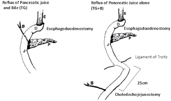
Figure 1: Surgical procedure: TG, TG+B.
Reflux of pancreatic juice and bile (TG): End-to-end esophagoduodenostomy with total gastrectomy (n=8) was performed to produce pancreatic juice and bile reflux. The gastroesophageal junction was ligated and the distal esophagus was transected 2 mm above the ligature. Moreover, the gastroduodenal junction was also ligated, and the proximal duodenum was transected 3mm distal to the pylorus. A total gastrectomy was performed with the removal of the entire stomach, and end-to-end anastomosis of the esophagus and duodenum.
Reflux of pancreatic juice (TG+B): End-to-end esophagoduodenostomy with total gastrectomy. Then, a bypass operation of the upper bile duct was made 25cm below the ligament Treitz to produce only pancreatic reflux. Choledocho-jejunostomy was performed. (n=6) the technique of biliary duct implantation in the small bowel was done as follows. The bile duct was exposed, divided and ligated distally with two 5-0 Vicryl ligatures. A vinyl chloride tube (length 30 mm, external diameter 0.61 mm, internal diameter 0.28 mm, Becton Dickinson and Company) was used as a stent. One end of the tube was inserted into the divided CBD 5mm distal to the hepatic confluence and ligated in place with 5-0 Vicryl (Figure 2A). The other end was inserted into the 18G needle which was inserted through the jejunum from the mesenteric side to the antimesenteric side of the jejunal lumen 25 cm distal to the ligament of Treitz (Figure 2B). After the 18G needle was removed from the jejunum, proximal end of the tube held in place with a purse-string suture with 5-0 nylon, which was ligated with 5-0 Vicryl in common bile duct. (Figure 2C). The ligated CBD tube brought out through the mesenteric side of the jejunum was cut just enough to re-establish bile flow. The cut end spontaneously retracted into the intestinal lumen. And the hole in the jejunum was closed with a stitch using 5-0 nylon (Figure 2D).
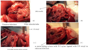
Figure 2: Procedure of choledocho-jejunostomy.
Sham group (n=5): Control Three weeks after operation, all rats were euthanized and the esophagus was evaluated histologically. Esophageal injury was evaluated by macroscopic and microscopic findings. Moreover, at the time of autopsy, blood was drawn for liver function.
Macroscopic examination: The esophagus was opened and gently rinsed with saline. Macroscopic ulcer lesions were scored by a person blind to the treatment as follows: normal glistening mucosal appearance (score 0), edematous mucosa with focal hemorrhagic spots (score 1), multiple erosions with hematin attached (score 2), linear ulcerations with yellowish exudates (score 3) or hemorrhagic coalesced ulcerations (score 4).
Microscopic examination: The entire area of damage was taken, and then fixed in 10% formalin for the histological evaluation. Where a mixed neutrophil/eosinophil inflammatory infiltrate was present, the severity of inflammation was graded as mild, moderate or severs. Sever inflammation denoted extensive ulceration; small foci of ulceration were seen in those graded moderate and no ulceration was present in mild inflammation. Mucosal squamous cell hyperplasia was scored from mild, a slight thickening, through moderate to marked, with a papillary appearance. Serum biological examination At the time of autopsy, blood was drawn from the abdominal aorta, and serum levels of total bilirubin, aspartate aminotransferase (AST), alanine aminotransferase (ALT) and alkaline phosphatase (Alp) were determined using standard auto analyser methods ( Hitachi Auto analyzer 736; Hitachi Tokyo, Japan).
Statistics
Comparisons between groups were made by the Mann-Whitney test. A difference between groups of p<0.05 was considered statistically significant. Statistical analysis was performed using the Stat View 5.0- J program (Abacus Concepts, Berkeley, CA, USA).
Results
Macroscopic finding
Compared to the sham-operated normal group, in TG rats, the esophageal wall was thickened and covered with whitish nodular patches that gave it a cobblestone appearance. Together with the changes, longitudinal ulcerations located primarily in the middle and lower thirds of the esophagus were observed. However, the gross appearance of the esophagus from TG+B group all showed only scattered erosions. The ulcer score in TG+B was significantly lowered compared to TG (Figure 3 and 4).
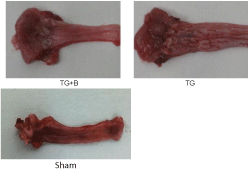
Figure 3: Macroscopic finding.
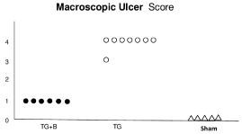
    Figure 4:  Macroscopic ulcer score. 1: edematous mucosa with focal
hemorrhagic spots 2: multiple erosions with hematin attached 3: linear
ulcerations with yellowish exudates 4: hemorrhagic coalesced ulcerations.
Microscopic finding
Compared to the sham-operated normal group, TG group developed large and long longitudinal ulcers; sever infiltration of inflammatory cells and hyperplasia of the epidermis and basal cell in the lower and middle portions of the esophagus. The inflammatory cell infiltration scores and hyperplasia scores were significantly decreased in the TG+B group compared to TG group (Figure 5, 6 and 7).
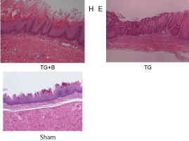
Figure 5: HE microscopic finding.
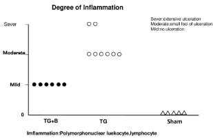
    Figure 6:  Degree of inflammation. Sever: extensive ulceration; Moderate:
small foci of ulceration; Mild: no ulceration.
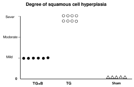
    Figure 7:  Degree of squamous cell hyperplasia Degree of Basal hyperplasia,
mitosis, balloon cells; mild, moderate, sever).
Serum biochemical examination
There is no difference between control, TG+B rat in T-Bil, AST, and ALT. Alp in TG+B rat was significantly higher than that in control (p<0.05) (Table 1).
Discussion
Helsigen [2] performed total gastrectomy and esophagoduodenostomy on rats, examined the esophageal mucosa from postoperative 4 days to 4 months, and reported that the mucosal epithelium was destroyed and inflammation occurred inside the lamina muscularis mucosa relatively early. Alkaline reflux esophagitis is a complication occurring after total gastrectomy. Reflux of the duodenal contents has been reported to contribute to the development of esophageal mucosal damage and inflammation. Esophagitis after total gastrectomy has been associated with biliary and pancreatic reflux into the esophagus. This study is to determine which fraction of the duodenum content reflux, pancreatic juice or bile acids contributes to the development of reflux esophagitis. The significant role of pancreatic juices in the pathogenesis of reflux esophagitis has previously been demonstrated. Trypsin, one of the proteases in the pancreatic juice, has in particular been reported to play an important role in the development of esophageal mucosal injury [3]. In our experiment on rats that underwent total gastrectomy and esophagoduodenostomy, erosion and ulceration of the esophagus were noted 2 weeks after operation. Hyperplasia, ulceration and inflammation of the mucosal epithelium were the histologic features of reflux esophagitis in rats, which were same features of Helsigen’s data. Cam stat mesylate (CMN) has been reported to be a serine protease inhibitor of various proteases, especially trypsin, kallikrein and plasmin. Imada, et al. [4] have performed total gastrectomy with rats, and found that treatment with CMN prevented the development of esophageal mucosal injury. Muds, et al. [3] have reported that the purpose of active trypsin in the esophagus contributes to the development of reflux esophagitis. They clearly showed that neither bile acid nor gastric juice alone induces esophageal mucosal damage, whereas combined with trypsin reflux, they both induce sever esophagitis. Gotley, et al. [5] trypsin activity in the esophageal reflux ate was reported to be associated with development of reflux esophagitis after gastrectomy. These studies clearly indicated that pancreatic trypsin is one of the important factors aggravating esophageal mucosal injury. On the other hand, bile reflux plays an important role in the etiology of reflux esophagitis. In patients with gastroesophageal reflux disease, the concentration of bile acids in the esophageal reflux ate correlates with the degree of esophageal mucosal injury [6]. Nahra, et al. [7] used prolong esophageal aspiration studies, followed by HPLC separation of bile acids for erosive esophagitis. He showed a significantly greater proportion of secondary bile acids, deoxycholic and taurodeoxycholic acids in patients with erosive esophagitis. A few studies [8,9] have attempted to measure the bile acid fractions using esophageal aspiration techniques and also found a predominance of the conjugated bile acids, taurocholic and glycocholic acids, in the esophageal reflux ate. These bile acids were significantly higher in the patients with esophagitis. Kono, et al. [10] investigated the relationship between the severity of esophagitis and gastroduodenal juice reflux, with particular focus on bile acids after distal gastrectomy with Billroth-1 anastomosis. Kono found patients with severe esophagitis had a higher amount of bile acid concentrations in the esophagus compared to patients with mild esophagitis. They advocate bile acids are important factor to contribute to the development of reflux esophagitis. Bili enteric anastomosis in rats is technically difficult, due to the small diameter of the bile duct. Lee [11] had described the method of choledochoduodenostomy in rats for orthotopic liver transplantation. The thread ligating the distal end of the open bile duct was withdrawn along a needle track into the intestinal canal, and was held by fixing the stump of the hepatic artery ligated to the duodenum to minimize tension. We used sutureless anastomosis in a rat experimental model by the modified Lee’s technique. The results of the present study indicate clearly that the sutureless anastomosis in a rat model is reliable and leads to long-term anastomotic patency at 4 weeks after surgery. From the serum biochemical examination, serum bilirubin, AST and ALT in TG+B rat was similar to those of control. Alp is an enzyme in the cells lining the biliary ducts of the liver. Therefore, Alp in TG+B rat was significantly increased compared to that in control due to stent tube. Therefore there is no problem about the bile flow via choledochojejunostomy in TG+B rat. Therefore, TG+B rat is the reflux of pancreatic juice alone to esophagus. In my data, the reflux of pancreatic juice alone is probably not significant development of reflux esophagitis after total gastrectomy compared to the reflux of bile and pancreatic juice.
Conclusion
The reflux of pancreatic juice alone is probably not significant development of reflux esophagitis after total gastrectomy compared to the reflux of bile and pancreatic juice.
References
- Tamura Y, Hirado M, Okamura K, Yoshihiro M, Fujji S. Synthetic inhibitors of trypsin, plasmin, kallikrein, thrombin, Cl r, and Cl esterase. Biochim Biophys Acta. 1977; 484: 417-422.
- Helsingen N. Oesophageal lesions following total gastrectomy in rats.1. Development and nature. Acta Chir Scand. 1960; 118: 202-216.
- Mud HJ, Kranendonk SE, Obertop H, Van HH, WestbroekDL. Active trypsin and reflux esophagitis: an experimental study in rats. Br J Surg. 1982; 69: 269-272.
- Imada T, Chen C, Hatori C Manabu S, Rino Y.Effect of trypsin inhibitor on reflux oesophagitis after total gastrectomy in rats. Eur J Surg. 1999; 165: 1045-1050.
- Gotley DC, Ball DE, Owen RW, Williamson RC, Cooper MJ. Evaluation and surgical correction of esophagitis after partial gastrectomy. Surgery. 1992; 111: 29-36.
- F Zhang, N K Altorki, Y C Wu, Soslow RA, Subbaramaiah K, Dannenberg AJ. Duodenal reflux induces cyclooxygenase-2 in the esophageal mucosa of rats; Evidence for involvement of bile acids. Gastroenterol. 2001; 121: 1391-1399.
- D Nehra, P Howell, C P Williams, JK Pye, Beynon J. Toxic bile acids in gastro-esophageal reflux disease: influence of gastric acidity. Gut. 1999; 44: 598-602.
- Johnsson F, Joelsson B, Floren GH, Nilsson A. Bile salts in the esophagus of patients with esophagitis. Scand J Gastroenterol. 1988; 23: 712-716.
- Iftikhar SY, Ledingham S, Steele RJC, Evens DF, Lendrum K, Atkinson M, et al. Bile reflux in columnar-lined Barrett’s esophagus. Ann R Coll Surg Engl. 1993; 75: 411-416.
- Kono K, Taklahashi A, Sugai H, Lizuka H, Fujji H. Trypsin activity and bile acid concentrations in the esophagus after distal gastrectomy. Digestive Disease Science. 2006; 51: 1159-1164.
- Lee S, Charters AC, Chandler JG, Orloff MJ. A technique for Orthotopic Liver transplantation in the Rat. Transplantation. 1973; 16: 664-669.