Clinical Image
Austin J Gastroenterol. 2017; 4(4): 1090.
A Very Case of Synchronous Gastrointestinal Stromal Tumor (GIST) and Primary Hepatocellular Carcinoma
Ghulam I¹*, Kagan J¹, Ahmad A¹ and Shao C²
¹Department of Pathology, SUNY Downstate Medical Center, NY
²Department of Pathology, Kings County Hospital Center, NY
*Corresponding author: Ghulam Ilyas, Department of Pathology, SUNY Downstate Medical Center, NY
Received: July 24, 2017; Accepted: August 08, 2017; Published: October 20, 2017
Clinical Image
A 67-year-old man presented to the emergency department with epigastric pain and abdominal distension. He had an unintentional weight loss of 10 lb. over the previous 2 months. Socially he endorsed excessive drinking. Physical exam was positive for right upper quadrant tenderness without rebound tenderness. The laboratory data showed hemoglobin (Hb) 9.4 g/dl (Normal 14-18 g/dl) and an elevated alpha fetu protein to 540 ng/ml (normal range < 8.3 ng/ml). The viral serology was negative for hepatitis B and C.
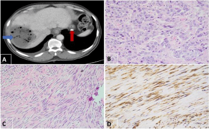
A CT scan was remarkable for a large right lobe hepatic mass (10 cm) (Figure 1A; Blue arrow) with areas of central necrosis and a calcified stomach wall mass (4 cm). (Figure 1A; Red arrow).

Figure 1:
The biopsy of the liver mass showed moderately differentiated hepatocellular carcinoma with large tumor cells, eosinophilic cytoplasm and distinct nucleoli. (Figure 1B) The cells were diffusely positive for Hep-par1 immunostain. (Figure 1B; inset) The biopsy of the stomach mass revealed predominantlyspindled cells with elongated nuclei that displayed blunt to pointed ends and occasional perinuclear vacuoles. The nuclei were focally palisaded. There was no necrosis and no mitotic figures. (Figure 1C) By immunohistochemistry, this neoplasm was immunoreactive for CD117 (Figure 1D) but negative for S100-protein.
Gastrointestinal stromal tumors (GISTs) are mesenchymal tumors that arise from the interstitial cells of Cajal or their derivatives, which are driven primarily by KIT and to a lesser degree PDGFRA mutations. The presence of mutations in KIT is pertinent because it enables GISTs to be treated with targeted tyrosine kinase inhibitors, such as imatinib mesylate [1].
The co-occurrence of a GIST and a primary hepatocellular carcinoma is a rarely documented event. Very few cases have been reported in the literature. As the management of a coexisting GIST with HCC versus a metastatic GIST would be quite different, an accurate diagnosis with histopathological examination must be performed. Metastatic hepatic lesions resulting from a GIST are treated with Tyrosine kinase inhibitors while the primary hepatocellular carcinomas are inherently refractory to such therapies [2].
References
- Miettinen M, Lasota J. Gastrointestinal stromal tumors review on morphology, molecular pathology, prognosis, and differential diagnosis. Arch Pathol Lab Med. 2006; 130: 1466-1478.
- RadoslawJaworski, Jastrzebski T, Swierblewski M, Drucis K, and Gulida GK. Coexistence of hepatocellular carcinoma and gastrointestinal stromal tumor: A case report. World J Gastroenterol. 2006; 12: 665-667.