Research Article
Austin J Gastroenterol. 2018; 5(1): 1094.
Gastrointestinal Tract Injury and Clinical Characteristics in 172 Children with Henoch-Schonlein Purpura Checked by Gastroscope
Zeng H*, Wang J and Li H
¹Department of Pediatric Allergy, Guangzhou Medical University, China
²Department of Pediatric Immunoloy, Guangzhou Medical University, China
³Department of Pediatric Rheumatology, Guangzhou Medical University, China
*Corresponding author: Zeng H, Pediatric Allergy, Guangzhou Women and Children’s Medical Center, Guangzhou Medical University, China
Received: February 18, 2018; Accepted: April 04, 2018; Published: April 25, 2018
Abstract
Objective: To investigate gastroscopic features and explore the relationship between clinical characteristics and Gastrointestinal tract injury of Henoch- Schonlein puepura (HSP) in children.
Methods: 172 cases of children with HSP in our medical center were checked by gastroscope, the gastrointestinal tract injury feature was summarized. All the case were divided into two groups by gastroduodenal mucosal bleeding or not. It was compared among the total time of abdominal pain, pain remission time, hospitalization time, fasting time and kidney injury cases were analysis in two groups. Results: Gastroscopic mainly revealed gastroduodenal mucosal congestion, edema, rough, erosion, bleeding and ulcer which involved 148 cases of gastric (86.0%), 158 cases of duodenal involvement (91.9%). Mucosal erosion and bleeding occurs mainly in duodenum, mostly in the descending duodenum. Duodenal bleeding accounted for 36 cases (20.9%) in the bulb and 92 cases (53.5%) in the descendant. Only five cases (2.9%) of ulcer occurred in the duodenum, where four cases of bulbar ulcer, one case of descending ulcer. Esophageal and gastric cardia mucosal just occurred in 1 case.
Conclusion: Gastroscopic features of HSP in children are characterized by bleeding, erosion of duodenal mucosa and occasional duodenal ulcer formation, which mostly involve the antral mucosa, rarely involving the esophagus, cardia. The HSP patients with gastrointestinal symptoms should be checked by gastroscope. It is important to make a right diagnosis for pediatric HSP especially to atypical cases.
Keywords: Gastrointestinal tract injury; Henoch-Schonlein puepura; Pediatric allergy
Introduction
HSP is a common IgA-mediated systemic vasculitis in children, which pathological changes are wide range of leukocytoclastic vasculitis, mainly in the capillaries, seldom involving venules and small arteries. Lesions can affect the skin, kidneys, joints, and gastrointestinal tract. Diagnosis of HSP relies on the typical rash. But it is certain difficult to diagnose some of the early stages of HSP in children with abdominal pain or gastrointestinal bleeding as the first symptom of existence [1], so prone to misdiagnosis and for the right treatment, this article aims to summarize gastroscopic feature of HSP in children and make some help for the assessment and treatment of the disease by exploring the correlation between the clinical characteristic and the degree to the injury of gastrointestinal mucosa [2,3].
Materials and Methods
Materials
172 pediatric cases with HSP hospitalized in our Pediatric Allergy, Immunology and Rheumatoloy Department in recent 9 months were explored, including male 104 cases (60.0%), female 68 cases (40.0%), whom ages are 2-12 (6.1 ± 2.3) years. 172 cases in children had abdominal pain, including 73 cases (42.4%) with rash as the first symptom, 57 cases (33.1%) with abdominal pain as the first symptom, 42 cases (24.4%) with abdominal pain and rash on the same day, 11 cases (6.4%) without rash, 6 cases (3.5%) without abdominal pain and 20 cases (11.6%) of kidney injury occurred during hospitalization. All cases were diagnosed HSP and finally cured after treatment in our department. Depending on gastroduodenal mucosal bleeding or not, 172 cases were divided into two groups o, one is severity of mucosal injury group, another is light mucosal injury group. These two groups had no significant differences (P> 0.05) in age and gender.
Method
172 cases were checked with gastroscope Electronics (OLYMPUS PCF-Q260JI), to observe the color of mucosal membrane from gastric to descending duodenum, the mucosal congestion, edema, rough, erosions, ulcers and bleeding.
According to the results of gastroduodenal mucosal bleeding or not, the 172 cases were divided into light (no bleeding) and heavy groups (bleeding). Total time of abdominal pain (days), abdominal pain, hospitalization time (days), fasting time (days) and kidney injury cases were record.
SPSS20.0 statistical software was used to analyses .T test, Χ² test were used, P <0.05 was considered statistically significant.
Results
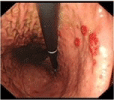
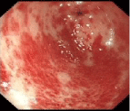
There were 169 cases (98.3%) in total 172 cases were found the gastrointestinal tract injury, those cases show that gastroduodenal mucosa with varying degrees of injury. The gastroscopic feature is mainly characterized by gastroduodenal mucosal congestion, edema, rough, erosion, bleeding and ulcer. The gastroscopic characteristic performed spotted rash-like hemorrhagic mucosa (Figure 1), parts of rash integrate into the mucosal surface (Figure 2), raising on the mucosal surface (Figure 3), which can be combined superficial erosion bleeding. In severe cases, ulcers with yellow moss surface were formed (Figure 4). Distribution of 172 cases of children with gastroscopic mucosal changes shown in Table 1.
    Mucosal manifestations
    Esophagus
    Cardia
    Gastric body
    Antral
    Pylorus
    Bulbar
    Descending
    Total


    Congestion
    1
    1
    115
    152
    1
    151
    144
    565


    Edema
    0
    0
    14
    21
    1
    16
    81
    133


    Coarse
    0
    0
    79
    76
    0
    98
    13
    266


    Erosion
    0
    0
    2
    7
    0
    63
    101
    173


    Ulcer
    0
    0
    0
    0
    0
    4
    1
    5


    Bleeding
    0
    0
    12
    24
    1
    36
    92
    165


    Total
    1
    1
    222
    280
    3
    368
    432
     -
Table 1: Distribution of 172 cases of children with gastroscopic mucosal changes.
As Table 1 shows, the mucosal injury distribute in each segment of gastro duodenum, mainly involving in duodenal mucosa .Gastric involvement is also common, which the frequency and severe degree are no more than and the duodenal mucosa. There were 148 cases (86.0%) had gastric mucosal involvement, performing for congestion and edema, mucosal rough, but it lack of specificity. Parts of the cases can be performed as a typical spotted rash-like changes in the mucous membranes, and even merge erosion, bleeding. Typical duodenal mucosal involvement showed in 158 cases (91.9%). Mucosal erosion and bleeding occurs mainly in the duodenum specially descending part. The mucosal bleeding of duodenal bulk and descending showed in 36 cases (20.9%), and 92 cases (53.5%) respectively. There were low incidence of ulcers, 5 patients (2.9%) were found ulcer in the duodenum, inducing 4 cases of bulk ulcer, and 1 case of gastric descending, rarely involving the pylorus, esophagus and cardia mucosa. There are 3 cases of pyloric involvement and only 1 case of esophagus and cardia mucosal involvement.

Figure 3: Fundus.

Figure 4: Descending.
Clinical features of two groups are compared. All 172 cases including 73 cases (42.4%) with rash as the first symptom, 57 cases (33.1%) with abdominal pain as the first symptom which time difference was 7.27 ± 7.14 days. There are 42 cases (24.4%) with abdominal pain and rash on the same day, 11 cases (6.4%) without rash, 6 cases (3.5%) without pain during the course. The no painful cases had gastric mucosal injury, including 2 cases of acute hemorrhagic duodenitis, 2 cases of erosive duodenitis which are typical mucosal changes of HSP, and the other 2 cases showed non-typical gastric mucosal inflammation. Because abdominal pain cannot remission, 2 cases were done abdominal surgery treatment in rural hospitals which made the wrong diagnosis as acute abdominal surgical diseases, then transferred to the pediatric surgery department of our medical center. There were total 8 patients with abdominal pain as the first symptom, who were the first diagnosis of acute abdomen and sent to surgery department in our medical center from rural hospitals, were finally made right diagnosis and transferred to our department. 11 cases had not rash during the course, but the gastroscope showed bleeding duodenal inflammation and gastrointestinal mucosal changes as HSP. During the hospitalization of 172 cases, 20 cases (11.6%) were diagnosed as nephritis. There were no significant difference (P> 0.05) in the total time of abdominal pain (days), abdominal pain remission time (days) after treatment, hospitalization (days), fasting time (days) and the incidence of kidney injury between two groups of patients Table 2.
    Groups
    Abdominal pain remission time (days) after treatment
    Total time pain(days)
    The days in hospital
    Fasting Days
    The incidence of nephritis


    Light group (n = 59)
    4.10 ± 4.97
    13.39 ± 12.10
    10.07 ± 5.32
    1.67 ± 2.19
    6 (10.2%)


    Severe group (n = 113)
    4.81 ± 5.07
    15.91 ± 23.01
    10.90 ± 5.36
    2.22 ± 2.10
    14 (12.4%)


    T
    0.87
    0.76
    0.98
    1.62


    ? 2
    -1.259
    -1.053
    -1.048
    -2.343
    0.19


    P
    > 0.05
    > 0.05
    > 0.05
    > 0.05
    > 0.05
Table 2: Comparison of the clinical characteristics of the two groups.
Discussion
HSP is a common childhood systemic vasculitis, mainly affects the small blood vessels, which is the most common type of vasculitis syndrome [1-3]. Most of children have a good prognosis and can be cured by clearing infection, anti-allergy, reducing vascular permeability and other symptomatic treatment. There were about 58% of the patients had abdominal pain [4]. In this study, most patients performed abdominal pain, part of the cases even appeared repeatedly severe abdominal pain, vomiting, gastrointestinal bleeding or even intussusceptions and other surgical cases. According to EULAR/PRINTO/PRES HSP diagnostic criteria [1], skin purpura is essential conditions of HSP diagnosis. However, we found some cases of HSP in children with neither part of the skin rashes in the whole course of the diseases. It is not difficult for most of the children cases to be diagnosed right because of typical rashes, but more difficult for the cases with non-typical rash or abdominal pain as the first symptom or even no rashes.
There are some different between adult and children in HSP cases [5]. In this study, there were 57 patients (33.1%) with abdominal pain as the first symptom, 11 patients (6.4%) without rash during the whole course. The gastroscopic findings showed that gastroduodenal mucosa with varying degrees of injury in 169 cases (98.3%) of total 172 cases. The gastroscopic features are mainly characterized by gastroduodenal mucosal congestion, edema, rough, erosion, bleeding and ulcer. The gastroscopic characteristic showed spotted rash-like hemorrhagic mucosa (Figure 1), parts of rash integrate into the film (Figure 2) raising on the mucosal surface (Figure 3), which can be combined superficial erosion bleeding. In severe cases, ulcers with yellow moss surface were formed. It is still visible spotted like congestion and hemorrhage between mucosal ulcers, which is similar to the skin purpura change. Typical lesions are more concentrated in antral and duodenal mucosa. Ulcer can only be found in duodenum. 11 cases with no rashes were found with typical gastroscopic mucosal changes, and more manifestations of acute hemorrhagic erosive duodenitis, including 1 case of multiple visible duodenal ulcer formation. All 11 cases were the clinical diagnosis of HSP, and given anti-infection, anti-allergy, acid suppression, protecting the gastric mucosa, reducing vascular permeability and glucocorticoid therapy. All 11 patients without rashes were cured. Therefore, we believe that in recent years there is a growing trend of non-typical HSP, such as have abdominal pain but without rashes. The possibility of HSP should be considered if this situation happens; gastroscope should be checked up quickly .it can be considered to diagnosis of HSP with typical mucosal changes under gastroscope, combining with other clinical and laboratory test.

Figure 1: Gastric Antra.

Figure 2: Gastric Antra.
Gastroscope contributes to the diagnosis of HSP [6], and can help determine the severity of gastrointestinal mucosa. The study also found that the severity of gastroscope have no significant correlation with clinical manifestations of patients. There was no significant difference in total time of abdominal pain, pain remission time after treatment, fasting time, hospitalization time between the light group without bleeding and the heavy group with bleeding. Maybe it need improved the research numbers.
In this study, there were 6 patients (3.5%) with no abdominal pain but showed gastroscopy mucosal injury, including 2 cases presented with acute hemorrhagic duodenitis, 2 cases of erosive duodenitis, all of that are typical mucosal changes of HSP . Those patients without abdominal pain may still exist serious gastrointestinal mucosal injury; they need a stronger and longer treatment.
Although gastrointestinal symptoms of HSP is more serious, but according to this statistics, 172 cases of patients were improved or cured, which suggest good prognosis of pediatric HSP with gastrointestinal symptoms. The right diagnosis, treatment and management are important [7,8]. The most important indicator of long-term prognosis depends on injury of kidneys [9]. In this study, 20 (11.6%) cases had kidney injury during the hospitalization. There was no significant difference in the incidence of kidney injury between the two groups which suggesting the severity of gastrointestinal tract lesions cannot be used as the indicators of recent kidney injury. But it remains to be further followed up to determine whether the severity of gastrointestinal tract lesions can be long-term indicators of renal injury by more cases study and following up.
In summary, the HSP patients with gastrointestinal symptoms should take gastroscope as soon as possible, it is important to make a right diagnosis for pediatric HSP especially to atypical cases. To understand the range and extent of gastrointestinal mucosal lesions is benefit to instructing for early diagnosis, assessment and treatment of HSP. The extent and time of abdominal pain cannot be used alone as the indicators of judging the severity of gastrointestinal injury in pediatric HSP.
Acknowledgement
Guangdong Natural Science Foundation, Guangzhou Municipal Science and Technology Innovation Committee, China, NO.201607010316.
References
- Ozen S, Pistorio A, Iusan SM, Bakkaloglu A, Herlin T, Brik R, et al. EULAR/PRINTO/PRES criteria for Henoch–Schönleinpurpura, childhood polyarteritisnodosa, childhood Wegener granulomatosis and childhood Takayasu arteritis: Ankara 2008. Part II: Final classification criteria. Ann Rheum Dis. 2010; 69: 798-806.
- Prathiba Rajalakshmi P, Srinivasan K. Gastrointestinal manifestations of Henoch-Schonlein purpura: A report of two cases. World J Radiol. 2015; 7: 66-69.
- Waelkens B, Van Moerkercke W, Lerut E, Sprangers B. Henoch-Schönlein Purpura complicated with major gastro-intestinal hemorrhage. Acta Gastroenterol Belg. 2014; 77: 379-382.
- Roberts PF, Waller TA, Brinker TM, Riffe IZ, Sayre JW, Bratton RL. Henoch-Schönlein purpura:a review article. South Med J. 2007; 100: 821-824.
- Gupta V, Aggarwal A, Gupta R, Chandra Chowdhury A, Agarwal V, Lawrence A, et al. Differences between adult and pediatric onset Henoch-Schonlein purpura from North India. Int J Rheum Dis. 2018; 21: 292-298.
- Nishiyama R, Nakajima N, Ogihara A, Oota S, Kobayashi S, Yokoyama K, et al. Endoscope images of Schönlein-Henoch purpura. Digestion. 2008; 77: 236-241.
- Hetland LE, Susrud KS, Lindahl KH, Bygum A. Henoch-Schönlein Purpura: A Literature Review. Acta Derm Venereol. 2017; 97: 1160-1166.
- Audemard-Verger A, Terrier B, Dechartres A, Chanal J, Amoura Z, Le Gouellec N, et al. Characteristics and Management of IgA Vasculitis (Henoch-Schönlein) in Adults: Data From 260 Patients Included in a French Multicenter Retrospective Survey. Arthritis Rheumatol. 2017; 69: 1862-1870.
- Buscatti IM, Casella BB, Aikawa NE, Watanabe A, Farhat SCL, Campos LMA, et al. Henoch-Schönlein purpura nephritis: initial risk factors and outcomes in a Latin American tertiary center. Clin Rheumatol. 2018.