
Clinical Image
Austin J Gastroenterol. 2020; 7(1): 1106.
Metastatic Melanoma in the Gastric Body
Borahma M*, Benelbarhdadi I, Berhili C, Lagdali N, Ajana FZ
Department of Gastroenterology C, Mohammed the Vth University, Ibn Sina Hospital, Rabat, Morocco
*Corresponding author: Borahma Mohamed, Department of Gastroenterology C, Mohammed the Vth University, Ibn Sina Hospital, Rabat, Morocco
Received: January 20, 2020; Accepted: February 06, 2020; Published: February 13, 2020
Clinical Image
A 65 year-old women presented with two months history of abdominal pain and obstructive jaundice. Fifteen years earlier, she had undergone malignant melanoma excision of the face. A computed tomographic scan showed Intrabiliary tree dilatation related to a nodular lesion (6cm of diameter) at the liver anterior segment. An ultrasonography-guided biopsy was carried out and the histological report confirmed the diagnosis of malignant melanoma metastasis. An Endoscopic Retrograde Cholangiopancreatography (ERCP) was performed for biliary stenting, during duodenoscopy, we found an umbilicate vegetating ulcerating lesion on the posterior wall of gastric body. The histological study of the vegetating lesion revealed gastric malignant melanoma metastasis. Patient transferred to oncology department for chemotherapy. Melanoma is an unusual predilection to metastasize to the gastrointestinal tract. Only 1-5% cases of metastatic melanoma [1] are diagnosed during life because they are generally clinically asymptomatic in the early stages. Diagnosis often takes place when an emergency complication occurs, such as intestinal perforation, obstruction, and acute gastrointestinal bleeding, or with non-specific symptoms like anaemia, weight loss, abdominal pain [2,3] (Figure 1).
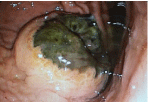
    Figure 1: Gastric Melanoma Metastasis: Black pigmented tumor with
ulceration in the gastric body.
References
- Ihde JK, Coit DG. Melanoma metastatic to stomach, small bowel, or colon. Am J Surg. 1991; 162: 208-211.
- Patti R, Cacciatori M, Guercio G, Territo V, Di Vita G. Intestinal melanoma: a broad spectrum of clinical presentation. Int J Surg Case Rep. 2012; 3: 395- 398.
- Klausner JM, Skornick Y, Lelcuk S, Baratz M, Merhav A. Acute complications of metastatic melanoma to the gastrointestinal tract. Br J Surg. 1982; 69:195- 196.