
Case Report
Gerontol Geriatr Res. 2019; 5(1): 1038.
The Effect of Whole Body Periodic Acceleration Combined with Anticoagulant on Lower Extremity Deep Venous Thrombosis
Luo Q1*, Tan J2*, Li G1, Zhuang B1, Luo L1, Huang S1, Ni Y1, Wang L1, Gong K3 and Shen Y1*
¹Departments of Cardiology, Tongji Hospital Affiliated to Tongji University, PR China
²Department of Sport Rehabilitation, Shanghai University of Sport, PR China
³Departments of Vascular surgery, Tongji Hospital Affiliated to Tongji University, PR China
*Corresponding author: Yuqin Shen, Qian Luo and Jingwang Tan, Department of Cardiology, Tongji Hospital Affiliated to Tongji University, 389 Xincun Road, Shanghai 200065, PR China
Received: April 22, 2019; Accepted: June 08, 2019; Published: June 15, 2019
Abstract
Deep Venous Thrombosis (DVT) is a life-threatening disease caused by a blood clot in a deep vein, the symptom of which may include swelling, pain, and tenderness, often in the legs. This study is the first report on the effect of whole body periodic acceleration on leg swelling caused by DVT. We report on a 72-year-old man with edema of lower extremity, who was diagnosed with thrombosis from left femoral vein to the left iliac vein by angiography and treated with surgery, including lower extremity venous filter implantation, iliac vein balloon dilatation and stent implantation. Meanwhile, he received long-term oral warfarin anticoagulation therapy for 40 days. During this period, the International Normal Ratio (INR) was maintained between 2.0 and 3.0, but the symptom of edema did not vanish. After 43 days’ treatment with warfarin combined with WBPA, the edema obviously subsided and the disappearance of left lower limbs DVT was confirmed by B-mode ultrasonography. Current study suggests that WBPA has positive effects on the treatment of edema caused by DVT.
Keywords: Whole body periodic acceleration; pGz; Anticoagulant; Deep venous thrombosis; DVT
Case Presentation
A 72-year-old man presented to the emergency room of Tongji Hospital affiliated to Tongji University, China, with a 1-day history of continuous pain and pitting edema extending from the root of left leg to the ankle. The patient reported no pain and oppression in chest, shortness of breath, hemoptysis, palpitation, and lipothymia. He had a remarkable medical history including 7-year Parkinson Disease, 3-year Type-2 diabetes mellitus and chronic nephropathy. He denied the history of tobacco, alcohol, illicit drugs, long distance aviation travel, family medical, infectious diseases and Deep Venous Thrombosis (DVT). Nevertheless, the patient admitted the history of sedentary behaviors due to the habit of playing Mahjong.
His vital signs: temperature 37°C, blood pressure 76/50mmHg, heart rate 80beats/min and respiratory rate 18breaths/min. The patient’s left leg was markedly red and swollen with edema extending from the groin to the ankle while the right leg was unaffected (Figure 1). Moreover, the left leg had no cyanosis and eczema-like changes in the skin. Dorsalis pedis arteries were symmetrical and could be normally palpated. Sensory and motor on the toes were normal. Homans’ sign and Neuhofs sign were both negative.
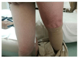
    Figure 1: Edema and erythema condition of left extremity and the normal
right leg.
Laboratory tests detected Mild anemia (hemoglobin 118g/L, red blood cell 3.82*10^12/L), mildly elevated C-reactive protein (13.62mg/L), dyslipidemia (TG 2.0mmol/L, HDL-C 0.49mmol/L), poor control of blood sugar (glycated albumin 20.15%; glycated hemoglobin 7.4%), and renal insufficiency (blood urine nitrogen 12.63mmo/L, creatinine 168ummol/L, epidermal growth factor receptor 35.011ml/min/1.73m2). Function of liver, as well as coagulation, sodium, potassium, chloride, and myocardial enzymes showed no specific results.
Ultrasonic examination showed that embolisms were found in the deep vein of left lower extremity (Figure 2). Enhanced-CT of hypogastrium presented non-uniform density of left external iliac vein and vascular sclerosis in abdominal aorta and its branches (Figure 3). Besides, angiography in lower extremity showed the thrombosis from left femoral vein to the left iliac vein.
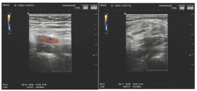
Figure 2: Angiography results in lower extremity.
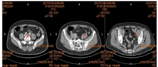
Figure 3: Enhanced-CT images of hypogastrium in the abdomen area.
In terms of the vascular thrombosis, surgery procedure was done as lower extremity venous filter plus iliac vein balloon dilatation plus stent implantation. After 7 days of anticoagulation using lowmolecular- weight heparin, the patient was discharged on August 5th and anticoagulation was continued with warfarin outside the hospital. After 40 days, this patient returned to the emergency room due to the problem of leg edema. Without any efficacy on low extremity edema using the therapy of warfarin, the patient was recommended to receive a therapy of whole body periodic acceleration (BodyGreenMB1, Taiwan, 20mm) on 8th September (Figure 4). To quantify the changes of the leg with edema during the WBPA period, measures of thigh circumference was done by therapist and the measurement point was positioned at 10cm above the left patella with a plastic tape measure. The WBPA treatment process was divided into two stages: Stage 1: 8th September to 27th September, 1time/day, 30min/time; 60cycles/min (frequency of the device). Stage 2: 27th September to 20th October, 1time/day, 30min/time,75cycles/min. Because of the long distance from home to hospital and the improvement of edema symptoms, the patient terminated the treatment and was followed up by therapist instead. During the intervention, there was no side effect and compliance of the patient is fine.

    Figure 4: Whole body periodic acceleration facility (BodyGreen MB1,
TaiWan).
After the treatment, it could be clearly seen that the edema and erythema vanish (Figure 5) compared with the symptoms presented before. Moreover, the thigh circumference has got back to the normal level and two legs were generally symmetrical. Figure 6 showed the exact change of thigh and calf circumference before and after the WBPA treatment. It could be easily seen that the numbers gradually decrease during the intervention period. Re-examination of ultrasonography showed the disappearance of deep venous thrombosis in lower extremities (Figure 7).
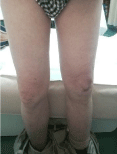
Figure 5: The condition of left and right leg after intervention.
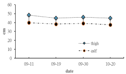
Figure 6: Circumference changesof thigh and calf.
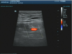
Figure 7: Angiography results in lower extremity.
Discussion
To our knowledge, this study is the first case report presenting the effect of whole body periodic acceleration on leg swelling caused by DVT. The results show that WBPA is effective in the treatment of the leg DVT.
DVT in lower extremities is mainly caused by blood stasis and hypercoagulability. It is more common in unilateral lower extremity and left-leg DVT is 3 to 8 times more common than right-leg DVT. Common causes include surgery, tumors, long-term bed rest, etc. DVT can easily lead to venous reflux obstruction, interstitial fluid deposition and in return cause swelling. Despite adequate anticoagulation, most iliofemoral thromboses do not completely re-canalize, resulting in post-thrombotic syndrome. Therefore, endovascular treatment is as an effective treatment for acute iliofemoral DVT.
After admission, the patient was diagnosed with femoral veiniliac deep vein thrombosis in the left lower extremity by B-mode ultrasonography and angiography. Facing this condition, surgery was done and certain tools were implanted to solve the acute problem. After discharge, the patient received long-term oral warfarin anticoagulation therapy for more than 1 month. During this period, the International Ratio (INR) was maintained between 2.0 and 3.0, but the symptom of edema did not vanish. After another month’s treatment with warfarin combined with WBPA, the edema obviously subsided and the disappearance of left lower limbs DVT was confirmed by B-mode ultrasonography.
Without former similar studies to refer to, mechanisms for the treatment effect may be speculated as follows. To begin with, thrombosis is likely to be eliminated based on the improvement of the function of fibrinolysis system brought by WBPA. Normally, plasminogen in plasma is inactive unless it is converted into the plasmin through Plasminogen Activator (PA). PA is an enzyme existing in blood, vascular endothelial cells, and many tissues, released in the condition of local injury, thrombosis, etc. Among activators, the most important one is tissue Plasminogen Activator (tPA) derived from vascular endothelial cells. Researches done by Adams [1] and Fujita [2] showed that WBPA is able to promote the release of vasoactive factor NO and tPA, which may promote the elimination of thrombosis. Apart from this mechanism, another one concerning the vanish of thrombosis is more likely to be related to the improved blood flow velocity, shear stress of vascular endothelium caused by WBPA [3].
The second possible mechanism may lie in the improvement of hemodynamics by WBPA. Previous studies [4,5] have shown that WBPA is capable of improving blood flow in peripheral artery, increasing organ blood perfusion. What is more, Matsumoto [6] suggests that WBPA can improve brachial artery endothelial function, increase vascular diameter and improve blood flowmediated vasodilation function. On the basis of this, it is possible for WBPA to promote the increase of venous reflux velocity of lower limbs and change blood stasis, contributing to the disappearance of edema through reducing venous and osmotic pressure.
However, since the absence of formerly reported studies, whether WBPA combined with anticoagulant of warfarin generates synergistic effect remain unclear.
Conclusion
The current study suggests that WBPA has positive effects on the treatment of edema caused by DVT. The possible mechanisms by which pGz induces the elimination of leg edema are more likely related to the changes of blood flow and endothelium function. It is also likely that some or all of these factors synergize to cause the treatment effects. Notwithstanding, the exact mechanism of this case need further investigation. This study provides a novel way for further studies to take advantage of pGz on leg edema caused by DVT.
Funding
This report was supported by grants from the National Natural Science Fund (No.81570359) and from Shanghai Municipal Commission of Health and Family Planning (No.2015ZB0502).
References
- Adams JA, Bassuk J, Wu D, Grana M, Kurlansky P, Sackner MA. Periodic acceleration: effects on vasoactive, fibrinolytic, and coagulation factors. Journal of Applied Physiology. 2005; 98: 1083.
- Fujita M, Tambara K, Ikemoto M, Sakamoto S, Ogai A, Kitakaze M, et al. Periodic Acceleration Enhances Release of Nitric Oxide in Healthy Adults. International Journal of Angiology. 2005; 14: 11-14.
- Sackner MA, Gummels EM, Adams JA. Nitric Oxide Is Released Into Circulation with Whole-Body, Periodic Acceleration. Chest. 2005; 127: 30-39.
- Adams JA, Mangino MJ, Bassuk J, Kurlansky P, Sackner MA. Regional blood flow during periodic acceleration. Critical Care Medicine. 2001; 29: 1983.
- Rokutanda T, Izumiya Y, Miura M, Fukuda S, Shimada K, Izumi Y, et al. Passive exercise using whole-body periodic acceleration enhances blood supply to ischemic hind limb. Arterioscler Thromb Vasc Biol. 2011; 31: 2872- 2880.
- Matsumoto T, Fujita M, Tarutani Y, Yamane T, Takashima H, Nakae I, et al. Whole-body periodic acceleration enhances brachial endothelial function. Circulation Journal Official Journal of the Japanese Circulation Society. 2008; 72: 139.