
Keywords
Hepatic adenomatosis; Adenomas; Liver transplantation
Clinical Image
Hepatic adenomatosis was first described in 1985 by Flejou as multiple adenomas, arbitrarily more than 10, in an otherwise normal liver parenchyma.
Several authors have suggested that it is a distinct entity from hepatic adenoma, which is predominantly seen in young women taking oral contraceptives [1].
The definitive treatment is still controversial; therapeutic options include clinical follow-up, liver resection, and liver transplantation [2]: it consists of close monitoring and imaging, resection of superficially located, large (4cm) or growing liver cell adenomas. Liver transplantation is the last resort in case of substantive concern about malignant transformation or for large, painful adenomas in liver cell adenomatosis after treatment attempts by liver resection [3].
We present the case of a 55-year-old female with no medical history of oral contraception who was under treatment for a pleural tuberculosis during which an abdominal ultrasound was made and has showed multiple diffuse small nodules. The patient had no abdominal symptomatology. Liver function tests were normal. The study was completed with CT and later with MRI which established the diagnosis of hepatic adenomatosis. Several hepatic nodules and masses were detected whose appearance would be rather evocative of hepatocellular lesions (multiple adenomas), the two largest of which were in V and VII segments measuring each 47 mm (Figure 1).
Percutaneous biopsy confirmed the diagnosis and showed no malignancy. The preserved parenchyma presented no disease.
We underwent resection of the 2 largest lesions like recommended [3] by performing a V and VII segmentectomies (Figure 2).
The postoperative course was uneventful and patient was discharged on postoperative day 5.
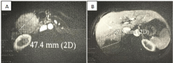
Figure 1: MRI: the two largest adenomas in segments V (A) and VII (B).
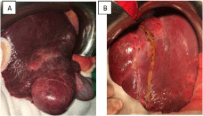
    Figure 2:  Preoperative view of the two largest adenomas in segments V (A)
and VII (B).
References
- Wesley OC, Bhattacharya B. Hepatic Adenomatosis. Arch Pathol Lab Med. 2008; 132: 1951-1955.
- Lucas E, Pareja E, Carvajal N, Pacheco A, Moya A. Hepatica adenomatosis: a disease of controversial treatment. Cir Esp. 2014; 92: 284-286.
- Ludger Barthelmes, Iain S. Tait. Liver cell adenoma and liver cell adenomatosis. HPB. 2005; 7: 186-196.