Abstract
Candida infection of gastrointestinal tract is frequent in immunocompromised patients, but rare in an otherwise healthy person. In the gastrointestinal tract, Candida frequently involves the oesophagus, followed by stomach and small bowel. Candida has been shown to colonize gastric ulcers, however its role in delaying gastric ulcer healing is controversial. We report a case of 45 years old Immunocompetent female who presented with non-steroidal anti-inflammatory drugs induced gastric ulcer, which did not heal despite taking proton pump inhibitors for 6 months. Exfoliative cytology obtained from the edge of the ulcer revealed spores and budding yeast forms of Candida species, and biopsy showed no evidence of malignancy or Helicobacter pylori. Anti-fungal treatment leads to healing of the gastric ulcer. This case highlights the fact that Candidiasis should be considered as a cause of non-healing benign gastric ulcer even in an immunocompetent host.
Keywords: Candida; Gastric ulcer; NSAIDS; Immunocompetent host
Introduction
Candida is a normal commensal of Gastro Intestinal (GI) tract but is infrequently isolated from healthy individuals. Candida infection of the GI tract is usually seen in immunocompromised hosts, though it has been reported in apparently healthy individuals also [1]. Oesophagus is the most commonly involved organ, followed by stomach and small bowel [2,3]. Gastric candidiasis has been classified into thrush, nodular and ulcerated types [2,3]. Large polypoidal growths of Candida in stomach called as yeast bezoars have also been described [4]. Candida-associated gastric ulcers have been reported in various studies [5]. However, the clinical significance of Candidaassociated gastric ulcer, its natural history and the need for antifungal treatment remain to be defined [6-8]. We describe a case of a middle-aged Immunocompetent female with Candida-associated non-healing gastric ulcer.
Case Report
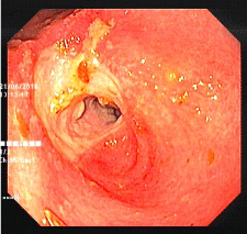
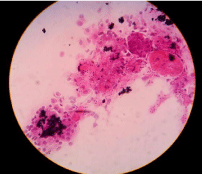
A 45-year-old female was admitted to our hospital with complaints of persistent epigastric pain and vomiting for one-month duration. There was history of significant weight loss (15 kg) in the last 10 months. There was history of misuse of NSAIDS (for joint pains) for 3 years, which she had stopped 6 months back. There was no history of GI bleed, corrosive intake, alcohol intake, smoking or any other addiction. An upper GI endoscopy performed 6 months back was suggestive of pyloric channel ulcer. Ulcer biopsy was negative for dysplasia and H. pylori. Patient was prescribed tablet Pantoprazole 40 mg twice a day. However, the patient had gradual worsening of symptoms since last one month. On general physical examination, she was grossly malnourished with B.M.I. of 16.5 kg/m2. Investigations revealed hemoglobin 9.6 g/L (microcytic hypochromic anemia), total leukocyte count 4.6 x 10⁹/L (polymorphonuclear leukocytes 69%, lymphocytes 23%, eosinophils 0%, and monocytes 7%), platelets 228 x 109/L and prothrombin time 14 s (control 13 s). Liver function tests revealed serum bilirubin 0.40 mg/dL, alanine and aspartate aminotransferase 20 and 31 U/L respectively, alkaline phosphatase 102 U/L, and total protein and albumin 37 and 17 g/L respectively. Serum creatinine was 1.05 mg/dL, postprandial blood sugar was 78 mg/dL, potassium was 2.36mEq/L, and sodium was 136 mEq/L. Chest radiography was normal. Enzyme-linked immunosorbent assay for human immunodeficiency virus was negative. Upper GI endoscopic examination showed a circumferential ulcer at pylorus extending into first part of duodenum (Figure 1). Exfoliative cytology obtained from the edge of the ulcer revealed fungal spores and budding yeast forms of Candida species (Figure 2). Biopsy from the ulcer showed mucosal ulceration with acute and chronic inflammatory infiltrate along with fibrosis with no evidence of Helicobacter pylori (H. pylori) infection. Patient was treated with fluconazole 200 mg once a day for 2 weeks. After one month of follow-up, the patient was asymptomatic, and repeat upper GI endoscopy showed a small healing clean based ulcer in the pyloric channel.

    Figure 1:  Upper GI endoscopic image showing circumferentia ulcer at
pylorus extending into first part of duodenum.

    Figure 2:  Exfoliative cytology from the edge of the ulcer revealing fungal
spores and budding yeast forms of candida species.
Discussion
Candida is a ubiquitous fungus, found even in the gut of healthy individuals. In a study by Ears et al., gut mycosis was observed in 109 (4.35%) of the 2517 total cases, which were studied from 1960–1964. Tsukamoto et al. from Japan reported that gut mycosis was present in 196 (5.9%) of the 3,339 cases which were reported from 1971 to 1983 [2]. In these reports, the most commonly affected organ was oesophagus, followed by stomach, small intestine and large intestine [3]. In a study by Scott et al, candidiasis was present in 27% of patients with oesophageal cancer, 20% of patients with gastric cancer, 16% of patients with benign gastric ulcers, and 15% of patients with esophagitis [2,3]. In prospective studies, fungal colonization has been found in 11%-54% of gastric and duodenal ulcers [9-11].
Candida is rarely pathogenic in the gut as low gastric pH hinders its growth. However, under certain conditions such as after administration of steroids, antibiotics, immunosuppressive drugs or anticancer drugs, and in cases of malignancy, uncontrolled diabetes or complicated gastric ulcer, normally saprophytic yeasts may grow abnormally, causing lesions of the digestive mucosa. The role of acid suppressive treatment in promoting candida infection of gastric ulcer remains controversial [12]. While an earlier study suggested predisposition to Candida overgrowth with H2-receptor antagonist therapy, another prospective study did not find higher Candida culture rates in patients receiving H2-blockers or Proton-Pump Inhibitors (PPI) compared to no treatment with acid suppressants. In our case, the patient was healthy and Immunocompetent, but she had a history of long-term use of PPIs, which might have been the predisposing factor for Candida infection of the gastric ulcer [13].
Normally, healing rates of gastric ulcer on treatment with any proton pump inhibitor are 80 to 93%. Non-healing benign gastric ulcers may be associated with smoking or may be due to unusual causes such as Zollinger-Ellison syndrome, tuberculosis, Crohn’s disease, polyarteritis nodosa, eosinophilic gastritis or low-grade lymphoma [5,14]. The role of Candida infection in delaying gastric ulcer healing is again controversial [15-17]. While there are case reports of delayed gastric ulcer healing due to Candida infection, other studies showed no difference in the healing rates [18-20]. In rats, fungal colonization of gastric ulcer is associated with hypochlorhydria and delayed healing [11,21]. Another study on experimental rats demonstrated delay in healing of duodenal ulcer infected with Candida [22]. The authors implicated low expression of Vascular Endothelial Growth Factor-A (VEGF-A) and Proliferating Cell Nuclear Antigen (PCNA) as the underlying pathogenic mechanism [23]. In the present case, patient didn’t have any one of the above-mentioned risk factors for non-healing; so, Candida was the likely culprit. It may be argued that the intractability of the disease may be affected by factors other than the fungus, such as H. pylori or NSAIDs, however, in the present case, patient had already stopped NSAIDs 6 months back and H. pylori was negative in biopsy specimen. Another uncommon case of recurrent gastric ulcer associated with Candida infection, in a H. pylori-negative patient with no history of NSAIDs intake, has been reported by Sazaki et al. [6].
While endoscopic features of Candida-associated gastric ulcer are mostly nonspecific, a summit-like lesion with a deep, central ulceration has been regarded as specific lesion by some Japanese authors. Large multiple ulcers associated with Candida have also been described [20,24]. In the present case, there was no definite endoscopic clue of Candida infection of the ulcer, except its large size [25].
Undiagnosed gastric candidiasis, especially when associated with other risk factors, can be associated with significant morbidity and mortality. Invasive Candida infections are characterized by fever and shock along with low blood pressure, an elevated heart rate, respiratory distress and multi-organ failure [8]. However, an early diagnosis and treatment can prevent these complications.
This case highlights a rare cause of non-healing gastric ulcer, i.e. Candida infection. Though Candida has not been established as a direct etiological agent for the development of gastric ulcer, its presence likely aggravates or perpetuates ulceration. Therefore, fungal infection should always be ruled out in cases of non-healing gastric ulcers, even in healthy patients, to reduce the mortality and complications associated with this disease.
References
- Cohen R, Roth FJ, Delgado E, Ahearn DG, Kalser MH. Fungal flora of the normal human small and large intestine. N Engl J Med. 1969; 280: 638-641.
- Ears P, Goldstein M, Sherlock P. Candida infections of the gastrointestinal tract. Medicine. 1972; 51: 367-379.
- Tsukamoto H. Clinicopathological studies on fungal infections of the digestive tract. Jpn J Gastroenterol. 1986; 83: 2341-2350.
- Minoli G, Teruzzi V, Butti G, Frigerio G, Rossini A. Gastric candidiasis: an endoscopic and a histological study in 26 patients. Gastrointestinal endoscopy. 1982; 28: 59-60.
- Ramaswamy K, Correa M, Koshy M. Non healing gastric ulcer associated with candida infection. IJMM. 2007; 25: 57-58.
- Sasaki K. Candida-associated gastric ulcer relapsing in a different position with a different appearance. World J Gastroenterol. 2012; 18: 4450-4453.
- Rai P, Chakraborty SB. Giant Fungal Gastric Ulcer in an Immunocompetent Individual. Saudi J Gastroenterol. 2012; 18: 282-284.
- Nalini Gupta. A Rare Cause of Gastric Perforation-Candida Infection: A Case Report and Review of the Literature. J Clin Diagn Res. 2012; 6: 1564-1565.
- Scott BB, Jenkins D. Gastro-oesophageal candidiasis. Gut. 1982; 23: 137-139.
- Ghoshal UC, Kochhar R, Goenka MK, Chakravorty A, Talwar P, Mehta SK. Fungal colonization of untreated peptic ulcer. Indian J Gastroenterol. 1994; 13: 115-117.
- Ramani R, Ramani A, Kumari GR, Rao SA, Chkravarthy S, Shivananda PG. Fungal colonization in gastric ulcers. Indian J Pathol Microbiol. 1994; 37: 389-393.
- Hirasaki S, Koide S, Ogawa H, Tsuji T. Benign gastric ulcer associated with candida infection in a healthy adult. J Gastroenterol. 1999: 34; 688-693.
- Wang K, Lin HJ, Perng CL, Tseng GY, Yu KW, Chang FY, et al. The effect of H2-receptor antagonist and proton pump inhibitor on microbial proliferation in the stomach. Hepatogastroenterology. 2004; 51: 1540-1543.
- Lanza F, Bardhan KD, Perdomo C, Niecestro R, Barth J. Efficacy of rabeprazole once daily for acid-related disorders. Dig Dis Sci. 2001; 46: 587-596.
- Battaglia G, Di Mario F, Piccoli A, Vianello F, Farinati F, Naccarato R. Clinical markers of slow healing and relapsing gastric ulcer. Gut. 1987; 28: 210-215.
- Chetri KK, Prasad KK, Jain M, Chouduri G. Gastric Tuberculosis presenting as non healing ulcer: case report. Trop Gastroenterol. 2000; 21: 180-181.
- Grubel P, Choi Y, Schneider D, Knox TA, Cave DR. Severe isolated Crohn's-like disease of the gastroduodenal tract. Dig Dis Sci. 2003; 48: 1360-1365.
- Neeman A, Avidor I, Kadish U. Candidal infection of benign gastric ulcers in aged patients. Am J Gastroenterol. 1981; 75: 211-213.
- Zwolinska-Wcislo M, Budak A, Bogdal J, Trojanowska D, Stachura J. Effect of fungal colonization of gastric mucosa on the course of gastric ulcers healing. Med Sci Monit. 2001; 7: 266-275.
- Morishita T, Kamiya T, Munakata Y, Tsuchiya M. Radiologic and endoscopic studies of gastric ulcers associated with Candida infection. Acta Gastroenterol Latinoam. 1993; 23: 223-229.
- Wu CS, Wu SS, Chen PC. A prospective study of fungal infection of gastric ulcers: Clinical significance and correlation with medical treatment. Gastrointest Endosc. 1995; 42: 56-58.
- Brzozowski T, Zwolinska-Wcislo M, Konturek PC, Kwiecien S, Drozdowicz D, Konturek SJ, et al. Influence of gastric colonization with Candida albicans on ulcer healing in rats: Effect of ranitidine, aspirin and probiotic therapy. Scand J Gastroenterol. 2005; 40: 286-296.
- Jin L, Yoshida M, Nakamura T, Ishikawa H, Wakabayashi G, Tanabe M, et al. Candida albicans infection delays duodenal ulcer healing in cysteamine-induced duodenal ulcers in rats. Dig Dis Sci. 2008; 53: 2878-2885.
- Nishimura S, Nagata N, Kobayakawa M, Sako A, Nakashima R, Uemura N. A case of candidal infection of gastric ulcers with characteristic endoscopic findings. Nihon Shokakibyo Gakkai Zasshi. 2011; 108: 1393-1398.
- Rajablou M, Ganz RA, Batts KP. Candida infection presenting as multiple ulcerated masses. Gastrointest Endosc. 2007; 65: 164-166.
