
Abstract
Nongranulomatous Chronic Idiopathic Enterocolitis (NCIEC) has been reported in association with celiac sprue, lymphoma, and hypogammaglobinemia [1], but there have been very few cases reported in the literature where it exists as a primary entity. Nongranulomatous Chronic Idiopathic Enterocolitis (NCIEC) is generally corticosteroid-responsive [1,2]. We present a case of a 65 yearold female with chronic diarrhoea who was diagnosed with NCIEC. Our case is remarkable as it represents this rare disease. In addition, literature review reveals no previous reports of the use of anti-TNF agent Adalimumab (Humira®, Abbott Laboratories, Abbott Park, IL) as the treatment for steroid-refractory NCIEC, which resulted in successful clinical outcome in our case.
Keywords: Nongranulomatous chronic idiopathic enterocolitis; Corticosteroid; Adalimumab; Diarrhoea
Case Presentation
A 65 year-old Caucasian female who presented with diarrhoea for the past 3 weeks. Her past medical history was significant for hypertension and hyperlipidemia on lisinopril and atorvastatin, respectively. She had a previous laparoscopic hysterectomy. Family history was significant for cardiovascular disease and COPD. She was a former smoker-she smoked 1 pack a day for 15 years and quit 30 years ago. She was a retired clerk and was married with 2 children. She described diarrhoea as non-bloody, non-foul smelling and occurring with a frequency of 3-4 episodes per day. She reported decreased oral intake and weight loss of 25 lbs since the onset of symptoms. She denied any sick contacts or any recent travels out of the state. Diarrhoea occurred even at night. She reported she tried BRAT diet with no improvement of the symptoms. She was prescribed lomotil (Diphenoxylate and atropine) at an outpatient clinic about two weeks before the presentation, with no improvement of symptoms. 3 days after starting lomotil, she discontinued it. She was started on metronidazole about a week before presentation, which she could not tolerate because of nausea and vomiting. 3 days before the presentation to the hospital, she was started on Ciprofloxacin which she eventually stopped as it caused her abdominal pain, nausea and vomiting. On the presentation to the hospital, her physical examination was remarkable for dehydration. Abdomen was non-tender and revealed normal bowel sounds. Other aspects of the physical examination were unrevealing.
Laboratory work-up was significant for mild hypokalemia and hyponatremia from dehydration. The lab work-up on presentation is illustrated in Table 1. An extensive work-up was performed including stool ova and parasites, stool cultures, stool Clostridium difficile toxin, celiac disease antibody (tissue transglutaminase antibody), human immunodeficiency virus antibodies, quantitative immunoglobulins, hepatitis panel, anti-nuclear antibody screen, anti-enterocyte antibody (for autoimmune enteropathy) and thyroid stimulating hormone. All studies were negative. She underwent colonoscopy on day 2. It showed non-specific findings of diverticulosis of the sigmoid colon and small external hemorrhoids. Colonic biopsy revealed colonic mucosa with increased acute and chronic inflammation within the lamina propria and focal cryptitis. Terminal ileum biopsies revealed extensive infiltration of the lamina propria and epithelium with a dense lymphocytic infiltrate with foci of acute inflammation within the superficial lamina propria and epithelium. She underwent Esophagogastroduodenoscopy (EGD) on day 7, which revealed corrugation of the oesophagus, antral erythema, and nodular appearance of the duodenum. Biopsy of the specimen from duodenum and gastroesophageal junction showed markedly increased acute and chronic inflammatory changes in lamina propria, whereas gastric specimen showed only chronic inflammation. In addition, duodenal biopsy showed focal villous blunting. There was no evidence of any organisms (negative for CMV and H. pylori), dysplasia, or malignancy. The diagnosis was NCIEC.
![]()
    Test
    Result (Normal Range)


    Sodium
    129(136-145) mmol/L


    Potassium
    2.9(3.4-5.1) mmol/L


    BUN
    10(6-23) mg/dL


    Creatinine
    0.92(0.5-1.1) mg/dL


    Albumin
    4.1(3.5-5.2) g/dL


    Bilirubin
    0.5(0.2-1.2) mg/dL


    ALP
    100(35-104) unit/L


    AST
    26(10-32) unit/L


    ALT
    35(8-33) unit/L


    WBC
    11.2(4-10) x 103/μL


    Neutrophils
    68(34-71) %


    Lymphocytes
    19(19-53) %


    ESR
    5(0-29) mm/hr


    CRP
    0.20(0.0-0.5) mg/dL


    Haemoglobin
    16.7(11.2-15.7) g/dL


    Hematocrit
    45(34-45) %


    MCV
    82(80-100) fL


    Platelets
    398(163-369) x 103/μL
Table 1: Blood test results on admission.
The time from the admission to the final diagnosis of NCIEC was 10 days. Patient was then started on an intravenous corticosteroid. She responded dramatically to steroid therapy within 24 hours with significant improvement of her diarrhoea. She was discharged on day 14 of her hospital stay. She was switched to 40 mg of prednisone upon discharge. One month follow-up after discharge, patient reported increased energy level. She was then started on Azathioprine 50 mg with plan to taper her prednisone. The dose of Azathioprine was escalated to 150 mg daily after 2 months, while the patient continued to taper off prednisone. However, when she was tapered to 15 mg of prednisone while she was already into her 3rd month of Azathioprine, her diarrhoea returned and she had to be re-admitted to the hospital. She was taken off Azathoprine and started on high-dose of an intravenous corticosteroid to which she responded well. She was then started on Adalimumab. Repeat EGD with biopsy was consistent with NCIEC. After a week of being in the hospital, she was finally discharged on Adalimumab (40 mcg every 2 weeks subcutaneously) and prednisone, with the plan to taper off prednisone as outpatient. She continued to feel better and continued to follow-up as an outpatient.
On her follow-up, which marked the 15th month of the diagnosis, she continued to be on Adalimumab, already into 9 months of treatment with it. She had been off prednisone for over 7 months.
Discussion
NCIEC is clinically characterized by sudden onset of severe watery diarrhoea and malabsorption. The etiology remains unknown [2]. It is usually responsive to steroids [1,2]. NCIEC was first reported in 1996 as a primary disease process in a case-series of 9 patients [1]. Later Soergel added 2 patients to the list [2]. Majority of the patients (90% or 10 patients) had responded to steroids partially to completely, whereas 27% (3 patients) had died.
There are many disease entities that share many clinical features with NCIEC and can pose a significant diagnostic challenge. Our patient’s clinical and histopathological features (no granulomas) were not consistent with Crohn’s disease. The antibody tests for celiac sprue and autoimmune enteropathy were negative. The patient didn’t report intake of any drugs, including nonsteroidal anti-inflammatory agents. Serum immunoglobulin levels were normal. Lymphoma and amyloidosis were ruled out with proper histopatholgical analysis. Further, the histopatholgical examination shared the features typical of NCIEC (Figure 1)- diffuse inflammatory infiltrate in the lamina propria with increased acute and chronic inflammatory cells, villous atrophy and crypt abscesses.
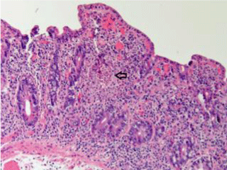
    Figure 1:  Duodenal biopsy shows severe villous atrophy. The lamina propria
shows increased acute and chronic inflammation. Arrow indicates crypt
abscesses.
A case of steroid-refractory NCIEC has been described in the literature in which cyclosporine resulted in almost complete clinical and histological resolution with cyclosporine, although the patient later died of infection [3]. To the best of our knowledge, our case is perhaps the first case of steroid-refractory NCIEC where anti-TNF agent Adalimumab was attempted, which resulted in the significant clinical improvement.
Adalimumab, a recombinant human monoclonal antibody, binds to and blocks the effects of the pro-inflammatory cytokine Tumor Necrosis Factor-α (TNF-α). Adalimumab is FDA-approved for the treatment of rheumatoid arthritis, Crohn’s disease, and Ulcerative Colitis. Our case might shed some light on the possible immune-mediated pathogenesis of NCIEC which might involve a dysregulation of the immune system with an over-expression of the pro-inflammatory cytokine TNF-α [4], as reflected by the therapeutic benefit obtained in our patient with Adalimumab, an anti-TNF agent.
Our case is significant for the following reasons: 1) It represents a rare disease. To the best of our knowledge, it is the 13th case of NCIEC reported in the literature [1-3], 2) It is the first case of steroidrefractory NCIEC where Adalimumab resulted in the significant clinical improvement, illustrating anti-TNF agent as a possible viable option for steroid-refractory NCIEC.
Conclusion
In conclusion, our case is a rare case of NCIEC which was refractory to steroids which was successfully treated with Adalimumab, an antitumor necrosis factor (TNF)-agent.
Acknowledgement
All the authors want to thank Kiyoko Oshima for her assistance with obtaining biopsy slide.
References
- Ruan EA, Komorowski RA, Hogan WJ, Soergel KH. Nongranulomatous chronic idiopathic enterocolitis: clinicopathologic profile and response to corticosteroids. Gastroenterology. 1996; 111: 629-637.
- Soergel KH. Nongranulomatous chronic idiopathic enterocolitis: a primary histologically defined disease. Dig Dis Sci. 2000; 45: 2085-2090.
- Sakoulas G, Anastopoulos H. Successful use of cyclosporine in the treatment of glucocorticoid-resistant nongranulomatous chronic idiopathic enterocolitis. Gastroenterology. 1999; 117: 1259-1260.
- Blandizzi C, Gionchetti P, Armuzzi A, Caporali R, Chimenti S, Cimaz R, et al. The role of tumour necrosis factor in the pathogenesis of immune-mediated diseases. Int J Immunopathol Pharmacol. 2014; 27: 1-10.