Abstract
Esophageal stricture due to Cytomegalovirus (CMV) infection is an uncommon pathology, with most reported cases occurring in patients infected with human immunodeficiency virus. We report a case who presented with Odynophagia followed Dysphagia to solids gradually progressive from last 3 weeks with Total Dysphagia to solids and liquids on presentation. Endoscopic examination revealed a long esophageal stricture with a necrotic lesion but no typical CMV esophageal ulcers, with foreign body impaction in form of beans. Histology was positive for CMV. Dysphagia resolved after treatment with ganciclovir and serial esophageal dilations. We are presenting the first case of esophageal stricture due to CMV esophagitis in a young adult with chronic alcoholism and are reviewing current literature.
Introduction
Cytomegalovirus (CMV) ulcerative esophagitis is well described in immunocompromised patients, but esophageal stricture is an exceedingly rare complication [1,2]. Most of the previous case reports of CMV esophagitis complicated with esophageal stricture have been described in patients with acquired immunodeficiency syndrome [1–5]. There is only 1 other reported case in a non-HIV-infected patient but that patient had undergone liver transplant and was on immunosuppressants [6]. This is the first report of esophageal stricture due to CMV in Young adult with Nil Co morbidities.
Case Report
A 30 year-old man presented with Odynophagia followed by Dysphagia to solids gradually progressive from the last 3 weeks with Total Dysphagia to solids and liquids on presentation Patient has chronic alcoholic from last 3 years. Daily alcohol intake in form of country liquor (4-5 Potli daily). His vital signs were blood pressure 114/80mmHg, heart rate 106bpm, temperature 37°C, and SpO2 95% in room air. Physical examination was unremarkable. Serum Hbsag, HCV and HIV testing was negative.
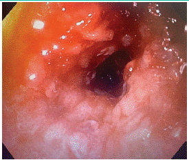
Esophagogastroduodenoscopy showed a severe distal esophageal stricture with near-complete luminal obstruction suggestive of malignancy (Figure 1) with foreign body impaction in form of beans. The stricture could not be traversed with a 6.5-mm pediatric gastroscope but allowed passage of biopsy forceps. There was also foreign body impaction just above stricture. Biopsies from the stricture site and the rest of the esophagus were taken.
Endoscopy Image-

Figure 1: Stricturing seen in lower esophagus.
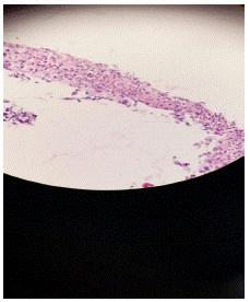
A computed tomography of the chest, abdomen, and pelvis showed a markedly dilated esophagus with probable stricture proximal to the gastro esophageal junction. Histopathology report (Figure 2) was suggestive of CMV esophagitis.
Biopsy Image (Figure 2)-

    Figure 2: Biopsy findings

Serial sections showed ulcerated squamous epithelium, necrosis, slough formation and inflammatory granulation tissue in the ulcer base endothelial cells and stromal cells shows intranuclear basophilic inclusion surrounded by clear halo.no evidence of dysplasia, fungal elements or malignancy is seen in received bit of tissue.
IV ganciclovir 5mg/kg/dose IV q12hr was initiated, and this was given for 7 days. The patient underwent serial dilations with SG dilator up to 14mm French under fluoroscopic guidance.
He was discharged on oral valganciclovir 450mg twice daily. He subsequently underwent 2 additional repeat endoscopic esophageal dilations with resolution in dysphagia. The patient was contacted 3 month later and denied current dysphagia.
Discussion
The most common esophageal strictures are peptic, anastomotic, iatrogenic (after Barrett's treatment, radiation, and nasogastric tube injury), and malignant [7]. CMV infection is a common complication in immunocompromised patients. The most common gastrointestinal manifestations of CMV infection are colitis and esophagitis. However, CMV esophageal stricture is a rare complication even in immunocompromised patients. After an extensive review of the literature, our case is the first to report CMV esophagitis in a young adult with Nil comorbidities.
In a series of 160 HIV-infected patients with esophageal ulceration, 13 (8%) developed stricture, with positive CMV in 5 and combined CMV and herpes simplex infection in 1. Our patient did not have HIV infection but was immunocompromised because of chronic alcoholism. The diagnosis of CMV esophagitis is suspected in patients with acute severe odynophagia and esophageal ulcerations on endoscopy and confirmed on histology (viral inclusion bodies and positive immunostaining) and/or viral culture. The organ-specific direct infection of CMV is poorly understood. However, CMV infection in our patient could be because of chronic alcoholism. The common macroscopic findings of CMV esophagitis are well-demarcated linear ulcers, erosions, and mucosal hemorrhages in the mid-to-distal esophagus [9]. However, in our patient, the endoscopic findings were not typical of CMV, rather there were findings stricture in lower oesophagus causing severe luminal narrowing with foreign body impaction over it giving impression of esophageal malignancy. This stresses the importance of diagnosis through histologic examination of biopsies with adequate sampling.
Without an obvious cause, such as caustic ingestion or radiation, we initially suspected gastrointestinal reflux disease or malignancy as the cause. Serum CMV quantification was negative. The final histopathological report confirmed the diagnosis of CMV esophageal stricture and was negative for malignancy. The stricture in the oesophagus was likely from the edema and inflammation from the CMV esophagitis, leading to fibrous healing [3]. This patient had an excellent clinical response to ganciclovir/valganciclovir and serial esophageal dilatations with no residual dysphagia.
In conclusion, this case is the first report of an esophageal stricture due to CMV in a young male patient. Thus, an esophageal stricture in an immunocompromised individual should raise the suspicion of CMV.
References
- Wilcox CM. Esophageal strictures complicate ulcerative esophagitis in patients with AIDS. Am J Gastroenterol. 1999; 94: 339–43.
- Mansfield BS, Savage-Reid MJ, Moyo J, Menezes CN. Cytomegalovirus-associated esophageal stricture as a manifestation of the immune reconstitution inflammatory syndrome. IDCases. 2020; 21: e00795.
- Sheth A, Boktor M, Diamond K, Lavu K, Sangster G. Complete esophageal obliteration secondary to cytomegalovirus in AIDS patients. Dis Esophagus. 2010; 23: E32–4.
- Goodgame RW, Ross PG, Kim HS, Hook AG, Sutton FM. Esophageal stricture after cytomegalovirus ulcer treated with ganciclovir. J Clin Gastroenterol. 1991; 13: 678–81.
- Olmos M, Sanchez Basso A, Battaglia M, Concetti H, Magnanini F. Esophageal strictures complicating cytomegalovirus ulcers in patients with AIDS. Endoscopy. 2001; 33: 822.
- Ahn J, Hirano Ikuo, Steven F. Cytomegalovirus infection associated with esophagitis and esophageal stricture after liver transplantation. Am J Gastroenterol. 2007; 102: 318–9.
- Baron TH. Management of benign esophageal strictures. Gastroenterol Hepatol (N Y). 2011; 7: 46–9.
- Rosolowski M, Kierzkiewicz M. Etiology, diagnosis and treatment of infectious esophagitis. Gastroenterol Rev. 2013; 6: 333–7.
- Li L, Chakinala RC. Cytomegalovirus esophagitis. In: StatPearls. StatPearls Publishing: Treasure Island, 2020.
- Kotton CN, Kumar D, Caliendo AM, Asberg A, Chou S, et al. Updated international consensus guidelines on the management of cytomegalovirus in solid-organ transplantation. Transplantation. 2013; 96: 333–60.
