Case Report
Austin Gynecol Case Rep. 2017; 2(1): 1012.
Successful Management of a Giant Ovarian Cyst: A Case Report
Kassidi F¹, Moukit M¹*, Ait El Fadel F¹, El Hassani ME¹, Guelzim K¹, Babahabib A¹, Kouach J¹, Moussaoui RD¹ and Dehayni M²
¹Department of Obstetrics and Gynecology, Military Training Hospital Mohammed V, Morocco
²Pole of Gynecology and Visceral Surgery, Military Training Hospital Mohammed V, Morocco
*Corresponding author: Mounir Moukit, Department of Obstetrics and Gynecology, Military Training Hospital Mohammed V, Hay Riyad, 10100, Rabat, Morocco
Received: January 23, 2017; Accepted: March 02, 2017; Published: March 10, 2017
Abstract
Giant ovarian cysts have become rare in current medical practice in both developed and developing nations. We report the case of a 32-yearold Moroccan woman, presented for progressive abdominal distension. Physical examination was difficult due to a massive abdominal pelvic swelling. Laparotomy was performed confirming the diagnosis of a giant ovarian cyst. Reporting such cases helps to increase the suspicion of its possibility and avoid any misdiagnosis or improper treatment.
Keywords: Giant ovarian cyst; Abdominal distention; Laparotomy
Introduction
Nowadays, ovarian cysts rarely grow immense due to availability of better imaging modalities permits early detection and appropriate treatment and only a few cases have been sporadically reported in the literature [1,2]. The massive size of tumour may splint the diaphragm and exert mass effect onto adjacent thoracoabdominal organs. Occasionally, they can have enormous dimensions without raising any symptom. We present a case of giant ovarian cyst, which was removed successfully without any complication despite a delay in diagnosis.
Case Report
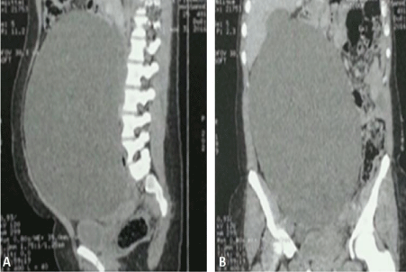
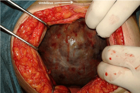
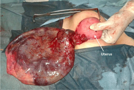
A 32-year-old Moroccan housewife presented in our department with chief complaints of a gradually increasing huge abdominal swelling noticed 5 months before. She denied any genito-urinary or gastrointestinal symptoms. On general examination, she was a febrile, with normal vital signs. There was no icterus or edema. Abdominal examination showed general distension with a dull note on percussion. The liver and spleen were not palpable. Based on sonographic examinations, a huge abdominal echogenic mass occupied the entire abdomen and pelvic cavity was seen. Abdominopelvic Magnetic Resonance Imaging (MRI) revealed a giant cyst, measured 62×45cm, displacing liver and spleen supero-laterally, kidneys posteriorly and bowel loops peripherally (Figure 1). Uterus was seen separately, but ovaries were not seen. There was no free fluid in peritoneal cavity. Tumor markers (CEA, a-fetoprotein and CA-125) were normal. An exploratory laparotomy was arranged for diagnostic and therapeutic purposes. Low midline incision extending up to the umbilicus was realized under general anaesthesia. After opening the parietal peritoneum, an irregular, hemorrhagic, necrotic and dark red in appearance cystic mass was noted (Figure 2). The mass was so large that it could not be excised without a large abdominal incision, so we drained the intra-cystic fluid (6 litres of hematic fluid) after creation of a small hole in the mass until we could excise the cystic mass (Figure 3). The left ovary was included in the mass and the left fallopian tube was adherent to the surface of the cyst. Complete excision of the cyst with left salpingo-oophorectomy was performed. Histopathological report revealed mucinous cystadenoma of the ovary. The postoperative period was uneventful and the patient was discharged on the fourth day after surgery.

    Figure 1:  Magnetic Resonance Imaging (A: Sagittal section, B: Coronal
section) showing large abdominal cystic lesion displacing spleen, liver and
bowel.

    Figure 2:  Peroperative view of the cystic mass just after opening the parietal
peritoneum.

Figure 3: Peroperative view after draining 6 litres of hematic fluid.
Discussion
The definition of giant ovarian cysts has not been well described in the literature. Some authors define large ovarian cysts as those more than 10 cm in diameter, other defined as those reaching above the umbilicus [3,4]. Our case well fits into the criteria of giant ovarian cyst, it being 62×45cm on MRI and extending up to xiphoid process. The clinical symptoms of giant ovarian cyst are usually progressive increase of abdominal circumference and pressure, non specific diffuse abdominal pain, and other symptoms related to organs compression. Physical examination usually shows abdominal distension mimicking ascites. There are many giant abdominal lesions which enter into the differential diagnosis with giant ovarian cysts. In this case, out of ascites, it was clinically confused with other gynaecologic masses (large uterine tumors), and non-gynecologic cysts (omental cysts, splenic cysts, mesenteric cysts, cysts arising from the retroperitoneal structures, cystic lymphangiomas and choledochal cysts). In the presence of abdominal cystic masses of unknown origin on ultrasound, the aspiration of fluid is to be avoided since it might produce clinical complications due to cyst perforation or possible dissemination of malignant cells. For this reason we did not perform any aspiration before investigating the precise nature of the abdominal cyst by MRI and tumor markers. The homogeneous content of the cyst, the absence of inner vegetations and the normality of all tumoral markers were suggestive of its benign nature. The most frequent complications of ovarian cysts are torsion, haemorrhage (as was our case), organs compression and rupture. Many cases of giant ovarian cysts are treated by surgery via laparoscopy or laparotomy, including cystectomy, oophorectomy, salpingo-oophorectomy, or total abdominal hysterectomy with bilateral salpingo-oophorectomy ± omentectomy. The choice between laparoscopy and laparotomy, conservative or radical treatments may be difficult and depends on the patient’s age, the size of the cyst and its histopathological nature [5]. A major factor that makes the gynecologic surgeon decide to perform laparotomy is definitely the size of the ovarian cyst. Some authors have limited laparoscopic surgery to women with an adnexal mass size less than 10 cm [6,7]. When confronted with extremely large, apparently benign ovarian cysts, only few surgeons advocate laparoscopic management [8,9]. In our patient, laparoscopy excision was not contemplated due to huge size of the cyst. We successfully decompressed and totally excised the large cyst with unilateral salpingo-oophorectomy through an infraumbilical midline laparotomy incision.
Conclusion
Ovarian neoplasms presenting with very large cystic masses can be mislead with ascites or other gynecologic and non-gynecologic lesions. Morphological examination using ultrasound and MRI appears fundamental to evaluate their precise nature and relationships with the other abdominal organs. Conservative management is well indicated for young patients; however, radical treatment is indicated for perimenopausal and menopausal women. With the advancement in minimal-access surgery, laparoscopy is a good alternative for removing moderate sized ovarian cysts.
Acknowledgment
We are thankful to the patient who permitted us to present his history in this report.
References
- Fobe D, Vandervurst T, Vanhoutte L. Giant ovarian cystadenoma weighing 59 kg. Gynecol Surg. 2011; 8: 177 179.
- Abbas AM, Amin MT. Brenner’s tumor associated with ovarian mucinous cystadenoma reaching a huge size in postmenopausal woman. J Can Res Ther. 2015; 11: 1030.
- Ou CS, Liu YH, Zabriskie V, Rowbotham A. Alternate methods for laparoscopic management of adnexal masses greater than 10 cm in diameter. J Laparo endoscopic Advanced Surgical Techniques. 2004; 11: 125-132.
- Salem HA. Laparoscopic excision of large ovarian cysts. J Obstetrics & Gynecology Research. 2002; 28: 290-294.
- Bhasin SK, Kumar V, Kumar R. Giant Ovarian Cyst: A Case Report. JK Science. 2014; 16: 131-133.
- Yuen PM, Yu KM, Yip SK, Lau WC, Rogers MS, Chang A. A randomized prospective study of laparoscopy and laparotomy in the management of benign ovarian masses. Am J Obstet Gynecol. 1997; 177: 109-114.
- Lin P, Falcone T, Tulandi T. Excision of ovarian dermoid cyst by laparoscopy and by laparotomy. Am J Obstet Gynecol. 1995; 173: 769-771.
- Murawski M, Golebiewski A, Sroka M, Czauderna P. Laparoscopic management of giant ovarian cysts in adolescents. Wideochir Inne Tech Maloinwazyjne. 2012; 7: 111-113.
- Alobaid A, Memon A, Alobaid S, Aldakhil L. Laparoscopic Management of Huge Ovarian Cysts. Obstetrics and Gynecology International. 2013; 2013: 380854.