Case Report
Austin Gynecol Case Rep. 2022; 7(1): 1031.
Struma Ovarii: A Rare Ovarian Teratoma and Follicular Adenoma of Thyroid Gland
Antovska V, Eva SB, Dabeski D, Malahova-Gjoreska I, Stojanovska MI* and Papastiev IA
University Clinic of Gynecology and Obstetrics, Medical Faculty, Skopje, Republic of North Macedonia
*Corresponding author: D-r Melita Ilievska Stojanovska, University Clinic of Gynecology and Obstetrics, Medical Faculty, Skopje, Republic of North Macedonia
Received: August 19, 2022; Accepted: September 20, 2022; Published: September 27, 2022
Abstract
Ovarian struma is a germ cell tumor in which thyroid tissue represents more than a half of the tumor and has incidence of 0,3-1% of all ovarian tumors. Most of the patients are asymptomatic and tumor is found incidentally during ultrasound, or with nonspecific symptoms, like pain or abdominal swelling.
We report a case of 49-years old woman with no symptoms and an ovarian mass, which was incidentally found during a routine ultrasound examination. On ultrasound she had a tumorous mass on the left ovary with central cystic part filled with dense fluid. Color Doppler showed resistance index RI - 0.64, which did not indicate malignant nature of the tumor, and CA-125 was slightly elevated (38,4 U/ml). The result of the scoring system ROMI was 11 ponts, i.e. low risk for ovarian carcinoma. The patient underwent hysterectomy and bilateral adnexectomy. The final histopathologic report was struma ovarii with well-differentiated neoplasm of uncertain malignant potential.
Keywords: Monodermal ovarian teratoma; Struma ovarii; Follicular adenoma on thyroid gland
Abbreviations
RI: Resistance Index; CA 125: Cancer Antigen 125; U/ml: Units Per Millilitre; ROMI: Risk of Ovarian Malignancy Index; Mcg: Microgram; TSH: Thyroid Stimulating Hormone
Introduction
Struma ovarii is a specialized monodermal teratoma, and is a germ cell tumor of the ovary in which the thyroid tissue represents more than a half of the tumor itself [1]. It is most commonly found in women between 40-60 years of age [2]. Struma ovarii is a rare tumor which comprises 1% of all ovarian tumors [3].
Case Report
A 49 year old woman was referred to our hospital without any symptoms and ovarian mass, which was found incidentally during the ultrasound examination. Her generative history included regular menstrual cycle before menopause, and three spontaneous deliveries. She had a medical history of arterial hypertension and no previous surgeries.
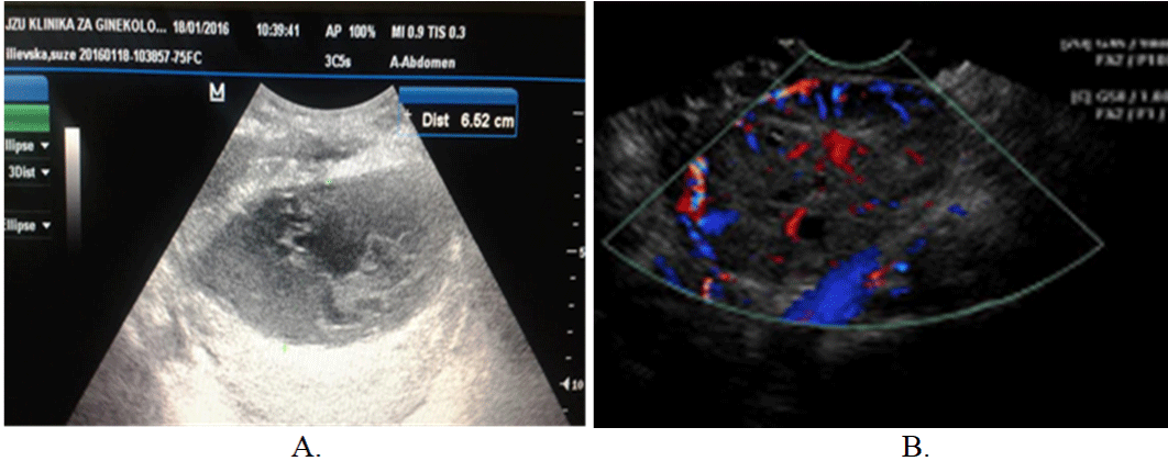
The patient was admitted to our department for additional evaluation and treatment. On ultrasound exam a tumorous mass of the left ovary was detected (D 66x60mm) with central cystic part filled with dense fluid, and peripheral areas containing abundant blood vessels and fibrous tissue. Color Doppler examination showed Resistance Index (RI) = 0,64, which did not indicate malignant nature of the tumor, and the vascular net was abundant and with regular vascular feature. The tumor marker CA-125 was slightly elevated (38,4 U/ml). Other laboratory analysis was in referent range. ROMI [5] scoring system showed low risk for ovarian carcinoma, e.g. ROMI 11 points = postmenopausal age (1 point), tumor size ≥6cm (1 point), partly cystic/solid tumor (1 point), dense capsule (2 points), dense septum (2 points), gross papillae (2 points), CA-125 level (2 points), absence of the ascites (0 points). The endometrium was thin, with postmenopausal characteristics, the right adnexa showed normal morphology and there was no fluid in pouch of Douglas (Figure 1).

Figure 1: Preoperative ultrasound findings, A-white-black scale ultrasonography; B-Color Doppler of the left ovary.
After preoperative preparation, the patient underwent surgical treatment. During surgery, a meticulous inspection of the abdomen and pelvis was done. The left ovary was with tangerine dimensions. Uterus and right adnexa were with normal appearance. The right ovary showed increased dimension and cystic structure, but its capsule was thick and intact. Total abdominal hysterectomy and bilateral adnexectomy, as well as abdominal washing for cytological sampling were performed. Postoperative period was stable and the patient was discharged on the 6th postoperative day in good condition.
The gross pathological examination revealed left ovary with dimensions of 60x55 mm with smooth, shiny and uninterrupted serosa. On the surface of the section, there was a multilocular cyst with yellowish, thick fluid. Microscopic examination showed cystic formation filled with eosinophilic colloid and solid zones of thyroid tissue. The immunophenotyping was consistent for thyroid tissue. The final histopathology report was struma on the left ovary with well-differentiated neoplasm of uncertain malignant potential. The cytopathology of pelvic fluid showed no tumor cells.
In view of her pathological findings, the patient was referred to an oncologist and according to his opinion there was no need for adjuvant therapy. Additionally, the nuclear medicine specialist performed control ultrasound of the thyroid gland and detected one hypoechogenic nodule and performed a fine needle aspiration. Cytopathology showed Atypia of undetermined significance/ Follicular lesion of undetermined significance (Bethesda Classification group III), (Classification group III), after winch the patient was referred to thoracic surgeon. Our patient underwent unilateral lobectomy with isthmetectomy. The histopathology result of the second surgery, showed follicular adenoma. Postoperatively, the patient was on continuous substitutive therapy with levothyroxine 5 mcg/day and has achieved normal serum levels of TSH and thyroxine.
Discussion
Less than 5-8% of patients with struma ovarii present with hyperthyroidism. Clinical manifestation of hyperthyroidism includes nervousness, disturbed sleep, heat intolerance, weight loss, sweating, palpitations, and diarrhea. On clinical examination there is tremor, tachycardia, warm and moist skin. If the hyperthyroidism is a result of struma ovarii, the blood examination would show hyperthyroidism, test results for TSH receptor antibody and thyroid stimulating antibody are negative, and thyroid scintigraphy shows no abnormal findings [6]. In our case, the patient had increased level of thyroxine.
Preoperative diagnosis of struma ovarii is difficult because the large majority of cases are subclinical. Yoo et al., [7] noted that 41.2% of cases were asymptomatic and tumors were discovered accidentally during routine ultrasound check-up. Clinical symptoms are nonspecific and include lower abdominal pain and/or a pelvic mass, and less frequently ascites, but only 5–8% of cases had clinical hyperthyroidism. Our patient did not show clinical signs of hyperthyroidism preoperatively, and struma ovarii was accidentally discovered during routine ultrasound check-up. Additionally, she had a follicular adenoma of the thyroid gland.
The imaging methods, such as ultrasound, CT and MRI give nonspecific findings. Their features could include multilobulated mass with multiple cysts and solid components containing abundant blood vessels and fibrous tissue [5]. Ultrasound demonstrates a complex appearance with multiple cystic and solid areas, findings that reflect the gross pathologic appearance of the tumor. The definitive diagnosis is made postoperatively after detecting thyroid follicles in resected ovary. In our case, ultrasonographic features of the tumor were in favor of malignant nature. On the contrary, ROMI scoring system [6] showed low risk in favor of benign nature, which was confirmed by histology. Our ROMI was very useful in predicting the nature of this tumor. We highly recommend ROMI scoring system as a useful tool in preoperative evaluation of all pelvic and abdominal tumors. Tumor marker CA-125, as a diagnostic tool could provide little value in diagnosis.
Definitive treatment for women with struma ovarii is surgery. In case of malignancy there is a need for long-term follow-up (at least 10 years), annual monitoring of stimulated serum thyroglobulin during the first two years, and if negative, unstimulated levels annually are recommended thereafter.
Conclusion
In this case report we endpoint the correlation between preoperative nonspecific findings and the specific postoperative diagnosis. There is no specific diagnostic test for all ovarian tumors, especially for more specialized tumors, like struma ovarii. In most cases, cancer presents with symptoms from local tumor growth or no symptoms at all.
References
- Dunzendorfer T, deLas Morenas A, Kalir T, Levin RM. Struma ovarii and hyperthyroidism. Thyroid. 1999; 9: 499-502.
- Kondi-Pafiti A, Mavrigiannaki P, Grigoriadis Ch, Kontogianni-Katsarou K, Mellou A, Kleanthis CK et al. Monodermal teratomas (struma ovarii). Clinicopathological characteristics of 11 cases and literature review. Eur J Gynaecol Oncol. 2011; 32: 657-9.
- Kim SJ, Pak K, Lim HJ, Yun KH, Seong SJ, Kim TJ, et al. Clinical diversity of struma ovarii. Korean J Obstet Gynecol. 2002; 45: 748-52.
- Goffredo P, Sawka AM, Pura J, Adam MA, Roman SA, Sosa JA. Malignant struma ovarii: a population-level analysis of a large series of 68 patients. Thyroid. 2015; 25: 211-5.
- Joja I, Asakawa T, Mitsumori A, Nakagawa T, Hiraki Y, Kudo T, et al. Struma ovarii: appearance on MR images. Abdom Imaging. 1998; 23: 652-6.
- Antovska V, Trajanova M. An original risk of ovarian malignancy index and its predictive value in evaluating the nature of ovarian tumour. SAJGO. 2015; 7: 52-9.
- Nagai K, Yoshida H, Katayama K, Ishidera Y, Oi Y, Ando N et al. Hyperthyroidism due to struma ovarii: diagnostic pitfalls and preventing thyroid storm. Gynecol Minim Invasive Ther. 2017; 6: 28-30.
- Yoo SC, Chang KH, Lyu MO, Chang SJ, Ryu HS, Kim HS. Clinical characteristics of struma ovarii. J Gynecol Oncol. 2008; 19: 135-8.