Clinical Image
Austin Gynecol Case Rep. 2023; 8(2): 1043.
The Mature Ovarian Teratoma: Sign of the Poké Ball
F Chait*; S Essetti; N Bahlouli; N Allali; S El Haddad; L Chat
Pediatric Radiology Service, Rabat Children’s Hospital, Avicenne University Hospital, Morocco
*Corresponding author: Fatima Chait Pediatric Radiology Service, Rabat Children’s Hospital, Avicenne University Hospital, Morocco. Email: fatima.chait1@gmail.com
Received: June 30, 2023 Accepted: August 10, 2023 Published: August 17, 2023
Clinical Image
The "Poké Ball sign" is a distinctive radiological feature observed in mature cystic ovarian teratomas. It is characterized by the presence of a spherical structure floating at the interface between fat and fluid, resembling a Poké Ball (Figure 1) [1]. In the Pokémon® series, a Poké Ball is a spherical device utilized for capturing and containing Pokémon [2].

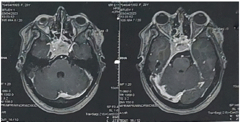
Figure 1: Cerebral MR angiography demonstrates a thrombus in the right lateral sinus with extension into the ipsilateral internal jugular vein, as well as the presence of a macroadenoma of the pituitary gland with extension into the right cavernous sinus.
Mature ovarian cystic teratomas are the most commonly encountered benign tumors of the ovary, comprising approximately 10 to 20% of all ovarian tumors. They typically occur in women of reproductive age and are often serendipitously discovered due to their frequently asymptomatic nature. Pathologically, these teratomas consist of mature tissues derived from the three embryonic germ layers: ectoderm, mesoderm, and endoderm [3].
The imaging characteristics of ovarian teratomas may vary on cross-sectional images depending on the embryonic growth pattern. While the presence of a Pokémon Ball sign is considered indicative, it exhibits relatively low sensitivity (reported incidence rate of 25 to 30% in certain case series), meaning its absence does not rule out the diagnosis of a mature cystic ovarian teratoma [1]. The visualization of a floating ball within a cyst containing a combination of fat and fluid is readily achieved through magnetic resonance imaging (Figure 2).
References
- Sahin H, Akdogan AI, Ayaz D, Karadeniz T, Sanci M. Utility of the ”foating ball sign” in diagnosis of ovarian cystic teratoma. Turk J Obstet Gynecol. 2019; 16: 118-23.
- Wagner-Greene VR, Wotring AJ, Castor T, Kruger J, Mortemore S, Dake JA. Pokémon GO: healthy or harmful? Am J Public Health. 2017; 107: 35-6.
- Saba L, Guerriero S, Sulcis R, Virgilio B, Melis G, Mallarini G. Mature and immature ovarian teratomas: CT, US and MR imaging characteristics. Eur J Radiol. 2009; 72: 454-63.