Clinical Image
Austin Gynecol Case Rep. 2023; 8(2): 1044.
Scar Formation after Unilateral Pectineal Suspension
Bolovis DI*; Brucker CVM
University Women’s Hospital, Paracelsus Medical University, Nuremberg, Germany
*Corresponding author: Bolovis DI University Women’s Hospital, Paracelsus Medical University, Nuremberg, Prof.-Ernst-Nathan-Str. 1, 90437 Nürnberg, Germany. Tel: +49-911-3982222; Fax: +49-911-3983399 Email: dimitrios.bolovis@klinikum-nuernberg.de
Received: September 22, 2023 Accepted: October 12, 2023 Published: October 19, 2023
Clinical Image
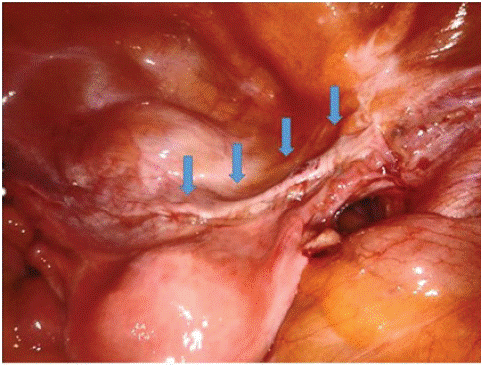
UPS is a minimally invasive endoscopic procedure using a single Ethibond suture to attach the anterior cervix or vaginal vault unilaterally to the pectineal ligament (Figure 1a). The procedure allows anatomical prolapse correction by repositioning the uterus or vaginal vault to its original place within the pelvis [1]. The clinical image demonstrates the effective creation of supportive scar tissue during laparoscopic re-evaluation (Figure 1b). The fibrous scar formation has the capacity to stabilize the permanent result.

Figure 1: UPS procedure: a single ethibond suture is applied to connect the anterior cervix with the pectineal ligament for anatomical prolapse correction.

Figure 2: 1 year after UPS a white scar delineates the area where an ethibond suture was previously placed between the pectineal ligament and the anterior cervix.
In mesh-free procedures for pelvic floor reconstruction such as sacrospinous fixation, the use of absorbable sutures does not affect outcome when compared to permanent sutures [2,3]. One logical explanation for this phenomenon would be the formation of a suture scar. However, the visualization of a suture scar with vaginal prolapse repair is anatomically not possible. One year after Unilateral Pectineal Suspension (UPS), the suture scar can be visualized within the pelvis, following the direction of the previously performed repair.