Case Report
Austin Gynecol Case Rep. 2023; 8(3): 1047.
Pregnancy-Associated Cancer: A Case Report of a Gastric Cancer Diagnosis in the III Trimester of Pregnancy and Subsequent Management
Sabattini A*; Morano D; Greco P
Department of Medical Sciences, Institute of Obstetrics and Gynecology, University of Ferrara, Italy
*Corresponding author: Sabattini A Department of Medical Sciences, University of Ferrara, Via Fossato di Mortara 64/B, 44124 Ferrara, Italy. Tel: 3495416311 Email: ariannasabattini.doc@gmail.com
Received: November 22, 2023 Accepted: December 18, 2023 Published: December 23, 2023
Abstract
The co-occurrence of gastric carcinoma and pregnancy is very rare event. Diagnosis is often delayed, frequently at an advanced stage due to the nonspecific symptomatology, which can be confused with the normal physiological symptoms of pregnancy. Therefore, a multidisciplinary approach is essential to initiate the treatment of maternal pathology as early as possible, with the goal of delivering at a moderately preterm gestation age to ensure favorable neonatal outcome.
We present a case of gastric carcinoma diagnosed during the third trimester, where the earliest presentation included liver metastases, and the primary tumor’s location was unknown. Throughout our observation period, we conduced clinical-laboratory monitoring, a preliminary radiological staging of the illness (considering the limitations imposed by pregnancy), and a histological diagnosis with biopsy sampling. An elective cesarean section was performed in the moderately preterm period, following the completion of Respiratory Distress Syndrome (RDS) prophylaxis.
Case Report
A 38-year-old female, in her third pregnancy and secondipara (with two previous C-sections), presented for detection during the third-trimester ultrasound of a vascularized intrahepatic lesion measuring approximately 6 cm. The lesion was tender to palpation and of unclear interpretation, prompting an urgent surgical evaluation. During anamnesis, the patient reported experiencing persistent nausea and vomiting since the beginning of pregnancy, along with cramp-like pains in the right hypochondrium that radiated to the epigastric region over the past 10 days. Additionally, the patient noted the development of a hard, wood-like nodule in the right hypochondrium that was tender to palpation.
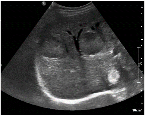
Our OB/GYN ER requested a complete abdominal ultrasound, hematological and chemical exams, and a gastroenterologic consultation. The abdominal ultrasound revealed a liver with altered echotexture due to multiple heterogeneous rounded iso-hyperechoic formations, some with a hypoechoic center, ranging in size from a minimum of 2 to a maximum of 5 cm in the right lobe. Focal biliary tract ectasis was noted, altering the hepatic profile at the VIII-VI segment. In the epigastrium, at the level of the celiac trunk, various rounded formations were observed, partly converging, with a maximum diameter of 6 cm.
The gastroenterologic consultation recommended an evaluation, with a thorough risk-benefit analysis, to determine the necessity for histological characterization through an ultrasound-guided hepatic biopsy. Hematochemical exams revealed microcytic anemia (Hgb 7.2g/dL; MCV 60 fl), average platelet levels (323000/microL), and a slight alteration in hepatic functioning (AST 44 U/L; ALT 33 U/L) with normal serum bilirubin.
Obstetric ultrasound and cardiotocographic monitoring yielded results within the normal range. The patient was admitted to the Obstetrics ward for the continuation of the diagnostic and treatment process.
During the hospitalization, tumor markers were measured, with the following results: alpha-fetoprotein 26510 ng/mL (n.v. in non-pregnant women <10); Ca 125 12565 U/mL (n.v. <35); CEA >1000 ng/mL (n.v. <5); NSE 19.6 microg/L (n.v. <18.3 microg/L); Ca 19.9 10 U/L (n.v. <37).
To obtain a histologic sample, an ultrasound-guided hepatic biopsy was performed using an intercostal approach on a lesion at the V hepatic segment, followed by histological analysis.
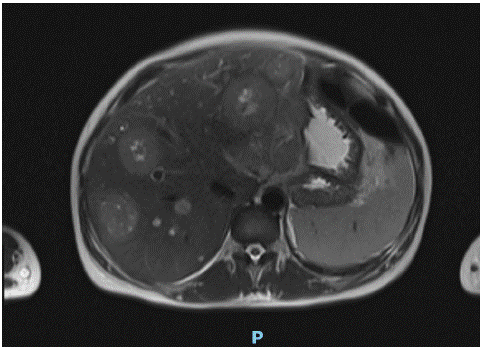
To identify the primary tumor, in consultation with Oncology, we conducted an abdominal MRI with/without contrast agents, revealing: “Numerous lesions of rounded appearance distributed throughout the liver parenchyma, some with central lesion necrosis, of probable substitute-replicative significance (the larger ones approximately 65x55 mm and 63x55 mm). In the retroperitoneal region, the gastrohepatic omentum, and along the mesenteric vascular pedicle, numerous and
sometimes voluminous solid, possibly neoplastic lesions were observed, some with internal necrotic components (with major elements respectively in the right paracaval site 80x64 mm, along the small gastric curvature 96x53 mm, and in the mesenteric adipose tissue 66x53 mm). These lesions appear to be indicative of lymphadenopathy. The gastric antrum wall appears significantly thickened and slightly hyperemic; these findings, though not unequivocal, seem to warrant an endoscopic targeted evaluation aimed at excluding the possibility of a primary tumor at this level.”

Figure 1: liver subverted in its echotexture by multiple rounded lesions.

Figure 2: MRI Axial T2-weighted image showing numerous lesions of rounded appearance distribuited throughout the liver parenchyma, some with central lesion necrosis, indicating probable substitute-replicative significance.

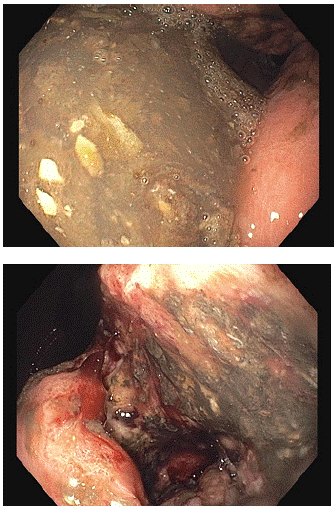
Figure 3: Esophagogastoduodenoscopy: at the stomach’s angular level, a large ulcerated lesion with a fibrinous base of approximately 40 mm, featuring raised borders and destructured appearance.
Additionally, a bilateral ultrasound and a chest x-ray were performed, both yielding negative results.
During her hospital stay, the patient received antenatal RDS prophylaxis with 12mg Betamethasone in two doses 24 hours apart, along with pain relief and antiemetic therapy as needed. To address microcytic anemia, likely due to iron deficiency (Hgb 6.7 g/dL), 2 units of packed red blood cells were transfused.
Given the compromised maternal clinical condition, the advanced gestational age (32 weeks + 4 days), and the completion of RDS prophylaxis, a C-section with tubal sterilization was deemed necessary during the collegiate assessment. The cesarean section was performed without complications, and the neonate was admitted to the NICU due to prematurity.
Due to the suspected gastric primary revealed in the MRI, the patient underwent an esophagogastroduodenoscopy, revealing: “At the stomach’s angular level, a large ulcerated lesion with a fibrinous base, approximately 40 mm in size, with raised borders and a destructured appearance. It is fragile and poses a high risk of bleeding, deforming the antrum and extending to the pyloric channel, which is difficult to pass and coated with a similar-looking mucous membrane.” Simultaneously, multiple urgent biopsies were performed.
Subsequently, consultation with the Emergency Surgery department did not recommend urgent surgical intervention.
- The histological investigations revealed: Hepatic needle biopsy: hepatic carcinoma metastasis with a predominantly solid architecture, partially necrotic, G3. Phenotypical description: CK (PAN) +/HEP-/CK7-/CDX2+/SABT2 +-/ Chromogranin -/ Synaptophysin-. The immunohistochemical profile indicates a primary likely located in the gastrointestinal tract.
- Biopses executed during EGD: infiltrating adenocarcinoma.
The patient was discharged with a diagnosis of poorly differentiated stage IV gastric adenocarcinoma, with the presence of secondary disease in the liver, lymph nodes, and the lung. She continued the therapeutic process in the Oncology department with two lines of Cisplatin and F-Fluorouracil.
This was followed by a series of Emergency Room admissions for intractable bilious vomiting and worsening anemia. Subsequently, she was transferred to Hospice with a diagnosis of bacterial and mycotic sepsis, further progression of the illness, pulmonary thromboembolism, resistant hypoglicemia, and hypokaliemia in advanced gastric carcinoma, leading to the patient’s demise.
Conclusions
The most commonly diagnosed malignancies during pregnancy are breast cancer and cervical carcinoma, followed by leukemia, melanoma, and thyroid cancers [1]. Gastric carcinoma diagnosed during pregnancy is extremely rare, with an incidence of approximately 1 in 1000 [2].
It’s estimate that gastric cancer associated with pregnancy, defined as a gastric cancer diagnosis given during pregnancy or up to a year after delivery, is a complication in 0.026-0.1% of all pregnancies3. Gastric carcinoma associated with pregnancy can be a devastating clinical situation for both the mother and fetus. Patients with this diagnosis often present with a dismal prognosis, primarily because the diagnosis frequently occurs at an advanced stage. The symptoms of gastric carcinoma are nonspecific, and many of them are considered physiological during pregnancy. Surgical treatment is administered in only 45-56% of cases [3-5].
Gastric cancer associated with pregnancy is often classified as type IV (according to the macroscopic classification proposed by Borrmann) and diffuse type (according to the classification proposed by Lauren) with signet-ring features [6]. These characteristics can overlap with gastric tumors in younger patients, who are tipically female [7,8]. The higher prevalence of females in the incidence of gastric tumors at a young age, along with the lower overall survival rate of women compared to men, suggests potential hormonal involvement. Estrogens, in particular, could heighten susceptibility to more aggressive gastric tumors [8].
The management of a pregnant patient with gastric carcinoma requires a multidisciplinary team. Two conflicting issues need consideration: the continuation of the pregnancy and prompt treatment of the neoplasm. Gastric carcinoma diagnosed during pregnancy often leads to a devastating clinical situation, with a very poor prognosis. Diagnosis is frequently delayed because symptomatic presentations are often attributed to pregnancy rather than the co-occurring illness.
The management of this pathology has two primary goals:
1. Early execution of histological diagnosis and initial radiological staging, considering the limitations imposed by pregnancy.
2. Early delivery to enable the patient to initiate the therapeutic process, preferably at a gestational age when prematurity would not significantly affect the neonatal outcome.
In the case reported here, a partial radiological staging was performed, along with a histological diagnosis that identified the primary site of the initially observed metastatic lesions. Delivery was carried out at 32 weeks + 4 days (moderate preterm) after completing RDS prophylaxis.
References
- Pavlidis NA. Coexistence of pregnancy and malignancy. Oncologist. 2002; 7: 279-87.
- Gonzalez-Mesa E, Armenteros MA, Molina A, Herrera J. Fulminant evolution of stomach cancer during pregnancy. Austin Journal of Clinical Case Report 1. 2014; 1051-3.
- Sakamoto K, Kanda T, Ohashi M, Kurabayashi T, Serikawa T, Matsunaga M, et al. Management of patients with pregnancy-associated gastric cancer in Japan: a mini-review. Int J Clin Oncol. 2009; 14: 392-6.
- Lee HJ, Lee IK, Kim JW, Lee KU, Choe KJ, Yang HK. Clinical characteristics of gastric cancer associated with pregnancy. Dig Surg. 2009; 26: 31-6.
- Ueo H, Matsuoka H, Tamura S, Sato K, Tsunematsu Y, Kato T. Prognosis in gastric cancer associated with pregnancy. World J Surg. 1991; 15: 293-7.
- Kodama I, Takeda J, Koufuji K, Yano S, Hanzawa M, Shirouzu K. Gastric cancer during pregnancy. Kurume Med J. 1997; 44: 179-83.
- Lai JF, Kim S, Li C, Oh SJ, Hyung WJ, Choi WH, et al. Clinicopathologic characteristics and prognosis for young gastric adenocarcinoma patients after curative resection. Ann Surg Oncol. 2008; 15: 1464-9.
- Isobe T, Hashimoto K, Kizaki J, Miyagi M, Aoyagi K, Koufuji K, et al. Characteristics and prognosis of gastric cancer in young patients. Oncol Rep. 2013; 30: 43-9.