Case Report
Austin Gynecol Case Rep. 2024; 9(1): 1049.
Retroperitoneal Cystic Vascular Malformation Impersonating Ovarian Tumor Accompanied with Stress Urinary Incontinence: A Case Report
Antovska V; Dabeski D; Aleksioska Papestiev I; Malahova Gjoreska I*; Pavlovski B; Zlateska Guric S; Dimitrova A
University Clinic of Gynecology and Obstetrics, Skopje, Republic of North Macedonia
*Corresponding author: Malahova Gjorevska I, University Clinic of Gynecology and Obstetrics, Skopje, Republic of North Macedonia. Email: ivamalahovagj@yahoo.com
Received: October 02, 2024; Accepted: October 23, 2024 Published: October 30, 2024
Introduction
Retroperitoneum is the anatomical space located behind the abdominal or peritoneal cavity. It is divided into three main spaces: the anterior pararenal, perirenal, and posterior pararenal space. The anterior pararenal space contains the head, neck, and body of the pancreas, ascending and descending colon, and the duodenum. The structures contained within the perirenal space include the adrenal gland, kidney, ureters, and renal vessels. The posterior pararenal space, which is surrounded by the posterior leaf of the renal fascia and muscles of the posterior abdominal wall, contains no major organs and is composed primarily of fat, blood vessels, and lymphatics.
Cystic lesions of the retroperitoneum can be subdivided into neoplastic and non-neoplastic lesions. Retroperitoneal cystic vascular malformations are rare benign lesions characterized by abnormal clusters of blood or lymphatic vessels within cystic spaces located in the retroperitoneal space. The exact cause is often unknown. They are thought to arise from developmental anomalies during fetal growth, resulting in abnormal growth of blood or lymphatic vessels and cystic spaces. Patients may present asymptomatically or with symptoms related to the size and location of the malformation. Symptoms can include abdominal pain, distension, urinary symptoms, or complications such as infection or rupture, which are almost the same with that one in ovarian tumors.
Stress Urinary Incontinence (SIU) is defined as a condition where involuntary leakage of urine occurs during physical activities that increase intra-abdominal pressure. It is divided into three degrees of severity: Grade 1 with urine loss when coughing, sneezing, and laughing, Grade 2 with urine loss when getting up, when walking, or under physical activity, and Grade 3 with urine loss while lying. It is caused by weakened pelvic floor muscles and the ligamentousconnective tissue in the pelvis that support the bladder and urethra.
Herein, we present a 60-year-old woman who was operated on with a preoperative diagnosis of right ovarian cystadenoma, only to be diagnosed as retroperitoneal mass intraoperatively.
Case Report
A 60-year-old woman presented with abdominal swelling for one-year duration. It was associated with lower abdominal pain for 6 months, which she described as cramping in nature, localized over the lower abdomen, and resolved by taking oral analgesics. She denied any history of biliary colic before. She had 2 successful vaginal deliveries. The patient has occasional urine leakage after coughing, sneezing or laughing. She noted no nocturia or urgency symptoms but described embarrassment and social discomfort due to her condition. Pelvic examination revealed mild urethral hypermobility during Valsalva maneuver. No significant pelvic organ prolapse noted, Marshall Test was high positive for leakage of urine upon coughing while lying and standing. Clinically, the abdomen was soft and no tender with a big palpable mass over the right side of the abdomen, which was mobile and firm in consistency.

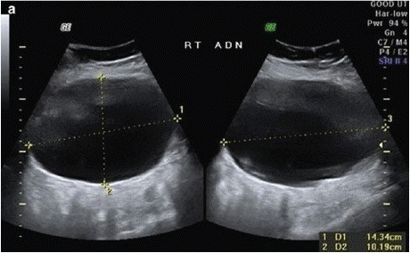
Figure 1: Ultrasound of the tumor in our patient.
Ultrasound of the pelvis and abdomen revealed a cystic formation over the right ovary with diameter 13cm with thick edges, and no solid component was seen.
Our original ROMI index [1] integrates: the serum CA-125 levels, the menopausal status, the patient’s familial and personal history and some ultrasound characteristics of the tumor (tumor size, multilocularity, solid areas in the tumor, dense or opalescent liquid, presence of septum or papillary vegetation, ascites, bilaterality, unclear margin with respect to the surrounding tissue and thickness of the capsule) to enhance the accuracy of malignancy risk assessment. The other originality of our ROMI is the three-stage gradation of serum cancer antigen 125 (CA-125), as well as the three-stage gradation of the quantity and location of the ascites. It is subdivided into three subgroups of ROMI = 11 (low risk), ROMI 12-14 (unclear risk) and ROMI = 15 (high risk). The data from the ultrasound characteristics of the tumor in our patient were: tumor size=6cm = 1 point; cystic tumor = 0 points; unilocular tumor = 0 points, dense and opalescent liquid=2 points; absence of septum or papillary vegetation = 0 points, unilaterality = 0 points, clear and jagged margin with respect to the surrounding tissue = 1 points and thickness of the capsule of 3-5 mm = 1 point, absence of ascites = 0 points, CA-125 within normal range = 0 points, age of the patient in the senium = 1 point. The final ROMI index was 6.
The patient was well-prepared preoperatively. The laparotomy was carried out in a usual manner. Intraoperatively, there was a huge mass arising from the retroperitoneal space attaching to the right ureter, which led to placement J-J sondage in the right ureter. The major organs were carefully identified and preserved. Excision of the retroperitoneal tumor was executed successfully without any intraoperative complications. An abdominal hysterectomy and our original modification of the Burch colposuspension, the Pleated Colposuspension after Antovska [2] followed.
On the 3rd postoperative day, bladder training was started by replacing continuous drainage with discontinuous drainage and then removing the urinary catheter. After several regular emptying of the bladder, the patient complained of a sensation of incomplete emptying and discomfort in the lower abdomen. Physical examination revealed a distended bladder palpable above the symphysis pubis. Ultrasound examination confirmed significant postvoid residual urine. Immediate insertion of a Foley catheter was performed to drain the retained urine. Subsequent monitoring of urine output and bladder function was undertaken. The patient was started on a prophylactic course of antibiotics to prevent urinary tract infection. Given the persistent urinary retention and discomfort, a decision was made to surgically remove some of the sutures placed during the previous colposuspension. Intraoperatively, we encountered difficulties in the dissection of the Retzius space and the visualization of the sutures, so only one-sided removal of the colposuspension sutures was performed, on the left side.
Intraoperatively, during the removal of sutures and preparation of the Retzius space, a 1 cm long lesion appeared on the front wall of the bladder, which was sutured in 2 layers. Postoperatively, the patient had continuous drainage with a Foley-catheter for 14 days. After removing the catheter, a cystoscopy was performed and the J-J tube was removed from the right ureter. Rest urine was 150 ml, which is why Tamsulosin 0.4 mg once daily was started for 4 weeks to improve bladder outlet. The patient was followed up after 4 weeks of tamsulosin therapy. The new measurement of the rest urine was 70 ml and the Marshall test was negative in lying and standing position.
At the 3-month postoperative control, there was no recurrence of stress incontinence, no urine retention; there were no signs of vesico-vaginal fistula, no retroperitoneal tumor recurrence. The patient reported significant improvement in quality of life and no recurrence of stress urinary incontinence. The residual urine was 60 ml. The patient complained of symptoms in addition to urgent incontinence, for which she was put on therapy with Utidry tab. once/ day. At the next follow-up, the patient stated that the symptoms of urgent incontinence completely disappeared with Utidry therapy, so the same therapy was continued. The histopathological analysis of the operative specimen was Cystic vascular malformation i.e. Lymphangioma retroperitoneale. The cytology of the peritoneal washing was Classification group I.
Despite the unilateral removal of the sutures from the colposuspension, we successfully resolved the patient's severe stress incontinence with unilateral placement of the sutures. Tamsulosin has been shown to be a successful therapy for postoperative urinary retention after corrective colposuspension in patients with stress incontinence.
Discussion
Retroperitoneal cystic masses are a rare surgical entity. They are usually asymptomatic, but they may grow and reach a certain size, large enough to cause symptoms especially a compression to the adjacent structures. Anterior displacement of the abdominal structures such as the aorta, colon, or kidneys may help us to identify the lesion site clinically.
Ultrasound is a non-invasive, rapid, inexpensive tool, and can easily be repeated as necessary. Transvaginal ultrasound remains a cornerstone in the initial assessment of ovarian masses due to its ability to provide detailed information on size, morphology, and vascularity. The ROMI index integrates ultrasound findings with serum CA-125 levels and menopausal status to enhance the accuracy of malignancy risk assessment. Our ROMI was the simple sum of points from the three-stage gradation of serum cancer antigen 125 (CA-125), data from the patient’s familial and personal history and the ultrasound characteristics of the tumour (i.e. tumour size = 6 cm, multilocularity, tumour with = ¼ solid areas, dense and opalescent liquid, septum or papillary vegetation = 3 mm), ascites, bilaterality, an unclear margin with respect to the surrounding tissue and thickness of the capsule = 3 mm. It is subdivided into three subgroups of ROMI = 11 (low risk), ROMI 12-14 (unclear risk) and ROMI = 15 (high risk). In our case ROMI index was 6, so there was a low risk for malignancy.
Yuki Matsuoka et al. [3] reported for a left retroperitoneal tumor which was incidentally detected in a 56-year-old Japanese man during Computed Tomography (CT) examination. The patient had no symptoms and medical history. CT revealed a 7 × 5 × 4-cmsized solid mass with an irregular margin that was located in the retroperitoneum just below the left kidney.
Retroperitoneal neoplasms are relatively rare [5]. Among retroperitoneal tumors, vascular malformations are extremely rare. The main organizational principle of the classification for vascular anomalies established by the International Society for the Study of Vascular Anomalies in 1996 is categorizing vascular lesions into vascular tumors (neoplastic), vascular malformations (non-plastic), and provisionally unclassified vascular anomalies [4]. Vascular malformations are usually benign but have vascular tumor-like growth patterns. Vascular malformations are histopathologically classified as capillary, lymphatic, venous, arteriovenous and combined types. In our case, vascular malformation was diagnosed as lymphatic, that is to say lymphangioma retroperitoneale.
The diagnosis of retroperitoneal vascular malformation is difficult because they are located deep within the trunk. CT, MRI, Doppler sonography, and angiography are usually used for the diagnosis and delineation of anatomy and planning of treatment. If vascular malformation was assumed before surgery, it may have been possible to avoid surgery, and choose transvenous embolization by performing angiography. However, accurate preoperative diagnosis of retroperitoneal tumors is difficult. Differential diagnosis includes other retroperitoneal tumors, such as: neurogenic tumors, sarcoidosis, amyloidosis. Watchful waiting is usually a common treatment strategy for asymptomatic vascular malformation [5]. Because such tumors are benign and asymptomatic, we could avoid invasive procedures and select watchful waiting as the treatment strategy. Unfortunately, we did not suspect a vascular malformation at all, but thought it was an ovarian cystic tumor.
Stress urinary incontinence is a common condition in women, often related to weakened ligamentous fascial elements of the pelvis due to factors such as childbirth or menopause. Surgical options, such as sling procedures, are considered for patients who do not respond adequately to conservative treatments. Tamsulosin is an alpha-1 adrenergic receptor antagonist commonly used to treat symptoms of Benign Prostatic Hyperplasia (BPH) by relaxing smooth muscle in the prostate and bladder neck. Its off-label use in women with stress urinary incontinence aims to reduce urethral resistance and improve bladder emptying. The mechanism of action involves relaxation of the smooth muscle of the urethra, which may help to reduce urethral resistance during urination.
In the placebo-controlled trial of Graham Chapmen et al [6], tamsulosin use was associated with a reduced risk of postoperative urinary retention in women undergoing surgery for pelvic organ prolapse. In our case, it helped to solve the postoperative urinary retention and to obtain residual urine of less than 100 ml after the anti-stress procedure.
Conclusion
Cysts originating outside the major organs of the retroperitoneum are incredibly uncommon. The integration of ultrasound imaging with the ROMI index offers a comprehensive approach to evaluating ovarian tumors, Surgery is still the essential factor in identifying the diagnosis. This article aims to raise awareness among doctors about retroperitoneal cysts, especially when they are masquerading as ovarian tumors.
The individualized treatment of stress urinary incontinence plans based on symptom severity, patient preferences, and surgical considerations is of great importance. If postoperative urinary retention occurs after an anti-stress procedure, unilateral removal of the sutures may be sufficiently effective in simultaneously resolving stress incontinence and urinary retention. This was confirmed in our case. This underlines the importance of individualized management and prompt intervention in surgical complications. Tamsulosin treatment is a promising new therapy in resolving postoperative urinary retention after an anti-stress procedure in women.
References
- Antovska V, Trajanova M. An original risk of ovarian malignancy index and its predictive value in evaluationg the nature of ovarian tumor. SAJGO. 2015; 7: 52-59.
- Antovska VS. Pleated colposuspension- a modification of Burch colposuspension. Indian J Urol. 2013; 29: 166-172.
- Matsuoka Y, Kato T, Sugimoto M. A case of retroperitoneal vascular malformation. Urol Case Rep. 2018; 21: 75–77.
- Mulliken JB, Glowacki J. Hemangiomas and vascular malformations in infants and children: a classification based on endothelial characteristics. Plast Reconstr Surg. 1982; 69: 412–422.
- Jackson IT, Carreno R, Potparic Z, Hussain K. Hemangiomas, vascular malformations, and lymphovenous malformations: classification and methods of treatment. Plast Reconstr Surg. 1993; 91: 1216–1230.
- Graham C Chapman, David Sheyn, Emily A Slopnick, Kasey Roberts, Sherif A El-Nashar, Joseph W Henderson, et al. Tamsulosin vs placebo to prevent postoperative urinary retention following female pelvic reconstructive surgery: a multicenter randomized controlled trial. American Journal of Obstetrics & Gynecology. 2021; 225: 274.e1-274.e11.
- https://www.ajog.org/action/showCitFormats?doi=10.1016%2Fj. ajog.2021.04.236&pii=S0002-9378%2821%2900466-X