Case Presentation
Austin Head Neck Oncol. 2017; 1(1): 1001.
Pleomorphic Sarcoma of the Head and Neck Region
Goswami M* and Tankshali RB
Gujarat Cancer Research and Institute, Civil Hospital Campus, Gujarat, India
*Corresponding author: : Goswami M, Gujarat Cancer Research and Institute, Civil Hospital Campus, India
Received: March 31, 2017; Accepted: July 11, 2017; Published: July 18, 2017
Abstract
Pleomorphic Sarcoma or Malignant Fibrous Histiocytoma is commonest soft tissue sarcoma but is rare in Head and neck. Commonest site in Head and neck being Nasal cavity and para nasal sinuses. It is more common in male. Tumor is diagnosed histologically consisting of both histiocytes and fibroblast cells. Pleomorphic Sarcoma is classified into primary and secondary types with mean age of presentation between 6th and 7th decade. The treatment of choice for this tumor is surgery with clear margins and adjuvant Radiotherapy. Prognosis remains poor even after surgery with local and distant metastasis seen commonly.
Keywords: Malignant Fibrous Histiocytoma; Pleomorphic Sarcoma; Head and Neck region
Introduction
Soft Tissue Sarcoma encompasses (STS) a broad array of malignant tumors that are derived from cells of mesenchymal origin at any anatomical site [1]. The originating tissue is diverse that includes bones, cartilage, muscular, fibrous, vascular, fatty and neural tissue [2]. Of all the soft tissue sarcomas only 5-20% occurs in the head and neck region [3].
The most common STS of the head and neck region are Rhabdomyosarcoma followed by Malignant fibrous Histiocytoma, Fibrosarcoma and Neuro-fibrosarcoma [4]. The incidence of MFH seems to be the highest among various types of adult malignant soft tissue sarcomas [5]. Pleomorphic Sarcoma or Malignant Fibrous Histiocytoma (MFH) is a rare primitive mesenchymal tumor showing both fibroblastic and histiocytic differentiation [6].
We report a case of Pleomorphic Sarcoma/ MFH and the review of literature in relation to Pleomorphic Sarcoma.
Case Presentation
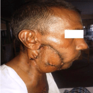
A 40 year old male presented with the chief complaint of gradually progressive, painless, irregular swelling over the right side of the face for the past 6 months. On examination it was a 10 x 8 x 8 cm large firm mass of the right parotid region with no evidence of intra oral lesion. There were no palpable neck glands.
On radiological evaluation, CT scan revealed a 98 x 113x 106 mm enhancing soft tissue lesion over the right parotid region. Lesion showed area of necrosis within. Lesion reaches up to the skin and involves the masseter muscle (Figure 1). Right submandibular gland is not seen separately from the lesion. Presence of few lymph nodes at level IA, II, III with the largest at IA measuring 20 x17 mm. No evidence of mandible erosion.

Figure 1:
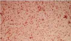
X-Ray chest, CT Thorax and USG (Abdomen + Pelvis) were normal. Core biopsy revealed – Desmin +ve, Vimentin +ve, Actin (nuclear stain) +ve, S-100 +ve, grade III Pleomorphic sarcoma (Figure 2).

Figure 2:
The patient eventually underwent wide-local excision with Supra Omohyoid Neck Dissection (SOHND) with distal mandibulectomy along with the reconstruction by Pectoralis Major Myo- Cutaneous and Delto-Pectoral flap. The post operative histology report revealed a 14 x 13 x 10 cm tumor, with infiltration of the overlying skin (Figure 3). All the surgical margins were >2cm away and free from the tumor; Total 14 lymph nodes were dissected out and all were free of tumor; mandible was not involved by tumor. Histological identified as a Pleomorphic sarcoma.

Figure 3:
Discussion
Head and neck MFH/PS has been reported to account for only 3-10% of all the MFH found in various parts of the body. In a series of 1215 STS, 128 (10.5%) were classified as MFH/PS of which only 9 cases (7%) were reported in the head and neck region [7].
PS/MFH is classified into primary and secondary types. 70% of the PS/MFH is of primary type that occurs in younger patients and the secondary tumors, which are seen mostly in 6th and 7th decade of life. The secondary tumors seem to be more aggressive than the primary ones. They are secondary to underlying conditions like Paget Disease, Fibrous dysplasia or prior radiation [8].
MFH was first described as a new malignant tumor by O’Brien and Stoutl in early 1960s. The detailed histopahtological features of MFH were described by Kempson and Kyriakos [9].
In the head and neck region, MFH has been reported to involve the nasal cavity and paranasal sinuses most frequently, accounting for 30% of all the cases. Of the sinonasal tract, it occurs most frequently in the maxillary sinus, followed by the ethmoid sinus, nasal cavity, sphenoid sinus and the frontal sinus. Other reported sites in the head and neck include the craniofacial bones (15-25%), larynx (10-15%), soft tissue of the neck (10-15%), major salivary gland (5-15%), and oral cavity (5-15%). Pharynx, ear and eyelid [10-13].
According to review done by Kanazawa et.al, among the MFH cases, 65% were male and 35% were female. The age distribution at diagnosis varied from 1.5 to 69 years, but the most common in the latter half of life, with a mean age of 41 [14]. Other studies reported a peak incidence in the 5th -7th decade of life [15].
With respect to the differential diagnosis of MFH from other malignant tumors in the head and neck- squamous cell carcinoma, malignant lymphoma, malignant giant cell tumors, fibro sarcomas and osteolytic osteosarcomas must be considered [16].
The treatment of choice for MFH is extended surgical resection. Radical surgical resection and clear surgical margins is very important in improving the survival [16,17].
Because of regional lymph node involvement in 10-18% of cases consideration should be given to elective neck dissection [17,18]. The decision for radiotherapy depends upon size, site, histopahtological grade and the achievable surgical clearance [2].
However, Kearney et.al. Conducted radiotherapy on 45 patients with a measurable tumor and reported that six of them showed partial response (13%). Many researchers have reported that radiotherapy for MFH was less effective [15].
Yamaguchi et al. reported that local recurrence was less common in patients with adjuvant therapy (RT with or without CT) compared to patients with surgery alone [2].
MFH metastasize to the lungs by heamatogenous routes. Sato et al. has reported distant metastasis in the lung, bone, skin and regional lymph nodes [9]. According to Rapidis et al. MFH has a 16-52% chance of developing local recurrence [18].
References
- Anavi Y, Herman GE. "Malignant fibrous Histiocytoma of the mandible." Oral Surg Oral Med Oral Pathol. 1989; 68: 436-443.
- Yamaguchi S, Nagasawa H, Suzuki T, Fujii E, Iwaki H, Takagi M, et al. "Sarcomas of the oral and maxillofacial region: a review of 32 cases in 25 years." Clin Oral Investig. 2004; 8: 52-55.
- Ohsawa, M, Tomita Y. "Angiogenesis in malignant fibrous Histiocytoma." Oncology. 1995; 52: 51-54.
- Pandey M, Thomas G, Mathew A, Abraham EK, Somanathan T, Ramadas K, et al. "Sarcoma of the oral and maxillofacial soft tissue in adults." Euro J Surg Oncol. 2000; 26: 145-148.
- Vargas A, Echevarria E. "Primary malignant fibrous Histiocytoma of the mandible: surgical approach and report of a case." J Oral Maxillofacial Surg. 1987; 45: 634-638.
- Bras J, JBatsakis GJ. "Malignant fibrous Histiocytoma of the oral soft tissues." Oral SurgOral Med Oral Pathol. 1987; 64: 57-67.
- Solomon MP, SuttonAL. "Malignant fibrous Histiocytoma of the soft tissues of the mandible." Oral Surg Oral Med Oral Pathol. 1973; 35: 653-660.
- Colmenero C, Garcia Rodejas E. "Osteogenic sarcoma of the jaws: malignant fibrous Histiocytoma subtype." J Oral Maxillofacial Surg. 1990; 48: 1323-1328.
- Sato T, Kawabata Y, Morita Y, Noikura T, Mukai H, Kawashima K, et al. "Radiographic evaluation of malignant fibrous Histiocytoma affecting maxillary alveolar bone: a report of 2 cases." Oral Surg Oral Med Oral Pathol Oral Radiol Endod. 2001; 92: 116-123.
- Barnes L, Kanbour A. Malignant fibrous Histiocytoma of the head and neck: a report of 12 cases. Arch Otolaryngology Head Neck Surg. 1988; 114: 1149-1156.
- Rodrigo JP, Fernandez JA, Suarez C, Gómez J, Herrero, Agustín, et al. Malignant fibrous Histiocytoma of the nasal cavity and paranasal sinuses. Am J Rhinol. 2000; 14: 427-431.
- Nagano H, Deguchi K, Kurono Y. Malignant fibrous Histiocy b toma of the uccal: a case report. Auris Nasus Larynx. 2008; 35: 165-169.
- Munk PL, Sallomi DF, Janzen DL, Lee MJ, Connell DG, O'Connell JX, et al. Malignant fibrous Histiocytoma of soft tissue imaging with emphasis on MRI. J Comput Assist Tomogram. 1998; 22: 819-826.
- Kanazawa H, Watanabe T. "Primary malignant fibrous Histiocytoma of the mandible: review of literature and report of a case." J Oral Maxillofacial Surg. 2003; 61: 1224-1227.
- Kearney MM, Soule EH. "Malignant fibrous Histiocytoma: a retrospective study of 167 cases." Cancer. 1980; 45: 167-178.
- Zohreh D, Nooshin M, Farnaz F. “Malignant Fibrous Histiocytoma of Mandible: A Review of Literatures and Report a Case ”Australian Journal of Basics and applied Sciences. 2011; 5: 936-942.
- Jamal, B, Tuluc TM. "A radiolucent lesion in the posterior mandible." J Oral Maxillofacial Surg. 2010; 68: 1371-1376.
- Rapidis AD, Andressakis DD. "Malignant fibrous Histiocytoma of the tongue: review of the literature and report of a case." J Oral Maxillofacial Surg. 2005; 63: 546-550.