Case Report
Austin Head Neck Oncol. 2018; 2(1): 1005.
A Rare Case of En Plaque Meningioma Causing an Effusive Chronic Otitis Media
Amadei EM* and Cola C
Department of Otorhinolaryngology and Audiology, Infermi Hospital, Rimini, Italy
*Corresponding author: Enrico Maria Amadei, Department of Otorhinolaryngology and Audiology, Infermi Hospital, Rimini, Italy
Received: August 31, 2018; Accepted: September 11, 2018; Published: September 18, 2018
Abstract
In daily practice, every otolaryngologist sees dozens of cases of chronic secretive otitis media. Suspicion must arise when the otitis occurs unilaterally. A rhinopharyngeal pathology will therefore be excluded. An en plaque meningioma can cause an unilateral serous otitis. This diagnosis is exceptional, but it is possible and rarely reported in Literature.
Keywords: En plaque meningioma; chronic monolateral otitis media
Case Description
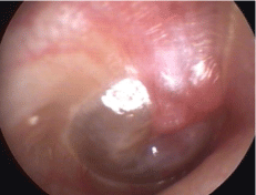
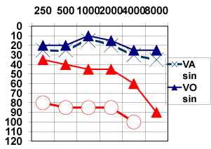
In this case we have a 64-year-old woman complaining of a right hearing loss for many years. At otomicroscopy we found a right effusive otitis media (Figure 1). With tonal audiometry we found a severe mixed right hearing loss and a left-handed normoacusia (Figure 2). The right tympanogram was flat (type B).

    Figure 1:  Otomicroscopy: Right tympanic membrane, with epitimpanic
bulging and middle chronic effusive otitis.

    Figure 2:  Tonal Audiometry: A severe mixed right hearing loss and a lefthanded
normoacusia.
We performed a negative rhinofibroscopy. So after three months of observation and local theraphy with nasal corticosteroid and tubal gymnastics with Otovent, we placed a transtimpanic tube, with good hearing recovery. After six months the tube was expelled spontaneously. One month later secretive otitis has returned.
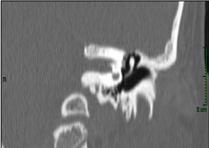
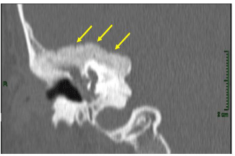
So we performed first a CT scan, finding a hyperostotic reshaped surface of the middle cranial fossa (Figure 3-4). Finally we executed a MRI, finding an en plaque meningioma with involvement of the bone surrounding the Eustachian tube (Figure 5).

Figure 3: CT left ear: Normal left middle ear.

    Figure 4:  CT right ear: Hyperostotic reshaped right temporal bone (see
arrows).

    Figure 5:  MRI: En plaque meningioma of right middle cranial fossa extending
to the middle ear with intraosseous expansive lesion.
Conclusions
An en plaque meningioma should be considered in the differential diagnosis of a chronic monolateral serous otitis media. In this case we would have reached a correct diagnosis in a short time by performing a MRI many years ago. So in case of monolateral chronic otitis, with normal nasopharynx, should we use imaging techniques routinely and without delay?
References
- Hyperostosis in meningiomas: MR findings in patients with recurrent meningioma of the sphenoid wings. Terstegge K, Scorner W, Henkes H, Heye N, Hosten N, Lanksch WR. Am J Neuroradiol. 1994; 15: 555-560.
- Sphenoid Wing en plaque meningiomas: Surgical results and recurrence rates. Nuno M. Simas, Joao Paulo Farias. Surg Neurol Int. 2013; 4: 86.
- Surgical management of meningioma en plaque of the sphenoid ridge. OrlandoDe Jesus, Mariia Mtolodo. Surgical Neurology, 2001; 55: 265-269.
- Serous Otitis Media Revealing Temporal En Plaque Meningioma. Denis Ayache, Franco Trabalzini, Philippe Bordure, Benoit Gratacap, Vincent Darrouzet et al. Otology & Neurotology. 2006; 27 : 992-998.
- Otitis Media with Effusion revealing Underlying Meningioma. Diane Evrard, Mary Daval, Denis Ayache. J Int Adv Otol. 2018; 14 : 155-156.
- Primary meningioma of the middle ear. Joseph G. Manjaly, Gillian MT Watson and Mary Jones. JRSM Short Rep. 2011; 2: 92.
- Temporal bone meningioma involving the middle ear: A case report. F. Ricciardiello, L. Fattore, M.E. Liguori, F. Oliva, A. Luce et al. Oncol Lett. 2015; 10: 2249-2252.
- Surgery of the skull base: an interdisciplinary approach. Madjid Samii, Wolfgang Draf. Springer book 2012.