
Case Report
Austin Head Neck Oncol. 2018; 2(1): 1008.
How to Manage a Buccal Space Mass – A Case Series
Franzen A, Glitzki S and Coordes A*
Department of Otorhinolaryngology, Head and Neck Surgery, Brandenburg Medical School, Campus Ruppiner Kliniken, Neuruppin, Germany
*Corresponding author: Coordes Annekatrin, Department of Otorhinolaryngology, Head and Neck Surgery, Charité Universitätsmedizin Berlin, Germany
Received: September 12, 2018; Accepted: October 04, 2018; Published: October 11, 2018
Abstract
Introduction: Patients presenting with masses in the cheek are common for head and neck specialists and present a diagnostic challenge against the backdrop of a wide variety of etiologies. Based on a case series the specific problems of differential diagnosis and management are discussed.
Case series: Six patients of our series presenting with a buccal mass suffered from a pleomorphic adenoma of an accessory parotid gland, an epidermoid cyst, a carcinoma of the Stensen`s duct, a carcinoma from the maxillary sinus, a secondary metastasis from oropharyngeal cancer and a distant metastasis of pulmonary cancer.
Discussion: Our case series underlines the vast origins of buccal masses. Important hints for malignancy are rapid and painful tumor development and a medical history of malignant disease. Clinical examination, sonography and CT/MRI scans are performed for diagnostic evaluation. Histologic examination is required if the proper diagnosis cannot be achieved and the tumor growth is not in spontaneous remission. The surgical management may be challenging depending on the location and tumor size.
Keywords: Cheek; Differential Diagnosis; Accessory Parotid Gland; Salivary Duct; Neoplasm; Metastasis
Introduction
Tumoral masses in the area of the cheek are at the same time, an aspect of the daily routine of the Ear Nose and Throat (ENT) specialist and a diagnostic and therapeutic challenge. The buccal region is bounded cranially by the zygomatic bone, caudally by the mandible, dorsally by the masseter muscle and towards the bottom by the buccinators muscle. The major part of the buccal volume is formed by the Bichat’s fat pad that is separated from the buccal fat by a connective tissue capsule. Regional lymph nodes of the buccal and parotid group, as well as 21% of the cases an accessory parotid gland can be identified. Usually, the gland measuring between 0.5 and 2.5 cm in diameter is located about 6mm from the anterior border of the parotid gland. The parotid duct (Stensen), the motoric branches of the facial nerve crossing the region and terminal branches of the second trigeminal nerve run through this region beside arterial and venous vessels (e.g. the facial artery and vein).
Knowledge about the incidence and clinical importance is based on single case reports or small series. Presenting our case series, we are in particular interested to underline the wide spectrum of tumor entities in the cheek, with regards to the aspects of diagnostic workup and therapy.
Case Reports
Patient 1
A 40-year old female patient presented with an indolent tumor mass of the left cheek that had been present for two years and that had slowly grown during this time.
Bimanual palpation revealed a rough, movable mass located in the buccal soft tissue. Sonography showed a tumor measuring about 2.5cm in diameter that was anechoic and polygonally limited – an organ relation, especially to the parotid gland, cannot be described. Tumor extirpation was performed from a preauricular skin incision with formation of a buccal skin flap displaying the parotid capsule. The findings were situated about 1.5cm in front of the anterior circumference of the parotid.
Histologic and immunohistochemical examination revealed the diagnosis of a pleomorphic adenoma– based on the location, the accessory parotid gland was considered as originating organ.
Patient 2
An 88-year-old female patient initially presented with a tumor of the left cheek that was rapidly growing and moderately painful. The subcutaneous tumor could easily be moved and had a diameter of about 1.5cm. The patient first declined to further diagnostic and therapeutic measures. Already three weeks later the tumor was meanwhile fixed and had nearly achieved the double size (Figure 1). CT scan and intraoperative aspect revealed a tumor infiltrating the masticatory muscles and subcutis. The diagnosis after intraoperative frozen section and definitive histology was a poorly differentiated squamous cell carcinoma. The tumor was located about 2cm in front of the anterior border of the gland originating from a dilated Stensen`s duct. According to the age of the patient no adjuvant therapy was performed. The lady died two years later without signs of local or systemic recurrence of the tumor.
Patient 3
A 49-year-old male patient presented with a history of a mass of the right lateral cheek that was inconstant in size and sometimes painful. Clinical examination showed a prominent mass of the lateral upper cheek. Palpation suggested close contact to the skin. Ultrasound examination revealed a hypoechoic oval lesion of less than 2 cm with well-defined margins to the underlying soft tissue. The tumor including a small area of skin was removed. The clinical diagnosis of an epidermoid cyst was confirmed by histological examination.
Patient 4
A 72-year old male presented with a one-month history of painful swelling of the left cheek. His medical history included tumor resection and adjuvant radiochemotherapy as treatment for squamous cell carcinoma of a left oro-hypopharyngeal cancer 38 month previously. CT- scan showed an exophytic tumor of up to 3,5 cm`s in diameter with ill-defined margins to the underlying tissues (Figure 2). Further tumor manifestations and a local return of the previously diagnosed pharyngeal cancer were excluded by staging investigations. Tumor resection was performed from a preauricular - inferior eyelid skin incision. Histopathological examination showed the R1-resection of a late metastasis of the pharynx cancer. The patient received adjuvant radiotherapy in combination with immunotherapy. He died 3 months after diagnosis of the metastasis following a brainstem insult.
Patient 5
A 54-year old male presented with a one-month history of rapidly growing, painful lump in the right buccal region. He reported nasal secretion exclusively from the right side for three months. CT-scan revealed an inhomogeneous tumor of the right sinus maxillaries infiltrating the button of the orbital and the cheek and enlarged cervical lymph nodes of the same side (Figure 3). Following nasal biopsy, histopathological examination suggested squamous cell carcinoma. The patient received radical open surgery of the primary, neck dissection and adjuvant radio chemotherapy. He has been in remission for eleven years to date.
Patient 6
10 month after initial diagnosis and first line chemotherapy of an adenocarcinoma of the left lung a 69-year old woman was presented with bilateral buccal swellings and a progressive trismus. The bimanual palpation revealed a rough, not movable and extremely painful mass located in the buccal soft tissue (Figure 4). Inspection of the mouth showed a small mucosal lesion of the left cranial cheek. Biopsy revealed a badly differentiated adenocarcinoma that was interpreted as distant metastasis of the formerly diagnosed lung cancer. CT-scan showed the extent of tumor in the buccal space and further brain metastases. The patient received radiotherapy of the buccal space and the brain to improve on local symptom control.
Discussion
Patients presenting with masses in the buccal region are common for head and neck specialists and present a diagnostic challenge against the backdrop of a wide variety of etiologies. If the buccal tumor is not spontaneously regressing, histological diagnosis is always required. Based on a case series the specific problems of differential diagnosis and management are discussed.
The origins of buccal tumors are manifold, the literature at disposition includes in particular casuistics and small series. Considering clinical observations and including our own patient populations, most of the tumor developments are supposed to originate from (post-) infectious enlarged lymph nodes. On the other hand lymph node swellings are also reported on in the context of the manifestation of specific infections, malignant lymphomas and metastatic infiltrations. Concerning to our presentation (patient 4) the latter situation may be given when medical history includes head and neck cancer. As a consequence of therapy the normal lymphatic pathways are altered and metastasis may occur in untypical locations. Metastases from primary tumors below the clavicle (patient 6) are extremely rare and often an expression of disseminated metastasis. Infraclavicular primary tumors seem to be mostly located in the lung, breast and kidney and carry a poor prognosis [1].
From the local fatty connective tissue including the Bichat’s fat pad and the buccal muscles, in particular lipomas develop, more rarely myomas or fibromas, and very rarely also malignant variations of those entities [2]. Further rarely occurring neoplasms are neuromas.
Quite frequently patients present with tumors originating from the deeper layers of the skin (patient 3). In the majority of cases there is a history of equal formations in other locations. Clinical diagnosis should not be difficult.
Differential diagnosis of a mid-cheek mass includes pathologies arising from the accessory parotid gland. The gland may be enlarged because of a sialadenitis or a glandular tumor (patient 1). The described entities correspond to those of parotid tumors as far as this can be evaluated based on the small number of cases. The rate of malignant tumors seems to be higher than that of the parotid gland [3].
Tumor formations originating from Stensen duct (patient 2) can develop based on benign ductal neoplasms such as diverticula or non-neoplastic lesions, e.g. calculi or stenosis. In rare cases, nonmalignant or malignant neoplasia can develop [3]. In the presented case, the parotid duct carcinoma developed from the middle third of the duct, infiltration of the duct from the oral mucosa could be excluded.
Tumor formations of the buccal space may also be secondary to neoplasms of the underlying anatomical structures. Rare nonmalignant or malignant tumors [4] of the maxillary bone frequently present as a buccal mass. In rare cases buccal tumors are described following infiltration of advanced paranasal sinus carcinoma [5]. In patient 5 the cheek mass was the first clinical manifestation of an infiltrating carcinoma of the maxillary sinus.
Most patients report about a slowly growing, indolent mass of the buccal region that could be touched either with their tongue or felt from the outside. Some patients discover a facial asymmetry that they had not seen before. History including periods of pain with or without infiltration of the skin may be a hint to tumor infection (patient 3). If the patient reports about a continuously or rapidly growing pane in context with a buccal mass, then this will be a strong clue to malignant disease (patients 4,5 and 6). Further immanent indications of malignant tumor growth are paresis of the facial nerve, skin or mucosal infiltration, or the obstruction of Stensen duct [2,6]. Moreover our results reveal that the history of malignant diseases may be an important clue to the origin of a tumor in the cheek.
Depending on the size of the buccal tumor and the patient’s constitution the inspection from the outside and inside happen to be inconspicuous. A better impression of the tumor location, size, consistence, and mobility is often found by bimanual palpation. Further information about tumor limits and probably also the tissue of origin (echogenicity) is provided by ultrasound. Additional findings are revealed by computed tomography and magnetic resonance imaging; especially for the differentiation between a benign and a malignant mass. The value of those procedures is limited [7]. The significance of Fine Needle Aspiration Cytology (FNAC) is judged differently because the risk of damaging Stensen duct and branches or the facial nerve has to be considered [6].
Finally each tumor of the cheek has to be carefully worked up. Lymph nodes that can be identified as clearly (post-) infectious or non-malignant masses like lipomas without aesthetic relevance may not be routinely removed. In these cases follow-up by clinical and ultra-sound investigations should be sufficient [2]. Any buccal mass of unclear origin, a tumor that is increasing in size should be respected. This is especially true for cases that show clinical indications for malignant tumor growth (see above).
Regarding the choice of the surgical approach, dignity, location and size of the tumor, important anatomic structures (facial nerve, Stensen duct) and the postoperative cosmetic result should be considered. Based on those requirements, an intraoral approach seems to be appropriate for certain sub mucous tumors located medially to the buccinators muscle [6]. Up to now there is rare experience in endoscope-assisted transoral approach to the buccal space [8].
In the majority of the cases, an external approach will be chosen. If the tumor originates from structures of the skin (patient 3), a skin incision above the tumor will be necessary. A preauricular skin incision provides adequate access to superficially located tumors from the accessory parotid gland and Stensen`s duct including the possibility of adequate exposure of the buccal branches of the facial nerve (patients 1 and 2). Larger tumors may demand an extended preauricular-suborbital incision (patient 4) and tumors of the lower buccal space a preauricular-submandibular approach. In patient 5 a paranasal access was used to resect a paranasal sinus carcinoma without identification of the facial nerve. In this case exposure of facial nerve was not necessary [3,6]. Exposure of branches of the facial nerve may be realized by lateral parotidectomy. A modification was introduced by Jung et al. [9] who bisected the superficial gland along the course of the buccal branches of the facial nerve. After removal of the tumor the gland was repositioned to obtain the facial contour. Xie et al. [10] presented an endoscopic approach via small preauricular incisions in selected cases of benign tumors of the accessory parotid gland.
Conclusion
Tumors of the buccal region present a diagnostic problem against the backdrop of a wide variety of etiologies. Clinical examinations and imaging procedures help to find the extension and location of the tumor, however, final diagnosis can only be made by excising the tumor and performing histologic examination. The surgical management may be challenging depending on location and size of the tumor and the complexity of buccal space anatomy.
Abbreviation
ENT: Ear Nose and Throat; FNAC: Fine Needle Aspiration Cytology.
Consent for Publication
The patients were rendered unrecognizable on the photos to protect their personal rights. Unfortunately, the patients shown on (Figure 1,2,4) died several years ago.
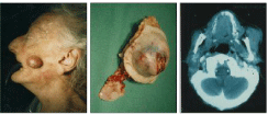
    Figure 1:  Stensen duct carcinoma. Preoperative picture (left picture), surgical specimen (middle picture) and CT scan of the tumor of the anterior border of the
gland (right picture).
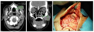
    Figure 2:  Metastasis of an oro-hypopharyngeal cancer. CT-scan showed an exophytic tumor of up to 3,5cm`s in diameter with ill-defined margins to the underlying
tissues of the left cheek (axial and frontal plane). Intraoperative situs (right picture). Tumor resection was performed from a preauricular - inferior eyelid skin incision.
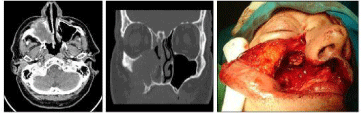
    Figure 3:  Paranasal sinus cancer. Axial and frontal plane of a CT-scan (left and middle picture). Intraoperative situs of the radical open surgery of the primary
(right picture).
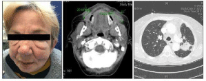
    Figure 4:  Metastases of a lung cancer. Picture of a patient with bilateral buccal swellings (left picture). CT-scan showing a tumor in the buccal space bilaterally
(middle picture). CT-scan of the primary tumor (right picture).
References
- Irani S. Metastasis to the oral soft tissues: A review of 412 cases. J Int Soc Prev Community Dent. 2016; 6: 393-401.
- De Wijn RS, van der Heijden EP, Kon M. On lipoma of the buccal fat pad: report of two cases and review of the literature. J Plast Reconstr Aesthet Surg. 2009; 62: 28-35.
- Newberry TR, Kaufmann CR, Miller FR. Review of accessory parotid gland tumors: pathologic incidence and surgical management. Am J Otolaryngol. 2014; 35: 48-52.
- Kammerer PW, Shabazfar N, Vorkhshori Makoie N, Moergel M, Al-Nawas B. Clinical, therapeutic and prognostic features of osteosarcoma of the jaws - experience of 36 cases. J Craniomaxillofac Surg. 2012; 40: 541-548.
- Perri F, Addeo R, Conson M, et al. Locally advanced paranasal sinus carcinoma: A study of 30 patients. Oncol Lett. 2017; 13: 1338-1342.
- Walvekar RR, Myers EN. Management of the mass in the buccal space. 2007; 17: 281-294.
- Kurabayashi T, Ida M, Tetsumura A, Ohbayashi N, Yasumoto M, Sasaki T. MR imaging of benign and malignant lesions in the buccal space. Dentomaxillofac Radiol. 2002; 31: 344-349.
- Woo SH. Endoscope-assisted trans oral accessory parotid mass excision. Head Neck. 2016; 38: E7-E12.
- Jung YH, Hah JH, Sung MW, Kim KH. Parotidotomy approach for a midcheek mass: a new surgical strategy. Laryngoscope. 2010; 120: 495-499.
- Xie L, Zhang D, Lu MM, Gao BM. Minimally invasive endoscopic-assisted resection of benign tumors in the accessory parotid gland: 5 case studies. Head Neck. 2012; 34: 1194-1197.