Case Report
Austin Head Neck Oncol. 2022; 5(1): 1013.
Multifocal Non-Hodgkin’s Lymphoma of the Testis and the Waldeyer’s Ring: A Rare Presentation
Zaffarullah MW and Eliyapura HN*
Department of ENT and Head and Neck Surgeon, District General Hospital Matale, Sri Lanka
*Corresponding author: Eliyapura HN, Department of ENT and Head and Neck Surgery, District General Hospital Matale, 166, MC Road, Matale, 21300, Sri Lanka
Received: January 04, 2022; Accepted: January 31, 2022; Published: February 07, 2022
Abstract
Waldeyer’s ring non-Hodgkin’s lymphoma (WR-NHL) is uncommon and of that, base of the tongue lymphoma is rare. Similarly testicular NHL too is rare.
We report an unusual case of multifocal base of the tongue and testicular B-cell NHL found simultaneously, which was confirmed by biopsy and immunohistochemistry. This article serves to illustrate the existence of multifocal primaries of NHL in extremely rare extra-nodal sites. Familiarity with such association will help detect multifocal NHL and staging, which has a direct impact on the management.
Keywords: Non-Hodgkin’s lymphoma; Waldeyer’s Ring; Malignant lymphoma; Head and neck; Extranodal; Testicular neoplasm
Abbreviations
NHL: Non-Hodgkin’s lymphoma; WR-NHL: Waldeyer’s Ring Non-Hodgkin’s Lymphoma; PTL: Primary Testicular Lymphoma; FDG-PET: Fluorodeoxyglucose-Positron Emission Tomography
Introduction
About a third of NHL cases occur at extra-nodal sites, but involvement of multiple non-contiguous extra-nodal sites at presentation without lymph node involvement is rare [1]. Moreover, primary testicular lymphoma (PTL) in addition to WR-NHL at presentation has not been reported in the English literature.
Case Presentation
A 62-year-old male presented with dysphagia and hot potato voice for 3 months’ duration. He was admitted for investigation and during his stay complained of a left testicular swelling, which was referred to the Surgical team. The patient has a past medical history significant for chronic obstructive airway disease. He is a regular smoker. The rest of his medical and surgical history was unremarkable.
He denies any weight loss, night sweats or fever. Fibreoptic nasolaryngoscopy examination revealed a large mass in the base of his tongue which appeared quite vascular. Testicular ultrasound scan and Doppler studies were suggestive of a malignant pathology. Tumour size was 55mm. The epididymis and spermatic cord were not involved by the tumour.
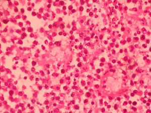
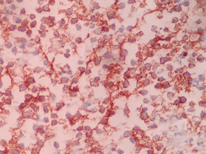
Combined direct laryngoscopy and biopsy and left side orchidectomy was done under general anaesthesia by the otolaryngologist and general surgeon respectively. Biopsy sample obtained through direct laryngoscopy of the base of tongue and testicular histology showed features of a high-grade NHL (B-cell type). Tumour cells were positive for CD 20 and negative for CD3, PLAP and AE1/AE3 (Figure 1 and 2).

Figure 1: H&E stain.

Figure 2: CD20.
Contrast-enhanced CT scan showed large soft tissue mass with central necrosis anterior to the aorta extending towards the left kidney encasing the superior mesenteric vessels and renal vessels. There was another enhancing lesion seen at the base of the tongue.
Patient was referred to the multidisciplinary team meeting for treatment planning.
Discussion
This patient’s case is unusual because he presented with multifocal NHL in his left testicle and in the Waldeyer’s ring region. While about 20-30% of NHL manifests at extra-nodal sites, it is even rarer to see the involvement of multiple in contiguous extra-nodal sites without any lymph node involvement at initial presentation [1,2].
According to our literature search, the last reported study on primary NHL in testis with secondary involvement of the Waldeyer’s ring or its adjacent structures was in 1980. This study observed a secondary lymphomatous involvement that developed in 5 of its 24 patients with extra-nodal NHL presenting in the testicle (22.5%). 4 of them were clinically staged as Stage I/II and 1 patient was in Stage III/ IV [3]. There was another case of primary WR-NHL who developed secondary involvement of the testis during the follow up period [4].
Primary testicular lymphoma (PTL) is an uncommon and aggressive form of extra-nodal NHL that accounts for less than 5% of testicular malignancies and 1% to 2% of NHL cases. Usually diagnosed at a median age of 66 to 68 years, PTL is the most common testicular malignancy in males older than 60 and the most common bilateral testicular malignancy which is seen in around 35% of the patients [3,5-7]. PTL demonstrates an affinity for extra-nodal sites such as the central nervous system and contralateral testis. Other extra-nodal sites include skin and pleura [5,6].
The head and neck region are also a common site for extra-nodal NHL, following the gastrointestinal tract, which is the most common site. Majority of head and neck NHL cases in our literature search were B-cell type, which coincides with our patient’s histology reports [6,7]. According to a review done in 2003 [8], subtypes of lymphoma tend to localise to specific locations within the head and neck region. The most common sites of NHL tend to be the Waldeyer’s ring (WRNHL), as observed with our patient [8-10].
Most patients with NHL of the head and neck usually present with symptoms attributable to a local mass rather than any “B” symptoms [10]. Our patient’s presenting complaint was dysphagia owing to the base of the tongue mass. His testicular mass was a secondary concern to the patient probably because it was painless and posed no acute problems to him.
PTL is an exceptional presentation of NHL. It has a poorer prognosis compared to gastrointestinal lymphoma and a high tendency to involve the central nervous system [5,7]. Younger age and earlier stage at diagnosis are independent prognostic factors that influence the overall and disease-free survival [3,4,6,7].
It has been observed in patients with WR-NHL, relapse could occur in locoregional sites, regional sites and distal sites notably abdominal lymphadenopathy, mediastinum, testis, central nervous system, and GI tract. If upper GI endoscopy was performed routinely, there’s a likelihood of diagnosing more cases [2,4,8-11].
Once diagnosed, it is important to map the extent of the disease. The initial workup includes routine haematological and biochemical blood work, CT scan of the neck, chest, abdomen and pelvis, bone marrow biopsy and ultrasound scan of the testis [4]. A lumbar puncture should be included due to the high tropism to the central nervous system [12].
The positive findings detected on staging will be followed in response evaluation. FDG-PET stands as part of response criteria in which contrast-enhanced CT scan can be complementary [13-15].
Conclusion
Diagnosis of extra-nodal diffuse large B-cell lymphoma at one site should alert the clinician to look for multifocal primaries at other sites. Clinicians irrespective of their specialty must be aware of the synchronous mode of presentation of such lesions. Immunohistochemical techniques play a vital role in the diagnosis because clinical characteristics may be misleading.
References
- Singh D, Sharma A, Mohanti BK, Thulkar S, Bahadur S, Sharma SC, et al. Multiple extranodal sites at presentation in non-Hodgkin’s lymphoma. Am J Hematol. 2003; 74: 75-77.
- Storck K, Brandstetter M, Keller U, Knopf A. Clinical presentation and characteristics of lymphoma in the head and neck region. Head Face Med. 2019; 15: 4-11.
- Duncan PR, Checa F, Gowing NF, McElwain TJ, Peckham MJ. Extranodal non-Hodgkin’s lymphoma presenting in the testicle: a clinical and pathologic study of 24 cases. Cancer. 1980; 45: 1578-1584.
- Ezzat AA, Ibrahim EM, El Weshi AN, Khafaga YM, AlJurf M, Martin JM, et al. Localized non-Hodgkin’s lymphoma of Waldeyer’s ring: Clinical features, management, and prognosis of 130 adult patients. Head Neck. 2001; 23: 547-558.
- Fonseca R, Habermann TM, Colgan JP, O’Neill BP, White WL, Witzig TE, et al. Testicular lymphoma is associated with a high incidence of extranodal recurrence. Cancer. 2000; 88: 154-161.
- Lantz AG, Power N, Hutton B, Gupta R. Malignant lymphoma of the testis: A study of 12 cases. J Can Urol Assoc. 2009; 3: 393-398.
- Cheah CY, Wirth A, Seymour JF. Primary testicular lymphoma. Blood. 2014; 123: 486-493.
- Weber AL, Rahemtullah A, Ferry JA. Hodgkin and non-Hodgkin lymphoma of the head and neck: Clinical, pathologic, and imaging evaluation. Neuroimaging Clin N Am. 2003; 13: 371-392.
- Wan Abdul Rahman WF, Mohamad I. The insights of head and neck nonhodgkin lymphoma. Medeni Med J. 2020; 35: 179-180.
- Shima N, Kobashi Y, Tsutsui K, Ogawa K, Maetani S, Nakashima Y, et al. Extranodal non-Hodgkin’s lymphoma of the head and neck. A clinicopathologic study in the Kyoto-Nara area of Japan. Cancer. 1990; 66: 1190-1197.
- Bajetta E, Buzzoni R, Rilke F, Valagussa P, Rovej R, Viviani S, et al. Non- Hodgkin’s lymphomas of Waldeyer’s ring. Tumori. 1983; 69: 129-136.
- Singh Boscán CS, Jurado DS, Unigarro AF, Corredor Silva CA. Testicular Lymphoma: Case Series. Findings in Ultrasound Mode B, Color and Spectral Doppler. Rev Colomb Radiol. 2020; 31: 5403-5407.
- Hokland P, Shah M, David K, Evens A, Auer R, Ledieu R, et al. How I treat advanced Hodgkin lymphoma-a global view. Br J Haematol. 2020; 190: 837- 850.
- Zanoni L, Cerci JJ, Fanti S. Use of PET/CT to evaluate response to therapy in lymphoma. Q J Nucl Med Mol Imaging. 2011; 55: 633-647.
- El-Galaly TC, Villa D, Gormsen LC, Baech J, Lo A, Cheah CY. FDG-PET/ CT in the management of lymphomas: current status and future directions. J Intern Med. 2018; 284: 358-376.