Clinical Image
Ann Hematol Oncol. 2014;1(2): 1010.
Erythroderma and Onychodystrophy in Sezary Syndrome
Manuel Neves1*, Isabel Pereira1, Maria Joao Costa1 and Jose Alves do Carmo1
1Serviço de Hematologia e Transplantaç&aTilde;o de Medula, Hospital Santa Maria, Centro Hospitalar Lisboa Norte
*Corresponding author: Manuel Neves, Serviço de Hematologia e Transplantaç&aTilde;o de Medula, Hospital Santa Maria, Centro Hospitalar Lisboa Norte, Avenida Professor Egas Moniz, 1649-035 Lisboa
Received: November 18, 2014; Accepted: November 21, 2014; Published: November 24, 2014
Clinical Image
Sezary syndrome is a rare T-cell non Hodgkin lymphoma characterized by the presence of lymphadenopathies and cutaneous disease with a dismal prognosis. Our patient is a 70 year-old female that presented with a generalized erythroderma and pruritus. CT scan showed mediastinic and abdominal adenopathies, as well as splenomegaly. Blood tests had Sezary cell count of 1200 cells/mm3 and elevated LDH. Cut aneous biopsy and bone marrow examintaion confirmed the diagnosis.
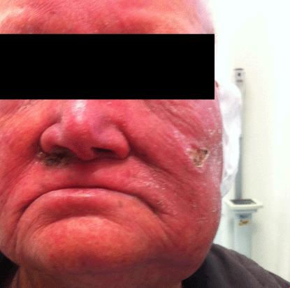
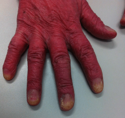
The patient started CHOP (cyclophosphamide, doxorubicin, vincristine and prednisolone) for 6 cycles with a partial response (reduction> 50% of adenopathies and a significative improvement of the skinlesions). However, lessthan 6 months after chemotherapy, the patient relapsed, with a significant worsening of the cut aneouslesions, that were predominantly in the face, including an ulcerative lesion (Panel A) and a very typical onychodystrophy (Panel B) and is now going to start treatment with alemtuzumab, an humanized monoclonal anti body anti-CD52.

Panel A: Facial erythroderma, with an ulceretive cutaneous lession.
